
Four weeks after a total vaginal hysterectomy for cervical dysplasia, a 57-year-old woman comes for a follow-up visit. A pathology report confirms the presence of cervical intraepithelial neoplasia (CIN) 3 with all surgical margins clear of disease. Which of the following is the most appropriate recommendation for future screening for recurrent squamous cell intraepithelial neoplasia in this patient
CIN3 hysterectomy vaginal cuff screening follow-up
post hysterectomy CIN3 clear margins vaginal cuff cytology screening guidelines ASCCP 2023
Post-Hysterectomy Screening After CIN 3 with Clear Margins
Most Appropriate Recommendation
- First, complete 3 consecutive annual HPV-based tests (since hysterectomy was performed as treatment for histologic HSIL/CIN 3).
- Then enter long-term surveillance: HPV-based testing (vaginal vault cytology +/- HPV co-test) every 3 years for 25 years total, regardless of whether the patient has since had a hysterectomy.
Why Surveillance Continues Even with Clear Margins
- Women treated for CIN 3 (histologic HSIL) remain at elevated risk for vaginal intraepithelial neoplasia (VAIN) - particularly VAIN 3 - and vaginal squamous cell carcinoma, because the oncogenic HPV field effect persists throughout the lower genital tract.
- The cervix has been removed, but the vaginal mucosa retains the same HPV exposure and susceptibility. The vaginal cuff/vault is sampled instead.
- Clear surgical margins reduce but do not eliminate future risk - residual HPV infection in adjacent vaginal epithelium can drive new dysplastic lesions.
- Per Berek & Novak's Gynecology: "Women with persistent abnormal cytology without evident cervical pathology and those with abnormal cytology after treatment of CIN should be examined carefully for vaginal intraepithelial neoplasia (VAIN)."
What is NOT Appropriate
| Incorrect Option | Reason |
|---|---|
| No further screening | Wrong - CIN 3 history mandates continued surveillance |
| Routine screening cessation at age 65 | The standard age-based exit rule does NOT apply to patients with a history of CIN 2+ - they require the full 25-year surveillance period |
| Annual Pap smear indefinitely without a defined endpoint | Close but outdated; modern guidelines favor HPV-based testing and a defined 25-year window |
| Colposcopy at fixed intervals | Colposcopy is reserved for abnormal test results, not routine surveillance |
Summary
Vaginal vault HPV-based cytology: 3 annual tests, then every 3 years - for a total surveillance period of 25 years from the time of treatment.
A 47-year-old woman comes for a routine health maintenance examination. She smoked one pack of cigarettes daily for 30 years, but she quit 1 year ago. She has had one lifetime sexual partner and has not been sexually active for 3 years. Pap smear and mammography showed normal findings 8 months ago. Her mother died of breast cancer at the age of 60 years. Physical and pelvic examinations show no abnormalities. Which of the following is the most appropriate screening test for this patient? 0 A) Pap smear Q B) X-ray of the chest Q C) Mammography 0 D) Colonoscopy 0 E) No screening indicated
Answer: D) Colonoscopy
Analysis of Each Option
- Goldman-Cecil Medicine: "for sporadic colorectal cancer, age is the major risk factor; for this reason, most patients should begin screening at age 45 years"
- Fischer's Mastery of Surgery: "recent changes in guidelines recommending a first screening colonoscopy at age 45"
- Goldman-Cecil (Women's Health chapter): "Screening is recommended beginning at age 45 years and continuing to age 75 years for both women and men"
Summary Table
| Test | Due? | Reason |
|---|---|---|
| Pap smear | No | Done 8 months ago; next due in ~2 years |
| Chest X-ray | Never | Wrong modality; LDCT is correct for lung screening |
| Mammography | No | Done 8 months ago; not yet due |
| Colonoscopy | Yes | Age 47, no prior colonoscopy mentioned; screening starts at 45 |
| No screening | No | Screening is clearly indicated |
The answer is D - Colonoscopy. She is 47 years old, meets the age threshold (≥45), and has no documented prior colorectal screening, making this the most appropriate next screening test at this visit.
what do you see in urine cytology
urine cytology urothelial carcinoma malignant cells microscopy
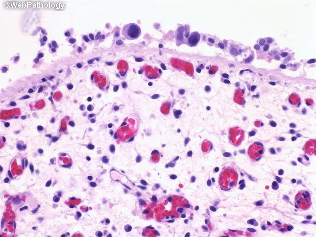
This bright-field light microscopy image depicts a bladder mucosal biopsy stained with Hematoxylin and Eosin (H&E) at high magnification (approximately 400x total). The surface urothelium is markedly reduced or denuded, revealing the underlying lamina propria with scattered cellular elements. Within the denuded field, clusters and individual cells exhibit high-grade cytologic features: enlarged, pleomorphic nuclei with hyperchromasia, irregular nuclear contours, and a high nuclear-to-cytoplasmic ratio. Occasional mitotic figures are present, supporting a neoplastic process. The background stroma contains mild to moderate inflammatory infiltrate, including lymphocytes and plasma cells, with scattered red blood cells in the interstitium. The combination of surface denudation and isolated malignant-appearing cells is characteristic of urothelial carcinoma in situ (CIS) with denudation, or denuding cystitis patterns described in bladder biopsies. In this context, urine cytology is frequently positive for malignant urothelial cells due to shedding from the abnormal surface. Accurate interpretation requires multiple histologic levels and correlation with clinical data, as reactive atypia can mimic CIS when surface epithelium is compromised. The image underscores the diagnostic challenge and the need for integration with adjunct tests (urine cytology, immunohistochemistry such as p53/Ki-67) and cystoscopic evaluation to confirm high-grade urothelial neoplasia and guide management (surveillance, intravesical therapy, or oncologic intervention).
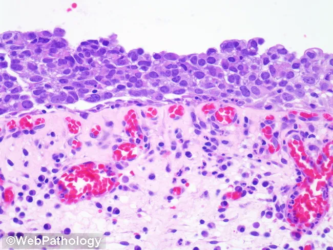
Imaging modality: Light microscopy of a hematoxylin and eosin stained bladder mucosa section. The lesion corresponds to urothelial carcinoma in situ (CIS) involving the urothelium of the bladder with preserved basement membrane but marked atypia. The urothelial layer shows widespread dysplastic, non-invasive malignant cells with marked nuclear pleomorphism and loss of polarity; nuclei are enlarged (approximately 4–6 times the size of surrounding small lymphocytes), hyperchromatic, with irregular nuclear contours. The architectural pattern is flat, without papillary fronds, consistent with CIS rather than invasive carcinoma. The lamina propria displays hypervascularized stroma with inflammatory infiltrate dominated by plasma cells and lymphocytes, indicating a reactive or inflammatory microenvironment accompanying CIS. Mitotic activity may be increased though not clearly described here. The differential includes dysplastic changes and benign reactive atypia; however, the combination of severe nuclear atypia, loss of polarity, full-thickness urothelial involvement, and dense inflammatory milieu supports a high-grade non-muscle-invasive urothelial carcinoma (CIS). Clinically this finding signals high risk of progression to muscle-invasive disease if untreated and warrants intravesical therapy and close surveillance. Correlation with cystoscopy, urine cytology, and patient risk factors (smoking, prior urothelial tumors) is essential. The image serves as a diagnostic teaching example for pathology education and oncology workflows.
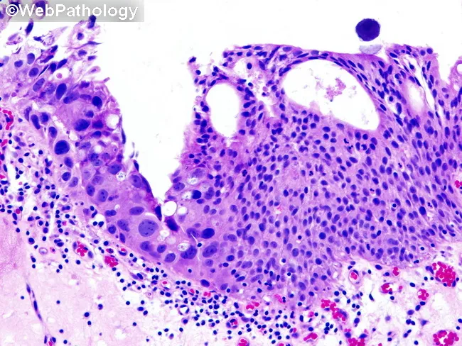
Imaging modality: Light microscopy of a hematoxylin and eosin stained urothelial biopsy from the urinary bladder. The section shows urothelium with carcinoma in situ (CIS) characterized by a flat, noninvasive lesion exhibiting extensive cytologic atypia throughout the thickness of the epithelium. The neoplastic cells display voluminous cytoplasm and markedly pleomorphic nuclei with prominent nuclear atypia and hyperchromasia, irregular contours, and frequent mitotic figures. This large cell, pleomorphic pattern is one of the most readily recognized forms of urothelial CIS. Adjacent right-hand mucosa demonstrates normal but hyperplastic urothelium, providing a contrast highlight of the malignant pattern. There is preservation of the basement membrane (as CIS is noninvasive) with crowding of atypical cells at the surface and disordered maturation. The stromal interface remains intact without invasion. Collectively, the image illustrates a high-grade urothelial carcinoma in situ occupying the epithelium, a critical diagnostic entity with significant risk of progression to invasive urothelial carcinoma if untreated. Clinically, CIS is associated with hematuria and is a marker of high-grade disease requiring immediate urological evaluation, cystoscopic surveillance, urine cytology, and potential intravesical therapy (BCG or chemotherapy). The pattern emphasizes the importance of recognizing large-cell pleomorphic CIS to prevent understaging and guide therapeutic decisions. This image serves educational and diagnostic planning use.
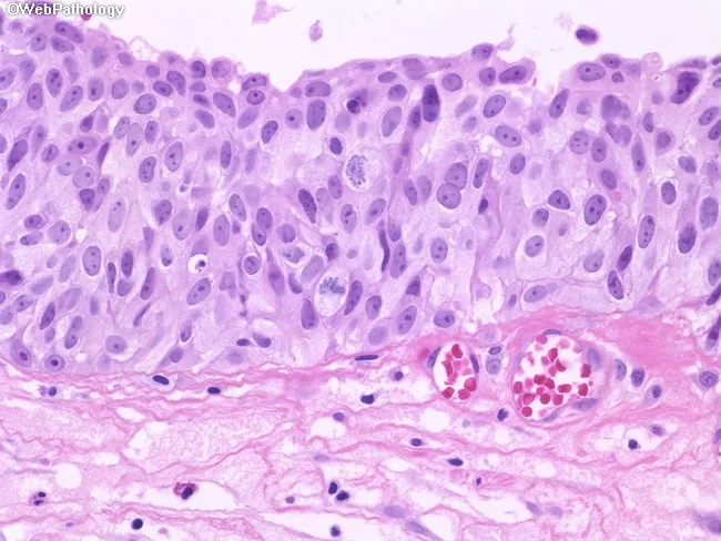
This histopathology image depicts a high-grade urothelial carcinoma in situ (CIS) of the urinary tract, captured by bright-field light microscopy on an H&E-stained section. The epithelium shows full-thickness neoplastic transformation of the urothelium with minimal pleomorphism but marked nuclear atypia. Neoplastic cells are relatively large, with abundant eosinophilic cytoplasm and enlarged, hyperchromatic nuclei; one or more prominent nucleoli are evident in several cells. Mitotic figures are readily visible, including atypical mitoses, reflecting high proliferative activity. A conspicuous finding is loss of normal basal polarity and disordered organization of cells relative to the basement membrane, consistent with CIS. The underlying lamina propria and stroma appear uninvaded at this level, though CIS is a known precursor to invasive urothelial carcinoma, underscoring oncologic significance. The sample demonstrates dense epithelial crowding with reduced cytoplasmic vacuolization and preserved cell borders in some areas, consistent with non-pleomorphic but malignant-appearing cells. Clinically, CIS signals a high-risk bladder cancer phenotype requiring definitive management and surveillance, including cystoscopy, urine cytology correlation, and possible intravesical therapy (BCG) or radical treatment if progression occurs. This image is valuable for educational teaching, pathology review, and research into urothelial neoplasia's in situ phase and its progression trajectory.
Urine Cytology: What You See
Normal / Background Elements
| Element | Appearance |
|---|---|
| Urothelial (transitional) cells | Umbrella cells on top (large, polygonal, abundant cytoplasm); intermediate and basal cells below |
| Squamous cells | From urethra or contamination |
| Renal tubular cells | Occasionally present |
| WBCs | Small numbers normal; increased = pyuria/infection |
| RBCs | May be present (hematuria) |
| Casts | Hyaline, granular - indicate renal origin |
| Bacteria/yeast | Indicate infection/contamination |
| Crystals | Uric acid, oxalate, etc. |
The Paris System - Cytologic Categories
1. Unsatisfactory
- Insufficient cells, poor preservation, or obscuring blood/inflammation
- Requires repeat specimen
2. Negative for High-Grade Urothelial Carcinoma (NHGUC)
- Normal urothelial cells
- Benign reactive changes (from stones, catheters, BCG therapy, infection)
- Cells with low N:C ratio, smooth nuclear contours, fine chromatin
- Decoy cells (polyomavirus-infected cells) - mimic malignancy, beware false-positive
3. Atypical Urothelial Cells (AUC)
- Some worrying features but not sufficient for HGUC
- Mildly increased N:C ratio, slight nuclear irregularity
- ~12-15% risk of underlying HGUC on follow-up
4. Suspicious for High-Grade Urothelial Carcinoma (SHGUC)
- Cells with marked atypia but quantitatively insufficient (fewer than the threshold for outright positive)
- High N:C ratio, irregular nuclear contours, hyperchromasia
5. High-Grade Urothelial Carcinoma (HGUC) - Positive
| Feature | Description |
|---|---|
| N:C ratio | Markedly increased (>0.7) |
| Nuclear hyperchromasia | Dark, deeply stained nuclei |
| Irregular nuclear contours | Angulated, jagged nuclear membranes |
| Coarse, clumped chromatin | "Salt and pepper" or smudged pattern |
| Prominent nucleoli | Especially in high-grade invasive tumors |
| Pleomorphism | Marked variation in cell and nuclear size |
| Necrosis/debris | Tumor diathesis in background |
| Mitotic figures | Abnormal mitoses may be seen |
6. Low-Grade Urothelial Neoplasm (LGUN)
- Papillary clusters of uniform cells with mild atypia
- Urine cytology has poor sensitivity for low-grade tumors (<30%) because they shed few cells and have subtle features
Key Diagnostic Points
- Cytology is highly sensitive and specific for HIGH-grade urothelial carcinoma and CIS (sensitivity 62-100%, specificity 94-100%)
- Poor sensitivity for low-grade papillary tumors (sensitivity ~31-40%) - these are better detected by cystoscopy
- CIS cells shed prolifically into urine
- Shows markedly atypical single cells with high N:C ratio, irregular chromatin, prominent nucleoli
- Urine cytology is often the FIRST clue to CIS (flat lesion, often missed on white-light cystoscopy)
| Mimic | Key differentiator |
|---|---|
| Decoy cells (BK polyomavirus) | Intranuclear viral inclusions, "owl-eye" nucleus; N:C ratio not markedly elevated |
| Reactive urothelial cells (stones, catheters) | Smooth nuclear contours, prominent nucleoli but fine chromatin |
| BCG therapy effect | Granulomatous inflammation + degenerative urothelial changes |
| Ileal conduit cells | Intestinal cells present if urinary diversion in place |
Practical Specimen Considerations
- Voided urine is preferred (avoid first morning void - cells degenerate overnight)
- Catheterized or barbotage specimens have more cells but may show instrument artifact
- Three consecutive specimens increase sensitivity
- Minimum adequate specimen: ≥10 well-preserved urothelial cells per Paris System criteria
Histologic Correlates (CIS Images)
low weight gain or weight loss in pregnancy
Low Weight Gain or Weight Loss in Pregnancy
What is Normal Gestational Weight Gain (GWG)?
| Pre-pregnancy BMI | Category | Recommended Total GWG (singleton) | Rate in 2nd & 3rd Trimester |
|---|---|---|---|
| <18.5 | Underweight | 28-40 lb (13-18 kg) | ~1 lb/week |
| 18.5-24.9 | Normal | 25-35 lb (11-16 kg) | ~1 lb/week |
| 25.0-29.9 | Overweight | 15-25 lb (7-11 kg) | ~0.6 lb/week |
| ≥30 | Obese | 11-20 lb (5-9 kg) | ~0.5 lb/week |
| First trimester (all) | - | 1.1-4.4 lb total | - |
Causes of Low / No Weight Gain or Weight Loss
1. Nausea and Vomiting of Pregnancy (NVP) - Most Common
- Affects up to 90% of pregnant women in the first trimester
- Usually mild and self-limiting
- Mild weight loss in the first trimester is common and generally acceptable
2. Hyperemesis Gravidarum (HG) - Most Severe
- Defined as severe NVP causing >5% weight loss from pre-pregnancy weight, with dehydration, ketonuria, and electrolyte imbalance
- Linked to elevated GDF15 (placenta-derived, suppresses appetite)
- Associated with: low birthweight babies, SGA fetuses, preterm birth, poor Apgar scores, and low total pregnancy weight gain (<7 kg)
- Maternal complications: Wernicke encephalopathy (thiamine deficiency), Mallory-Weiss tears, retinal hemorrhage, muscle wasting, psychological distress
3. Other Medical Causes
| Cause | Notes |
|---|---|
| Gestational thyrotoxicosis | HCG-driven hyperthyroidism; presents with palpitations, anxiety, weight loss, tachycardia; transient |
| Hyperthyroidism (Graves' disease) | Persistent, requires treatment |
| Inflammatory bowel disease flare | Especially Crohn's disease |
| Gastroparesis / peptic ulcer | Pre-existing GI dysmotility worsened by pregnancy |
| Eating disorders | Anorexia nervosa, bulimia - often underdiagnosed in pregnancy |
| Depression / severe anxiety | Appetite suppression |
| Chronic illness | Malignancy, HIV, renal disease |
| Multiple gestation | Higher demands but more NVP |
| Socioeconomic factors | Food insecurity, poor access |
Consequences of Inadequate GWG
| Consequence | Detail |
|---|---|
| Fetal growth restriction (FGR) | Primary concern - reduced substrate delivery |
| Small for gestational age (SGA) | Birth weight <10th percentile |
| Low birthweight (LBW) | <2,500 g |
| Preterm birth | Especially with HG |
| Poor 5-minute Apgar scores | Marker of fetal compromise |
| Increased perinatal mortality | Severe restriction |
| Reduced neonatal kidney size | Fewer nephrons, long-term hypertension risk |
| Fetal coagulopathy | From vitamin K deficiency in HG |
Assessment
- Serial fundal height measurements and ultrasound biometry - detect FGR
- Weight trend at every antenatal visit - plot on gestational weight gain chart
- Labs if HG suspected: electrolytes (hypokalemia, hyponatremia), urine ketones, LFTs, thyroid function (TSH/T4), CBC
- Thiamine (B1) level if prolonged vomiting - risk of Wernicke encephalopathy
Management
Mild-Moderate NVP
- Dietary changes: Small, frequent meals; avoid fatty/spicy foods; protein-predominant meals; salty liquids (electrolyte drinks)
- Ginger: Only non-pharmacologic intervention recommended by ACOG; reduces nausea (not vomiting)
- Vitamin B6 (pyridoxine) ± doxylamine - first-line pharmacotherapy
- Antihistamines: Promethazine, dimenhydrinate
- Ondansetron: Effective for refractory cases
Hyperemesis Gravidarum (Severe)
- IV fluid resuscitation: Normal saline or lactated Ringer's
- IV antiemetics: Ondansetron, metoclopramide, promethazine
- Thiamine 100 mg IV before any dextrose-containing fluid (prevents Wernicke encephalopathy)
- Electrolyte correction: Potassium, magnesium
- Monitor for refeeding syndrome when reintroducing nutrition
- Enteral nutrition (NG/NJ tube) for refractory cases
- Parenteral nutrition (TPN) as last resort
General Inadequate GWG
- Nutritional counseling - increase caloric intake by 340-452 kcal/day in 2nd-3rd trimester
- Address underlying cause (treat thyroid disease, IBD, depression)
- Enhanced fetal surveillance with serial growth scans if FGR suspected
- Multidisciplinary care if eating disorder suspected
no weight gain in pregnancy
No Weight Gain in Pregnancy
Why Zero Weight Gain is Always Abnormal (Except One Situation)
| Component | Weight Added |
|---|---|
| Fetus | ~3.3 kg |
| Placenta + membranes | ~0.7 kg |
| Amniotic fluid | ~1.0 kg |
| Uterus (growth) | ~0.7 kg |
| Blood volume expansion | ~1.3 kg |
| Breast tissue | ~2.0 kg |
| Adipose tissue + interstitial fluid | ~5.0 kg |
| Total (normal BMI woman) | ~11.5-16 kg (~25-35 lb) |
"Failure to gain weight is an ominous sign; birth weight parallels maternal [weight gain]."
The One Exception: Morbidly Obese Women
- Some authorities have recommended weight loss during pregnancy for obese women (BMI ≥30) to reduce risks of preeclampsia, cesarean delivery, GDM, and large-for-gestational-age (LGA) neonates
- Even so, the IOM minimum recommendation for obese women is 11-20 lb (5-9 kg) - not zero
- A key study by Catalano et al. found that in overweight/obese women with GWG ≤5 kg, neonates had significantly lower birth weight, lean body mass, fat mass, head circumference, and length, even after adjusting for GDM - showing that even minimal gain carries fetal cost
What Happens to the Baby When Mother Gains Nothing
| Fetal/Neonatal Outcome | Risk |
|---|---|
| Fetal growth restriction (FGR) | Primary risk - substrate deprivation |
| Small for gestational age (SGA) | <10th percentile birth weight |
| Low birthweight (LBW) | <2.5 kg |
| Preterm birth | Especially with HG-related zero gain |
| Poor 5-minute Apgar scores | Marker of perinatal compromise |
| Reduced brain growth | Head circumference reduction documented |
| Reduced lean body mass | Impaired muscle development |
| Long-term metabolic risk | "Thrifty phenotype" - adult T2DM, hypertension (Barker hypothesis) |
What to Do When a Patient Has No Weight Gain
Step 1 - Establish the Trimester Context
- 1st trimester zero gain: Often acceptable if due to NVP - can catch up in 2nd trimester
- 2nd/3rd trimester zero gain: Always investigate and manage actively
Step 2 - Identify the Cause
- Severe NVP / Hyperemesis gravidarum (most common)
- Hyperthyroidism - TSH, free T4
- Eating disorder - anorexia nervosa, bulimia (often hidden)
- Depression / severe anxiety
- Chronic illness - IBD, renal disease, malignancy
- Food insecurity / socioeconomic deprivation
- Gestational diabetes with extreme dietary restriction
Step 3 - Investigate the Fetus
- Serial growth ultrasounds every 2-4 weeks
- Doppler velocimetry (umbilical artery) if FGR suspected
- Amniotic fluid assessment (oligohydramnios = placental insufficiency)
- Fetal kick counts from 28 weeks
Step 4 - Nutritional Intervention
- Dietitian referral - individualized caloric goals (minimum +340-452 kcal/day in 2nd-3rd trimester)
- Treat underlying cause aggressively (antiemetics, thyroid treatment, etc.)
- Thiamine supplementation if prolonged vomiting (prevent Wernicke encephalopathy)
- Consider enteral nutrition (NG feeding) if oral intake is impossible and significant weight loss/zero gain persists
- TPN only as last resort in refractory cases
Key Clinical Principle
The fetus gains ~3.3 kg by term regardless of maternal reserve - it takes what it needs. A mother with zero net weight gain is essentially catabolizing her own tissues (muscle, fat, bone mineral) to support the pregnancy. This is physiologically unsustainable and directly harms fetal growth. Zero gestational weight gain at any BMI warrants urgent investigation and intervention.
A previously healthy 23-year-old primigravid woman at 10 weeks' gestation has had nausea and vomiting for 4 weeks. She can tolerate small amounts of food intermittently but usually vomits 3 to 4 times daily. She is 157 cm (5 ft 2 in) tall; she has lost 1.8 kg (4 lb) and currently weighs 54 kg (119 lb). Ultrasonography shows a viable fetus consistent with a 10-week gestation. Which of the following is the most likely effect of her condition on the fetus?
Answer: No significant effect on the fetus (favorable/normal fetal outcome)
Why This is NOT Hyperemesis Gravidarum
| Feature | This Patient | Hyperemesis Gravidarum |
|---|---|---|
| Weight loss | 1.8 kg (~3.3% of body weight) | >5% of pre-pregnancy weight |
| Vomiting frequency | 3-4x/day | Persistent, intractable |
| Oral tolerance | Can tolerate some food | Cannot keep anything down |
| Ketonuria | Not mentioned/likely absent | Present |
| Dehydration | Not present | Present |
| Electrolyte disturbance | Not present | Hypokalemia, hyponatremia |
What Happens to the Fetus with NVP?
"Nausea and vomiting of pregnancy is associated with a favorable outcome for the fetus, with no increase in risk for congenital abnormalities, miscarriage, or perinatal mortality."
Contrast: What Would Happen with True Hyperemesis Gravidarum
- Low birthweight (<2,500 g)
- Small for gestational age (SGA)
- Preterm birth
- Poor 5-minute Apgar scores
- Fetal coagulopathy (from vitamin K deficiency)
- Congenital malformations (more common with severe HG)
Answer to the Question
No effect - the fetus will have a normal outcome.