
Give me PBS with CBC interpretation and likely disease, entire topic for consultant
Reading File
Finding Sources
Finding Sources
Finding Sources
Finding Sources
Finding Sources
Finding Sources
Reading File
Finding Sources
Reading File
Reading File
Reading File
Reading File
Loading Image
Loading Image
Loading Image
I now have comprehensive content from Harrison's, Goldman-Cecil, and Henry's. I have enough for a thorough consultant-level answer. Let me now compose the full response.
Peripheral Blood Smear (PBS) with CBC Interpretation - Consultant-Level Reference
Sources: Harrison's Principles of Internal Medicine 22E (2025); Goldman-Cecil Medicine; Henry's Clinical Diagnosis and Management by Laboratory Methods
NORMAL PBS - Reference Point
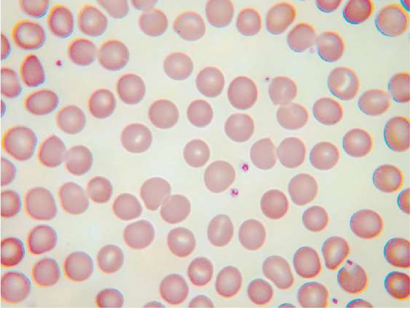
Normal RBC: Biconcave disc, 6-8 µm, central pallor ≤1/3 of cell diameter, normochromic, normocytic (MCV 80-100 fL). Compare RBC size to nucleus of a small lymphocyte - both ~8 µm. Normal WBC differential: Neutrophils 50-70%, Lymphocytes 20-40%, Monocytes 3-8%, Eosinophils 1-4%, Basophils 0-1%. Normal platelets: 150,000-400,000/µL; 1 platelet per 10-30 RBCs on smear; 7-20 per oil-immersion field.
"The examination of a peripheral blood smear is one of the most informative exercises a physician can perform." - Harrison's, Ch. 65
PART 1: RED CELL ABNORMALITIES ON PBS
A. SIZE (MCV-Based Classification)
1. MICROCYTIC HYPOCHROMIC ANEMIA (MCV < 80 fL)
| PBS Finding | Features | Likely Disease |
|---|---|---|
| Microcytes | Small cells, MCV <80, large central pallor >1/3 | Iron deficiency anemia, Thalassemia, Anemia of chronic disease |
| Target cells (codocytes) | Bull's eye appearance, increased SA:V ratio | Thalassemia, HbC disease, iron deficiency, liver disease |
| Pencil cells (elliptocytes) | Cigar-shaped elongated cells | Iron deficiency anemia (classic) |
| Basophilic stippling | Coarse blue dots (precipitated ribosomes) | Thalassemia, lead poisoning, sideroblastic anemia |
CBC Correlation:
- Iron deficiency: Low MCV, low MCH, high RDW (anisocytosis), low ferritin, high TIBC
- Thalassemia trait: Low MCV, normal/high RBC count, Mentzer index (MCV/RBC) <13 = thalassemia; >13 = iron deficiency
- Anemia of chronic disease: Low/normal MCV, low iron, low TIBC, high ferritin
2. NORMOCYTIC NORMOCHROMIC ANEMIA (MCV 80-100 fL)
| PBS Finding | Features | Likely Disease |
|---|---|---|
| Echinocytes (burr cells) | 10-30 evenly spaced spicules, crenated | Uremia, liver disease, pyruvate kinase deficiency |
| Acanthocytes (spur cells) | 3-12 irregular spicules of varying length | Abetalipoproteinemia, severe liver disease, McLeod syndrome |
| Spherocytes | Small, dark, dense, no central pallor | Hereditary spherocytosis, AIHA, ABO incompatibility |
| Bite cells (degmacytes) | "Bitten out" chunk missing | G6PD deficiency (Heinz body removal by spleen) |
| Polychromasia | Blue-grey tinge (reticulocytes) | Hemolytic anemia, acute blood loss (marrow regeneration) |
| Nucleated RBCs (nRBCs) | RBCs with nucleus | Extramedullary hematopoiesis, severe hemolysis, hypoxia, leukemia |
CBC Correlation:
- Hemolytic anemia: Low Hb, elevated reticulocyte count, elevated LDH, low haptoglobin, elevated indirect bilirubin
- AIHA: Positive direct Coombs test; spherocytes on smear
- Aplastic anemia: Pancytopenia (all 3 cell lines low), no reticulocytosis
3. MACROCYTIC ANEMIA (MCV > 100 fL)
| PBS Finding | Features | Likely Disease |
|---|---|---|
| Macro-ovalocytes (oval macrocytes) | Large oval RBCs + hypersegmented neutrophils (>5 lobes or ≥1 with 6 lobes) | Megaloblastic anemia (B12/folate deficiency) |
| Round macrocytes | Large but round RBCs, no hypersegmentation | Liver disease, hypothyroidism, alcohol, reticulocytosis |
| Dimorphic picture | Both microcytes and macrocytes, high RDW | B12 + iron deficiency combined, sideroblastic anemia post-treatment |
CBC Correlation in Megaloblastic Anemia:
- MCV often >110-115 fL, sometimes >130 fL
- Pancytopenia in severe cases (hypersegmented neutrophils = first clue)
- Low B12 (<200 pg/mL) or low folate; elevated homocysteine; elevated methylmalonic acid (B12 deficiency specific)
4. FRAGMENTED RED CELLS (SCHISTOCYTES) - HIGH-PRIORITY FINDING
| PBS Finding | Features | Likely Disease |
|---|---|---|
| Schistocytes (helmet cells, triangle cells) | Fragmented RBCs, irregular sharp edges | TTP, HUS, DIC, HELLP syndrome, mechanical heart valve hemolysis, malignant hypertension |
| Microangiopathic hemolytic anemia (MAHA) | Schistocytes + thrombocytopenia | TTP (low ADAMTS13), HUS (Shiga toxin), DIC (PT/APTT prolonged), HELLP |
Consultant pearl: >1% schistocytes on smear with thrombocytopenia is a hematological emergency until TTP is excluded. TTP = pentad: MAHA, thrombocytopenia, renal failure, fever, neurological changes. Immediate plasma exchange is life-saving.
5. SICKLE CELL AND HEMOGLOBINOPATHIES
| PBS Finding | Features | Likely Disease |
|---|---|---|
| Sickle cells (drepanocytes) | Crescent/sickle shaped | Sickle cell disease (HbSS) |
| Target cells + sickle cells | Mixed morphology | HbSC disease |
| Target cells + microcytes | Thalassemia intermedia/major | Hb Barts, HbH disease |
6. OTHER IMPORTANT RBC INCLUSIONS
| Inclusion | Stain | Disease |
|---|---|---|
| Howell-Jolly bodies | Wright stain - dark purple dots | Asplenia, post-splenectomy, megaloblastic anemia, hyposplenism |
| Heinz bodies | Supravital stain (crystal violet) | G6PD deficiency, unstable Hb, oxidant stress |
| Pappenheimer bodies | Iron-containing granules | Sideroblastic anemia, post-splenectomy |
| Basophilic stippling | Fine or coarse blue dots | Thalassemia, lead poisoning, sideroblastic anemia |
| Cabot rings | Ring/figure-8 remnants of spindle fibers | Megaloblastic anemia, lead poisoning, severe anemia |
| Malaria parasites | Ring forms, trophozoites, schizonts | Plasmodium spp. (confirmatory test) |
7. RED CELL DISTRIBUTION ABNORMALITIES
| Finding | Description | Disease |
|---|---|---|
| Rouleaux formation | Stacks of coins appearance | Multiple myeloma, Waldenstrom macroglobulinemia, chronic inflammation (raised fibrinogen, ESR) |
| Agglutination (clumping) | Irregular clumps | Cold agglutinin disease, AIHA, paraproteinemia |
PART 2: WHITE CELL ABNORMALITIES ON PBS
A. NEUTROPHIL ABNORMALITIES
| PBS Finding | Features | Likely Disease |
|---|---|---|
| Left shift (bands >10%) | Immature neutrophils, band forms | Bacterial infection, inflammation, recovery from bone marrow suppression |
| Hypersegmented neutrophils (>5 lobes or ≥1 with 6+ lobes) | Nuclear hypersegmentation | Megaloblastic anemia (B12/folate deficiency) |
| Toxic granulation | Prominent dark primary granules | Severe bacterial infection, sepsis |
| Döhle bodies | 1-2 µm pale blue cytoplasmic inclusions (rough ER aggregates) | Infection, burns, pregnancy, May-Hegglin anomaly |
| Vacuolated neutrophils | Cytoplasmic vacuoles | Bacterial sepsis (highly specific), alcohol toxicity |
| Pelger-Huet anomaly | Bilobed "pince-nez" nucleus | Benign hereditary (or pseudo-Pelger in MDS) |
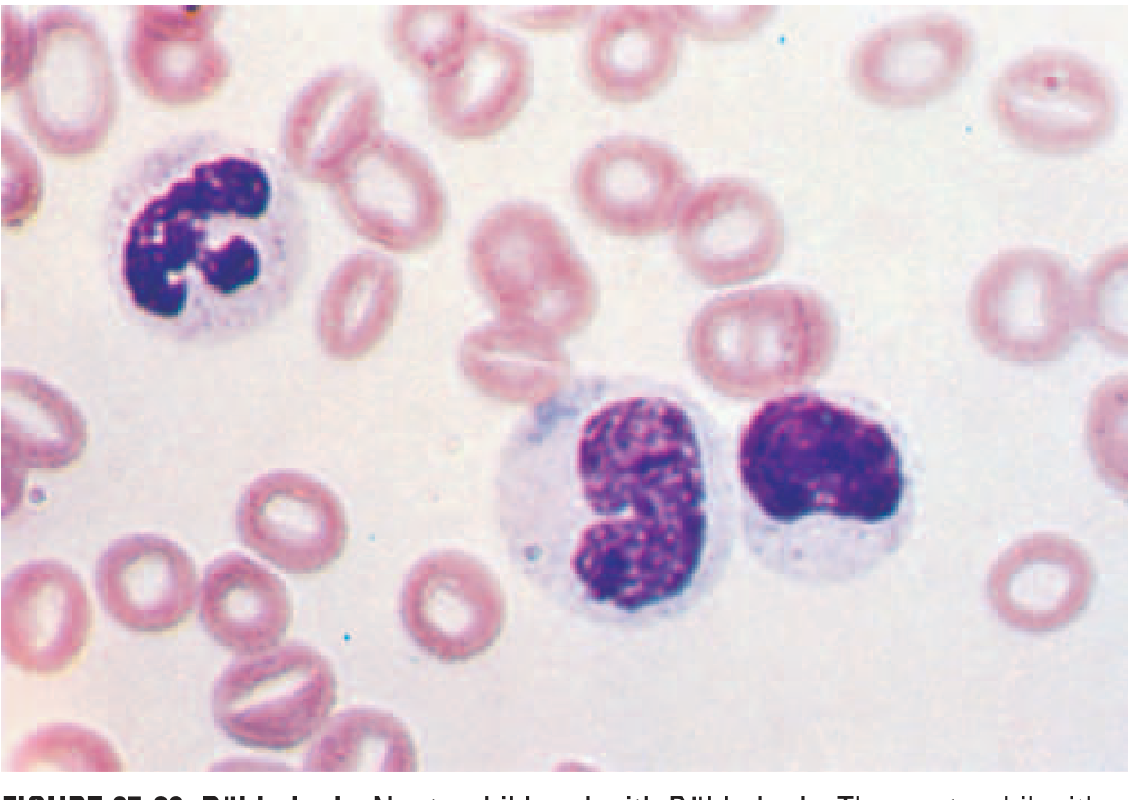
| Leukoerythroblastic reaction | Immature WBCs (myelocytes, promyelocytes) + nRBCs in peripheral blood | Bone marrow infiltration: metastatic cancer, myelofibrosis, lymphoma, granulomas |
| Blast cells | Large cells with high N:C ratio, prominent nucleoli, fine chromatin | Acute leukemia (AML, ALL) - urgent referral |
| Hypergranular neutrophils | Massive azurophilic granules | AML-M3 (APL) - DIC risk, oncological emergency |
B. LYMPHOCYTE ABNORMALITIES
| PBS Finding | Features | Likely Disease |
|---|---|---|
| Reactive (atypical) lymphocytes | Large, irregular, abundant pale blue cytoplasm that molds around RBCs (Downey cells) | EBV (infectious mononucleosis), CMV, viral hepatitis, drug reactions |
| Smudge (basket) cells | Fragile lymphocytes that smear during slide preparation | CLL (pathognomonic) |
| Small mature lymphocytes | Monotonous small lymphocytes, clumped chromatin | CLL, SLL |
| Cleaved/irregular lymphocytes | "Buttock cells" - bilobed nucleus | Follicular lymphoma, mantle cell lymphoma |
| Hairy cells | Cytoplasmic projections (TRAP-positive) | Hairy cell leukemia |
| Sezary cells | Cerebriform nuclei | Sezary syndrome (cutaneous T-cell lymphoma) |
| Large granular lymphocytes | Large cells with azurophilic granules | LGL leukemia, NK cell disorders |
| Plasma cells in blood | Eccentric nucleus, "clock-face" chromatin, basophilic cytoplasm | Plasma cell leukemia, advanced myeloma |
C. MONOCYTE ABNORMALITIES
| PBS Finding | Features | Likely Disease |
|---|---|---|
| Monocytosis (>1,000/µL absolute) | Increased monocytes | CMML, TB, bacterial endocarditis, inflammatory bowel disease, myeloid neoplasms |
| Promonocytes | Immature monocytes, irregular nucleus | AML-M4/M5 (AMML/AMoL) |
D. EOSINOPHIL ABNORMALITIES
| PBS Finding | Features | Likely Disease |
|---|---|---|
| Eosinophilia (>500/µL) | Bilobed cells with orange-red granules | Parasitic infections, allergic disorders, drug reactions, Addison's disease |
| Hypereosinophilic syndrome | Eosinophils >1500/µL persistently | Idiopathic, FIP1L1-PDGFRA mutation (imatinib-responsive), cardiac/pulmonary involvement |
| Eosinophils with blast cells | Mixed population | Eosinophilic leukemia (CEL) |
E. BASOPHIL ABNORMALITIES
| PBS Finding | Features | Likely Disease |
|---|---|---|
| Basophilia (>100/µL) | Dark purple-black granules, bilobed nucleus, can obscure nucleus | CML (very characteristic), polycythemia vera, myelofibrosis |
Consultant pearl: Basophilia >2% in a patient with leukocytosis, left shift, and possible splenomegaly = suspect CML until BCR-ABL FISH/PCR done.
PART 3: PLATELET ABNORMALITIES ON PBS
| PBS Finding | Features | Likely Disease |
|---|---|---|
| Thrombocytopenia (<150,000/µL) with large platelets | Young platelets = rapid turnover | ITP, TTP, hypersplenism, peripheral destruction |
| Thrombocytopenia without large platelets | Marrow production failure | Aplastic anemia, chemotherapy, B12/folate deficiency, leukemic infiltration |
| Giant platelets | >3 µm diameter | Bernard-Soulier syndrome, MYH9-related disorders, ITP (reactive), MPN |
| Hypogranular platelets | Reduced granularity | Gray platelet syndrome, MPN (especially MF), MDS |
| Thrombocytosis (>400,000/µL) | Platelet clumps may be seen | Essential thrombocythemia, CML, reactive thrombocytosis (infection, iron deficiency, post-splenectomy) |
| Platelet clumping | Pseudothrombocytopenia | EDTA-induced platelet agglutination - repeat in citrate tube |
PART 4: INTEGRATED CBC + PBS PATTERNS - DISEASE RECOGNITION
Pattern 1: Pancytopenia
- All 3 lineages low (Hb, WBC, platelets)
- PBS: Hypocellular smear, macro-ovalocytes if megaloblastic
- DDx: Aplastic anemia, megaloblastic anemia, hypersplenism, bone marrow infiltration (leukemia, myeloma, metastatic), MDS, HIV
Pattern 2: Bicytopenia (2 lineages) + Dysplastic cells
- PBS: Pseudo-Pelger-Huet, hypogranular neutrophils, oval macrocytes, hypolobated megakaryocytes
- DDx: Myelodysplastic syndrome (MDS) - gold standard is bone marrow biopsy + cytogenetics
Pattern 3: Leukocytosis + Differential Clue
| WBC Diff Pattern | Likely Diagnosis |
|---|---|
| Neutrophilia + left shift + toxic granulation | Bacterial infection, CML |
| Lymphocytosis + smudge cells + mature lymphocytes | CLL |
| Lymphocytosis + atypical lymphocytes + pharyngitis/splenomegaly | EBV mononucleosis |
| Eosinophilia (>1500/µL) + organomegaly | HES, CEL, parasites |
| Basophilia + neutrophilia + left shift + splenomegaly | CML (BCR-ABL PCR) |
| Blasts >20% | Acute leukemia (AML or ALL) |
| Monocytosis (>1000/µL) + dysplasia + anemia | CMML |
Pattern 4: Hemolytic Anemia - Key PBS Features
| Feature | Intravascular Hemolysis | Extravascular Hemolysis |
|---|---|---|
| Schistocytes | Yes | No |
| Spherocytes | No (or few) | Yes (AIHA, HS) |
| Haptoglobin | Very low/absent | Low |
| Hemoglobinuria | Yes | No |
| LDH | Very elevated | Elevated |
| Causes | TTP, HUS, DIC, mechanical valves, G6PD (acute) | AIHA, HS, hypersplenism |
Pattern 5: The Leukoerythroblastic Picture
- PBS: Immature myeloid cells (myelocytes, promyelocytes) + nucleated RBCs + teardrop cells (dacrocytes)
- This triad = bone marrow space-occupying lesion
- DDx: Primary myelofibrosis, metastatic carcinoma to marrow, lymphoma, granulomatous disease (TB, sarcoid), mastocytosis
- Next step: Bone marrow trephine biopsy
PART 5: KEY CBC INDICES AND THEIR CLINICAL MEANING
| Index | Normal Range | Significance |
|---|---|---|
| MCV | 80-100 fL | <80 = microcytic; >100 = macrocytic |
| MCH | 27-33 pg | Low = hypochromic (iron deficiency, thalassemia) |
| MCHC | 32-36 g/dL | High >36 = spherocytosis; Low = iron deficiency |
| RDW | 11.5-14.5% | High = anisocytosis; distinguishes mixed deficiencies |
| Reticulocyte count | 0.5-2.5% | Elevated = regenerative anemia; Low in aplasia/megaloblastic |
| Reticulocyte Production Index (RPI) | >2 = adequate response | <2 with anemia = hypoproliferative (marrow problem) |
| Mentzer Index (MCV/RBC) | <13 = thalassemia trait; >13 = iron deficiency | Quick bedside discriminator |
PART 6: SPECIFIC DISEASE PROFILES - CONSULTANT QUICK REFERENCE
Chronic Myeloid Leukemia (CML)
- CBC: WBC markedly elevated (often 50,000-500,000/µL), basophilia, eosinophilia, thrombocytosis
- PBS: Full myeloid spectrum (myeloblasts to mature neutrophils), basophilia, no Auer rods
- Confirm: BCR-ABL1 PCR or FISH (Philadelphia chromosome t(9;22))
Acute Myeloid Leukemia (AML)
- CBC: Variable WBC, anemia, thrombocytopenia
- PBS: Blasts (often >20%), Auer rods (pathognomonic - pink rod-like inclusions in cytoplasm), blast "hiatus" (gap between blasts and mature cells)
- APL (AML-M3): Hypergranular blasts, Auer rod bundles ("faggot cells"), DIC - emergency!
Chronic Lymphocytic Leukemia (CLL)
- CBC: Lymphocytosis (usually >5,000/µL), mild anemia and thrombocytopenia in advanced stages
- PBS: Small mature lymphocytes + smudge (basket) cells (fragile CLL cells crushed during smear prep) - pathognomonic
- Confirm: Flow cytometry (CD5+, CD19+, CD23+, weak surface Ig)
Multiple Myeloma
- CBC: Normocytic normochromic anemia, normal/low WBC, normal/low platelets
- PBS: Rouleaux formation (strongly), plasma cells may appear, background protein staining (blue tint)
- Confirm: Serum protein electrophoresis (M-protein), free light chains, bone marrow biopsy
Myelodysplastic Syndrome (MDS)
- CBC: Cytopenias in 1-3 lineages, macrocytosis common
- PBS: Pseudo-Pelger-Huet cells, hypogranular neutrophils, oval macrocytes, hypersegmented or ring-shaped platelets, dimorphic RBCs
- Confirm: Bone marrow biopsy + cytogenetics (del(5q), monosomy 7, etc.)
Iron Deficiency Anemia
- CBC: Low Hb, low MCV, low MCH, high RDW
- PBS: Microcytic hypochromic cells, pencil cells (elliptocytes), anisocytosis, poikilocytosis
- Confirm: Low ferritin (<12 µg/L diagnostic), low serum iron, high TIBC, low transferrin saturation
Megaloblastic Anemia (B12/Folate Deficiency)
- CBC: High MCV (often >110), pancytopenia in severe cases
- PBS: Oval macrocytes (macro-ovalocytes) + hypersegmented neutrophils (hallmark); occasional nRBCs
- B12 deficiency additionally: neurological symptoms (subacute combined degeneration)
- Confirm: Low B12 or folate, elevated methylmalonic acid (B12 specific), elevated homocysteine
Thalassemia Major vs Trait
| Feature | Major | Trait |
|---|---|---|
| Hb | <7 g/dL | Near normal or mildly low |
| MCV | Very low (<65) | Low (65-75) |
| PBS | Target cells, nucleated RBCs, basophilic stippling, teardrop cells | Mild microcytosis, occasional target cells |
| RBC count | Low | Normal or HIGH |
| Mentzer index | <13 | <13 |
| Diagnosis | HbF elevated, Hb electrophoresis | Hb A2 elevated (>3.5%) |
Hereditary Spherocytosis
- CBC: Normocytic or slightly macrocytic (if reticulocytosis), elevated MCHC (>36 g/dL - key clue)
- PBS: Spherocytes, polychromasia, no central pallor
- Confirm: Osmotic fragility test (or EMA binding flow cytometry - more sensitive)
Sickle Cell Disease
- CBC: Anemia (Hb 6-9 g/dL), reticulocytosis, leukocytosis, Howell-Jolly bodies (functional asplenia)
- PBS: Sickle cells, target cells, Howell-Jolly bodies, polychromasia, nRBCs during crisis
- Confirm: Hb electrophoresis (HbS >80%, no HbA)
Polycythemia Vera (PV)
- CBC: Elevated Hb (>16.5 g/dL M, >16 g/dL F), elevated RBC, elevated hematocrit, often elevated WBC and platelets
- PBS: Plethoric picture, may show eosinophilia and basophilia
- Confirm: JAK2 V617F mutation (present in >95%), low serum EPO
Primary Myelofibrosis
- CBC: Anemia, variable WBC, variable platelets
- PBS: Leukoerythroblastic pattern + teardrop cells (dacrocytes) - classic combination
- Confirm: Bone marrow biopsy (fibrosis, reticulin/collagen), JAK2/CALR/MPL mutations
TTP (Thrombotic Thrombocytopenic Purpura)
- CBC: Anemia, severe thrombocytopenia, normal PT/APTT (differentiates from DIC)
- PBS: Schistocytes + thrombocytopenia (MAHA picture)
- Confirm: ADAMTS13 activity <10%, inhibitor present
- Treatment: Plasma exchange - do NOT wait for ADAMTS13 result
PART 7: SYSTEMATIC APPROACH TO READING A PBS (Consultant Protocol)
Step 1 - Technical assessment: Is the smear well-made? Correct feathered edge, uniform staining, right area examined (just behind feathered edge where RBCs barely touch).
Step 2 - Platelets first (smallest elements): Count per oil-immersion field (x1000), assess size, granularity, clumping. Estimate count: avg per field × 20,000.
Step 3 - Red cells:
- Size vs. small lymphocyte nucleus (both ~8 µm)
- Chromia (central pallor: normal ≤1/3 diameter)
- Shape (list all abnormal poikilocytes)
- Color (polychromasia = reticulocytes = regeneration)
- Inclusions (Howell-Jolly, Heinz, Pappenheimer, parasites)
- Distribution (rouleaux, agglutination)
Step 4 - White cells:
- Estimate count
- Differential (neutrophils → lymphocytes → monocytes → eosinophils → basophils)
- Morphology (toxic changes, dysplastic features, blasts, atypical lymphocytes)
- Any left shift
Step 5 - Integrate with CBC indices: MCV, MCH, MCHC, RDW, reticulocyte count, absolute cell counts.
Step 6 - Formulate hypothesis and order confirmatory tests.
PART 8: INDICATIONS FOR REQUESTING A BLOOD SMEAR (Goldman-Cecil)
Automated CBC flags warranting smear review:
- Anemia (any), polycythemia
- Macrocytosis, microcytosis
- Neutrophilia, lymphocytosis, eosinophilia
- Thrombocytopenia, thrombocytosis
- Any "blast" or "atypical cell" flag from the analyzer
- Clinical suspicion overriding normal automated counts (fever + travel + malaria risk, hemolytic symptoms)
Sources: Harrison's Principles of Internal Medicine 22E, Ch. 65 (Dan L. Longo) | Goldman-Cecil Medicine, Ch. 143 | Henry's Clinical Diagnosis and Management by Laboratory Methods, Ch. 31
This is a shared conversation. Sign in to Orris to start your own chat.