
APTA Proper protocol for TKR in detail proper structured evidence based protocol
"total knee arthroplasty"[MeSH Terms] AND "physical therapy" AND rehabilitation protocol
"knee arthroplasty"[MeSH Terms] AND "physical therapy modalities"[MeSH Terms]
APTA clinical practice guideline TKR total knee replacement physical therapy protocol 2024 2025
PMID: 32542403
https://www.guidelinecentral.com/guideline/300177
"knee arthroplasty"[MeSH Terms] AND "rehabilitation"[MeSH Terms] AND exercise
https://pubmed.ncbi.nlm.nih.gov/32542403
"total knee arthroplasty" AND "preoperative" AND "physical therapy"
total knee arthroplasty rehabilitation exercises physical therapy
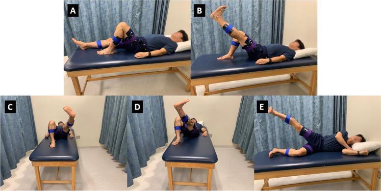
A composite of five clinical photographs (labeled A–E) illustrating a series of open-chain lower extremity rehabilitation exercises commonly used in post-total knee arthroplasty (TKA) physical therapy. A: Supine knee flexion with the foot planted. B: Straight leg raise (SLR) showing hip flexion with full knee extension. C: 'V out' exercise involving hip flexion, abduction, and external rotation. D: 'V in' exercise involving hip flexion, adduction, and internal rotation. E: 'Jane Fonda' side-lying hip abduction with the upper leg extended. The subject is equipped with a marker-based motion capture system, featuring reflective sensors placed at key anatomical landmarks: the greater trochanter (hip), medial/lateral femoral epicondyles (knee), and malleoli (ankle). Blue and purple compression bands secure the sensor arrays. These images demonstrate standardized postures for quantitative kinematic analysis of joint angles and range of motion (ROM) to assess neuromuscular control and strength during physical therapy protocols.
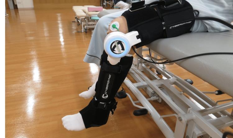
A clinical photograph depicting a patient undergoing postoperative rehabilitation following a Total Knee Arthroplasty (TKA). The image shows a single-joint Hybrid Assistive Limb (HAL-SJ) robotic exoskeleton attached to the patient's lower extremity. The device is secured to the thigh and calf with black padded 'CYBERDYNE' straps, with its central motor unit aligned precisely over the lateral aspect of the knee joint. Surface electromyography (sEMG) electrodes are visible on the distal vastus medialis, utilized to detect bio-electrical signals (BES) for cybernic voluntary control. The patient is positioned on a medical treatment table in a rehabilitation gym setting, performing knee extension and flexion exercises. This robotic intervention is used in physical therapy to assist active-assisted mobilization and neuromuscular gait re-education by providing torque support triggered by the patient's muscle signals. The photograph illustrates a procedural application of advanced medical robotics in orthopedic recovery for conditions like knee osteoarthritis.
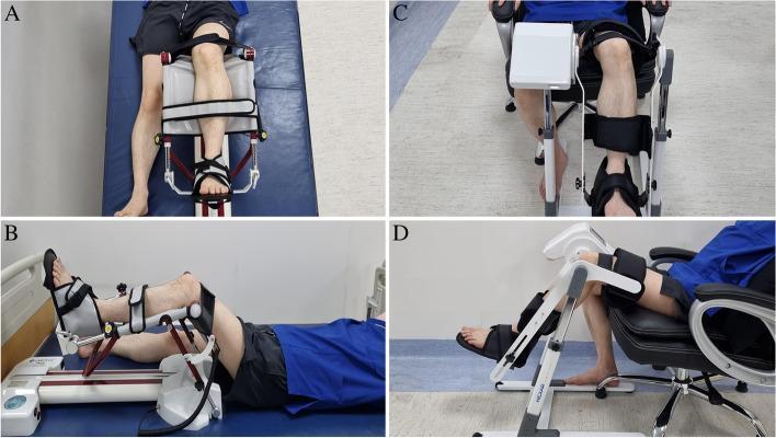
This composite clinical photograph illustrates two different Continuous Passive Motion (CPM) machine configurations used for postoperative knee rehabilitation, specifically following Total Knee Arthroplasty (TKA). Panels A and B demonstrate a conventional CPM setup (RCF1121®) where the patient is in a supine position on a hospital bed. The device frame rests horizontally, with the limb secured via adjustable straps at the thigh, mid-calf, and foot cradle to guide flexion and extension. Panels C and D show a newer CPM model (Resilion K20P®) designed for a seated position in a chair. This vertical-oriented mechanism features a distinct leg brace and foot plate system, supporting the limb while the patient remains upright. Both systems utilize padded stabilizers and mechanical hinges to automate range of motion (ROM) exercises. These images highlight the ergonomic differences between traditional supine therapy and modern seated rehabilitation protocols in orthopedic physical therapy.
APTA Evidence-Based Physical Therapy Protocol for Total Knee Replacement (TKR/TKA)
Evidence Grade Key
| Grade | Meaning |
|---|---|
| S | Strong - High confidence evidence |
| M | Moderate - Moderate confidence evidence |
| W | Weak - Low confidence evidence; clinical judgment required |
| BP | Best Practice - Recommended despite limited direct evidence |
Phase 1: PREOPERATIVE ("Prehabilitation")
Timing
- Begin 4-8 weeks before scheduled surgery
Goals
- Optimize lower extremity strength, flexibility, and ROM pre-surgery
- Reduce anxiety; establish patient-provider relationship
- Set realistic expectations for recovery trajectory
Interventions
| Intervention | Recommendation | Grade |
|---|---|---|
| Preoperative exercise program (strength + flexibility) | Recommended | M |
| Patient education (procedural expectations, home exercise instruction, pain management, discharge planning) | Recommended | BP |
| Continuous Passive Motion (CPM) | NOT recommended preoperatively | W |
Exercise Focus
- Quadriceps sets
- Straight leg raises (SLR)
- Hip abductor/extensor strengthening
- Active knee ROM (flexion and extension)
- Aerobic conditioning (stationary bike if tolerated)
- Balance and proprioception training
Phase 2: ACUTE POSTOPERATIVE (Day 0-7)
Timing
- Begin PT within 24 hours of surgery and prior to discharge (Grade M)
- Same-day ambulation with trained PT or RN on day of surgery
Goals
- Safe mobilization and weight bearing
- Pain and edema control
- Early ROM recovery
- Prevent postoperative complications (DVT, pneumonia, contracture)
- Safe discharge planning
Immediate Interventions
| Intervention | Recommendation | Grade |
|---|---|---|
| Start PT management within 24 hours of surgery | Required | M |
| Cryotherapy for pain management | Recommended | M |
| Neuromuscular Electrical Stimulation (NMES) | Recommended | M |
| High-intensity strengthening programs within 7 days post-surgery | Recommended | M |
| Motor function training (functional movement patterns) | Recommended | S |
| Passive, active-assistive, and active ROM exercises | Recommended | BP |
| Position knee in 30-90° flexion while resting (reduces blood loss and swelling, first 7 days only) | Recommended | W |
| CPM devices | NOT recommended | W |
| Supervised PT - individualize setting to patient safety, mobility, environment | Recommended | M |
| Group OR individual PT sessions (either is appropriate) | Either acceptable | W |
Exercise Program (Days 0-7)
- Ankle pumps (every 1-2 hours for DVT prophylaxis)
- Quadriceps sets (isometric)
- Gluteal sets (isometric)
- Straight leg raises (initiate once patient can perform with minimal quad lag)
- Heel slides (active-assistive knee flexion/extension)
- Short arc quads (terminal knee extension, 0-40°)
- Sit-to-stand transfers with rolling walker
- Ambulation with rolling walker - weight bearing as tolerated (WBAT) unless surgeon specifies otherwise
- Stair negotiation training (step-to pattern) before discharge
Discharge Criteria from Acute Setting (Campbell's Orthopaedics)
- Safe ambulation on a rolling walker
- Ability to perform a straight leg raise
- Pain appropriately controlled
- Independent voiding
- Diet tolerance
Phase 3: EARLY OUTPATIENT (Weeks 1-6)
Timing
- Outpatient PT starts 3-5 days after surgery (for ambulatory surgical center patients)
- Frequency: 1-2 sessions/week over a 6-8 week period (Campbell's Operative Orthopaedics, 15th Ed 2026, p. 535)
Goals
- Achieve knee flexion ≥90° by week 4-6
- Achieve full active knee extension (0°)
- Restore functional gait pattern (no assistive device by 6-8 weeks if appropriate)
- Progress strengthening
- Normalize ADL function
ROM Targets
| Timepoint | Flexion Target | Extension Target |
|---|---|---|
| 1 week | 70-80° | 0-5° |
| 2 weeks | 90° | 0° |
| 4 weeks | 100-110° | 0° |
| 6 weeks | ≥110° | 0° |
| 3 months | 110-120°+ | 0° |
Exercise Progression
- Mini squats (0-45°)
- Step-ups (forward and lateral)
- Leg press (limited arc)
- Sit-to-stand progressions
- Terminal knee extension with resistance band
- Short arc quads
- Progressive SLR with ankle weights
- Quadriceps: primary target - address quad inhibition/weakness (typically 30-50% deficit vs. contralateral limb)
- Hip abductors and extensors
- Hamstrings
- Calf complex
- Progress from rolling walker → cane (typically by week 4-6)
- Address antalgic gait deviations
- Weight bearing as tolerated throughout
- Single-leg stance progression
- Eyes-open to eyes-closed progression
- Unstable surface training (foam pad)
Adjunct Modalities
- Cryotherapy: continue post-exercise for swelling/pain management
- NMES to quadriceps: continue if significant quad inhibition present (Grade M)
- Manual therapy: patellar mobilization, scar tissue mobilization, joint mobilization as needed
- Soft tissue mobilization to incision (after wound healing is confirmed)
Phase 4: INTERMEDIATE STRENGTHENING (Weeks 6-12)
Goals
- Achieve functional quadriceps strength (≥80% limb symmetry index)
- Normalize gait without assistive device
- Return to community ambulation
- Resume driving (typically 4-6 weeks post-surgery, right knee; confirm with surgeon)
Exercise Progression
- Progress resistance training intensity (APTA CPG supports high-intensity training)
- Blood flow restriction (BFR) training: emerging evidence shows benefit for quad strength with low mechanical load (PMID 39461901 - RCT, 2024)
- Full squat progression as ROM allows
- Step-down eccentric control
- Functional balance challenges
Phase 5: RETURN TO FUNCTION (Weeks 12+)
Goals
- Return to all ADLs and recreational activities
- Community ambulation on varied surfaces and terrain
- Stair negotiation reciprocally (if appropriate)
- Return to low-impact sport/activity (cycling, swimming, golf, walking)
- High-demand activities individualized to patient goals
Activity Guidance
- Encouraged: walking, cycling, swimming, golf, low-impact aerobics
- Cautious/Individualized: hiking, doubles tennis, light dancing
- Generally discouraged: high-impact running, jumping, contact sports
- Returning to running: a 2025 systematic review (PMID 39987394) supports multimodal progressive protocols for select patients
Outcomes Assessment (APTA CPG - Best Practice)
| Measure | Type | Tool |
|---|---|---|
| KOOS JR | Patient-reported outcome (PRO) | Self-report questionnaire |
| 30-Second Sit-to-Stand Test (30s-STS) | Functional performance | Timed test |
| Timed Up and Go (TUG) | Functional mobility + fall risk | Timed test |
Discharge Planning (Acute Setting - Grade M)
- Provide objective guidance to care team and patient on:
- Functional status at discharge
- Assistive equipment needed (rolling walker, raised toilet seat, shower chair)
- Home vs. inpatient rehab vs. SNF placement
- Home health PT (if patient cannot safely attend outpatient)
- Criteria for safe discharge are based on mobility, safety, environmental factors, and personal/social support
What NOT to Do (Evidence-Based)
| Do NOT | Grade |
|---|---|
| Use CPM devices routinely | W (against) |
| Delay mobilization past 24 hours without clinical justification | M |
| Prescribe prolonged bed rest | BP |
| Use continuous flexion positioning >90° in first 7 days (increases risk of blood loss) | W |
Visual Reference
Key Citations
- Primary CPG: Jette DU et al. "Physical Therapist Management of Total Knee Arthroplasty." Physical Therapy. 2020. PMID 32542403 - the official APTA CPG
- Surgical/Postoperative context: Campbell's Operative Orthopaedics, 15th Ed 2026, p. 535
- Clinical outcomes/recovery: Goldman-Cecil Medicine, International Ed, Vol 2, p. 1907
- BFR prehabilitation: Jorgensen SL et al. Scand J Med Sci Sports. 2024. PMID 39461901 - RCT
- Return to running: Faldini C et al. Musculoskelet Surg. 2025. PMID 39987394 - Systematic Review