
Taresiam by ak Khurana book
Reading File
Do you mean the lacrimal (tear) system from A. K. Khurana’s Comprehensive Ophthalmology?
What would you like: anatomy and physiology, examination, diseases, or exam notes?
Pterygium
Reading File
Finding Sources
Finding Sources
Searching PubMed
pterygium AND treatment
Searching the Web
pterygium American Academy Ophthalmology management treatment
Searching Images
clinical photograph nasal pterygium eye conjunctival fibrovascular growth cornea
Reading File
Finding Sources
Reading Web Page
https://www.aao.org/eyenet/article/management-of-pterygium-2
Pterygium
A pterygium is a triangular fibrovascular growth of bulbar conjunctiva that crosses the limbus and extends onto the cornea. It is usually nasal and occurs in the interpalpebral area.
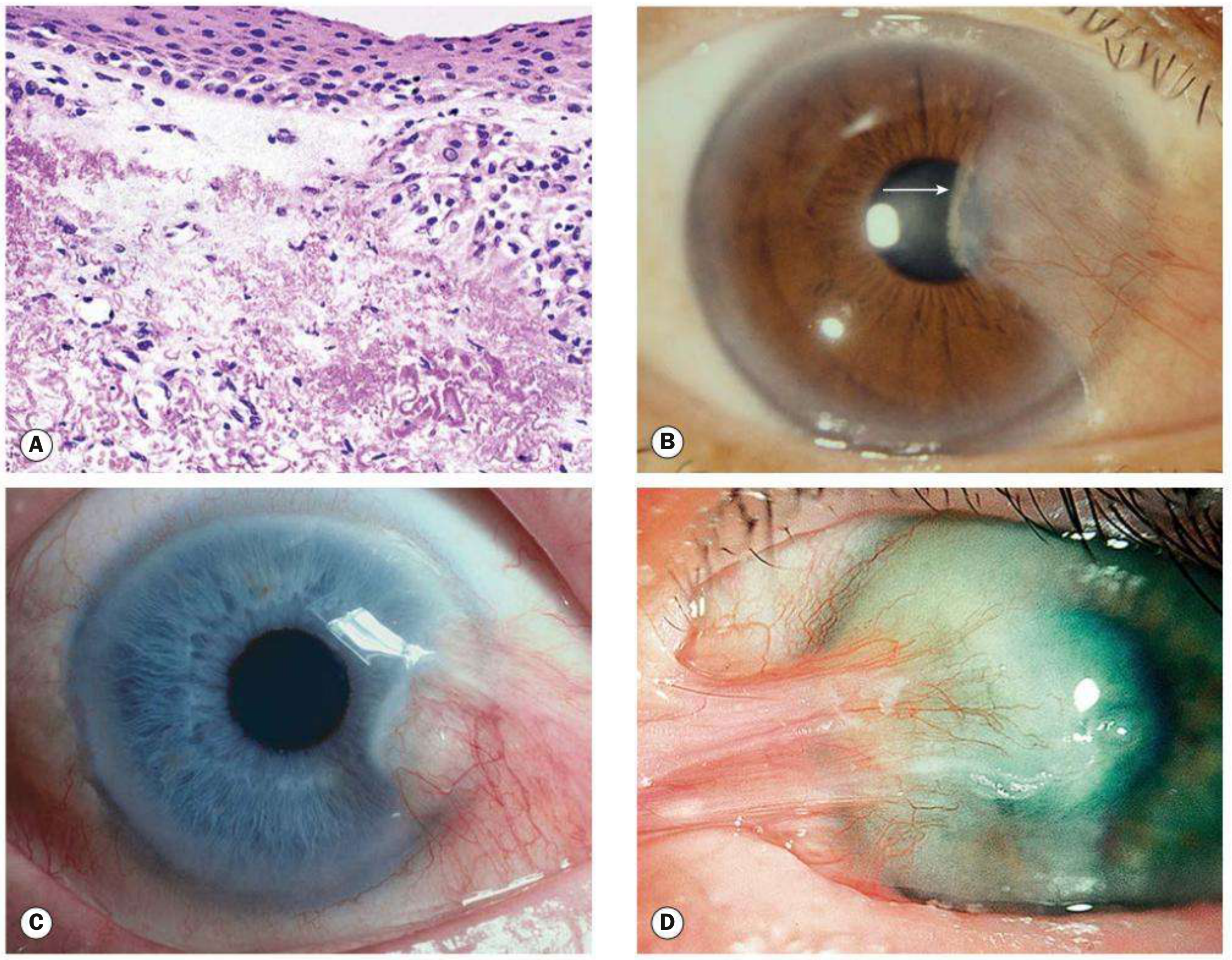
Etiology and risk factors
- Chronic ultraviolet-B exposure, especially sunlight
- Dust, wind, dry and hot climates
- Chronic ocular-surface irritation and dryness
- Outdoor work
- Familial predisposition
Parts
From apex to base:
- Cap: Grey, avascular, superficial zone ahead of the head.
- Head: Firm adherent portion attached to cornea.
- Neck: Area at the limbus.
- Body: Triangular, vascular conjunctival part.
- Stocker line: Brown iron deposition line just anterior to the head in long-standing lesions.
Symptoms
Small lesions may cause no symptoms. Larger or inflamed lesions cause:
- Foreign-body sensation, grittiness, irritation
- Redness and watering
- Cosmetic concern
- Contact-lens intolerance
- Blurred vision from induced astigmatism or, rarely, direct encroachment on the visual axis
Signs
- Usually a nasal, triangular, elevated fibrovascular lesion
- The apex points toward the pupil and grows over cornea
- May become inflamed and congested
- Chronic lesions can produce corneal astigmatism
Differential diagnosis
Pinguecula
- Yellowish conjunctival degeneration near the limbus
- Does not cross onto the cornea
Pseudopterygium
- Conjunctival adhesion to cornea after trauma, chemical burn, corneal ulcer, or inflammation
- May occur at any meridian
- A probe can usually be passed beneath the neck, unlike true pterygium
Treatment
Conservative
- UV-blocking sunglasses and broad-brimmed hat
- Lubricating eye drops for irritation
- Short supervised course of topical anti-inflammatory treatment for significant inflammation
Indications for surgery
- Progressive growth threatening or involving the visual axis
- Significant induced astigmatism or decreased vision
- Recurrent inflammation or persistent irritation not controlled conservatively
- Restriction of ocular motility, rarely
- Significant cosmetic concern
- Suspicious or atypical lesion, where excision biopsy may be needed
Preferred operation
- Excision with conjunctival autograft or limbal-conjunctival autograft. This has a lower recurrence risk than simple bare-sclera excision. The AAO review also lists visually significant astigmatism, visual-axis threat, severe irritation, and cosmesis as surgical indications.
Complications
- Induced astigmatism
- Visual-axis obstruction
- Recurrence after excision, particularly in young people and after bare-sclera techniques
- Rarely, diplopia due to subconjunctival fibrosis in extensive or recurrent lesions
A pterygium crosses the limbus and invades cornea, whereas a pinguecula remains confined to conjunctiva. Kanski's Clinical Ophthalmology: A Systematic Approach, p. 215.
This is a shared conversation. Sign in to Orris to start your own chat.