
Different ventilator modes for different surgies in Anaesthsia and their advantage n disadvantage
ventilation modes intraoperative anaesthesia
ventilator modes mechanical ventilation diagram pressure volume control
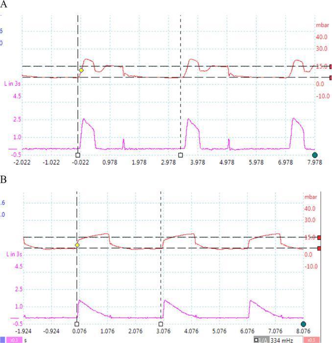
A comparison chart consisting of two sets of mechanical ventilation waveforms (pressure-time and flow-time curves) demonstrating different control variables. The upper panel (Ventilator 1) shows characteristics of volume-controlled ventilation, where the pressure curve (red) reaches a peak and then maintains a plateau, while the flow curve (purple) rises instantaneously and remains constant before falling, indicating a square flow waveform. The lower panel (Ventilator 2) illustrates pressure-controlled ventilation. In this mode, the pressure curve (red) rapidly rises to a set level (approximately 15 mbar) and maintains a plateau, while the flow curve (purple) shows a peak followed by a decelerating ramp pattern. Both panels show three respiratory cycles over an approximately 8-second window, with vertical dashed lines marking the start of inspiration. These waveforms are critical for respiratory therapy education to distinguish between volume-preset and pressure-preset mechanical ventilation strategies.
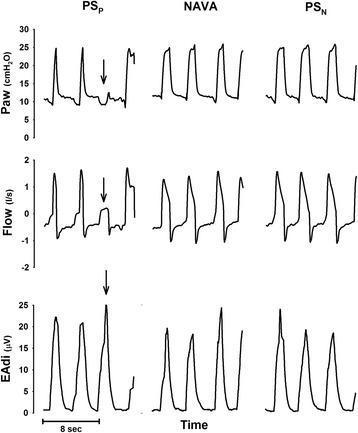
This physiological tracing compares three mechanical ventilation modes in a human patient: pneumatically triggered pressure support (PSP), neurally adjusted ventilatory assist (NAVA), and neurally controlled pressure support (PSN). The data is organized in a 3x3 grid across columns (modes) and rows (parameters). The vertical axes measure airway pressure (Paw in cmH2O), respiratory flow (l/s), and electrical activity of the diaphragm (EAdi in μV) as a function of time. In the PSP column, an arrow identifies an 'ineffective effort' where a significant spike in EAdi (diaphragmatic contraction) fails to trigger a corresponding ventilator-supported airway pressure rise, demonstrating patient-ventilator asynchrony. In contrast, the NAVA and PSN columns show improved synchrony, where neural signals (EAdi) directly correlate with the onset of pressure delivery. PSN shows a more rapid pressurization profile compared to the proportional ramp-up seen in NAVA. This comparison illustrates how neural-based triggering and control mechanisms can mitigate triggering delays and missed efforts common in traditional pneumatic pressure support.
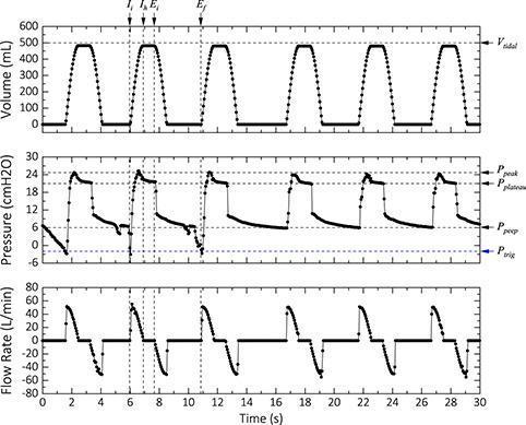
This diagnostic graphic displays three stacked ventilator waveforms (Volume, Pressure, and Flow Rate) over a 30-second interval, demonstrating mechanical ventilation in assisted mode. The top graph shows Tidal Volume (Vt) pulsing consistently to approximately 500 mL with a distinct plateau indicating an end-inspiratory hold. The middle Pressure-time waveform illustrates critical respiratory parameters: a negative deflection reaching the trigger pressure (Ptrig) of -2 cmH2O initiates each breath, followed by a rapid rise to a peak inspiratory pressure (Ppeak) of ~25 cmH2O. An inspiratory hold reveals a plateau pressure (Pplateau) of ~22 cmH2O, with the cycle concluding at a positive end-expiratory pressure (PEEP) of ~5 cmH2O. The bottom Flow Rate graph displays corresponding inspiratory and expiratory flow pulses. Annotations mark key temporal points: initial inspiration (Ii), inspiratory hold (Ih), end-inspiration (Ei), and end-expiration (Ef). This visualization is essential for understanding ventilator-patient synchrony, mechanical lung properties (compliance and resistance), and the functioning of pressure-triggered assisted ventilation modes.
Ventilator Modes in Anaesthesia: Surgery-Specific Applications, Advantages & Disadvantages
Foundational Concepts
The Core Ventilator Modes
1. Volume-Controlled Ventilation (VCV) / Assist-Control Volume Control (AC-VC)
| Independent (set) | Vt, RR, inspiratory flow, PEEP, FiO2 |
| Dependent (monitored) | Peak airway pressure, plateau pressure |
- Guarantees minimum minute ventilation - reliable CO2 clearance
- Predictable tidal volumes regardless of patient effort
- Simplest to set up; most commonly used in operating rooms
- Good control in deeply sedated/paralysed patients
- If lung compliance decreases (pneumoperitoneum, obese patient, Trendelenburg position), airway pressures rise unpredictably - risk of barotrauma
- Constant inspiratory flow can cause patient-ventilator asynchrony in lightly sedated, spontaneously breathing patients
- Does not adapt to changes in compliance during surgery
Bailey & Love's Short Practice of Surgery (28e): "In volume control ventilation, a preset volume is delivered by the machine irrespective of the airway pressure. In laparoscopic surgery requiring the Trendelenburg position, morbidly obese patients and those with lung disease, this may result in excessive pressures being developed, which may lead to barotrauma."
2. Pressure-Controlled Ventilation (PCV) / Assist-Control Pressure Control (AC-PC)
| Independent (set) | Inspiratory pressure, RR, PEEP, FiO2, inspiratory time |
| Dependent (monitored) | Tidal volume, minute ventilation |
- Limits peak and plateau airway pressures - protects against barotrauma
- Decelerating inspiratory flow pattern - more homogeneous distribution of tidal volume, reduced dead-space ventilation
- Better for patients with poor compliance (ARDS, post-thoracotomy, post-pneumonectomy)
- Useful when airway or lung parenchyma suture lines must be protected
- Tidal volume varies with lung compliance - must monitor Vt and minute ventilation closely
- If compliance improves suddenly (e.g., release of pneumoperitoneum), tidal volumes can become dangerously large (volutrauma)
- Does not guarantee ventilation
Morgan & Mikhail (7e): "Although there is no unequivocal evidence that one mode of ventilation may be more beneficial than the other, pressure-controlled ventilation may diminish the risk of barotrauma by limiting peak and plateau airway pressures, and the flow pattern results in a more homogenous distribution of the tidal volume and reduced dead space ventilation."
3. PCV-Volume Guaranteed (PCV-VG) / Pressure-Regulated Volume Control (PRVC)
- Best of both worlds: guarantees minute ventilation (like VCV) while limiting airway pressure (like PCV)
- Adapts automatically to changes in compliance (e.g., shifting surgical positions, pneumoperitoneum)
- Recent 2025 network meta-analysis (Liu Z et al., Ann Med 2025, PMID 41157953) supports improved intraoperative respiratory mechanics and reduced postoperative pulmonary complications vs. conventional modes
- More complex to understand and troubleshoot
- Available only on newer generation ventilators
- Can over-deliver volume if patient makes strong spontaneous efforts while ventilator is "targeting" the set Vt
4. Pressure Support Ventilation (PSV)
- Maintains patient respiratory effort - prevents diaphragmatic atrophy
- Reduces work of breathing during weaning
- Promotes patient-ventilator synchrony
- Lower mean airway pressures - less haemodynamic impairment
- No guaranteed respiratory rate or minute ventilation - completely depends on patient effort
- Apnoea risk if sedation/opioids cause respiratory depression
- Not appropriate for deeply anaesthetised or paralysed patients
- Tidal volumes can be excessive if pressure support is too high
5. Synchronized Intermittent Mandatory Ventilation (SIMV)
- Gradual weaning by lowering mandatory rate while increasing PSV component
- Maintains some spontaneous breathing, reducing haemodynamic impact
- Safer than pure PSV - has a backup rate
- Increased work of breathing compared to AC or PSV alone at equivalent ventilation levels
- More complex to manage
- Evidence for superiority over AC + PSV weaning is lacking
- If rate set too low without adequate PSV, can lead to respiratory muscle fatigue
6. Airway Pressure Release Ventilation (APRV) / BiLevel
- Recruits and maintains alveolar volume continuously
- Minimises peak alveolar pressures
- Allows spontaneous breathing throughout - better haemodynamics
- Useful in ARDS and severely reduced compliance
- Complex to set up; requires experienced providers
- Not practical as a primary intraoperative mode in routine surgery
- Risk of auto-PEEP and CO2 retention if release time (TL) too short
- Not easily adapted to rapidly changing surgical conditions
7. High-Frequency Oscillatory Ventilation (HFOV)
- Minimal alveolar pressure swings - ultra-lung protective
- Can recruit without overdistending
- Clinical trials in adults have not shown survival benefit in ARDS
- Impractical in the operating theatre
- Requires specialty equipment and expertise
- Haemodynamic compromise at high mean airway pressures
Surgery-Specific Ventilation Recommendations
Laparoscopic / Robotic Surgery (Trendelenburg, Pneumoperitoneum)
- Pneumoperitoneum raises intra-abdominal pressure, reduces diaphragm excursion, reduces functional residual capacity (FRC), and increases airway pressures
- Steep Trendelenburg (e.g., robotic prostatectomy, gynaecological surgery) further worsens respiratory mechanics
- Preferred: PCV or PCV-VG to limit peak pressures
- Apply PEEP 5-8 cmH2O + periodic recruitment manoeuvres
- Increase RR to compensate for CO2 absorption from insufflation
- Watch: VCV in this setting risks barotrauma
Thoracic Surgery (One-Lung Ventilation)
- One-lung ventilation (OLV) with double-lumen tube or bronchial blocker; ventilated lung receives all tidal volume
- Use PCV with low Vt (<6 mL/kg, ideally 4-5 mL/kg per lung), limit plateau pressure <25 cmH2O, peak pressure <35 cmH2O
- FiO2 50-80% (avoid 100% oxygen - risk of absorption atelectasis and oxygen toxicity)
- PEEP to dependent lung to prevent atelectasis
- CPAP to operative (non-dependent) lung if hypoxaemia refractory
- Permissive hypercapnia acceptable
Cardiac Surgery
- Patients often on CPB (cardiopulmonary bypass) - ventilation suspended during CPB; resume with VCV or PCV on coming off bypass
- Lung-protective ventilation (low Vt 6-8 mL/kg, PEEP 5 cmH2O) reduces post-cardiac surgery pulmonary complications
- After sternotomy, high pressures risk disruption of sternal wires - avoid excessive Vt
- Post-bypass: watch for decreased compliance from pulmonary oedema/reperfusion injury
Neurosurgery / Head-Elevated Position
- Goal: Normocapnia (PaCO2 35-40 mmHg) to maintain cerebral blood flow
- In raised ICP: mild hyperventilation (PaCO2 30-35 mmHg) for acute ICP control - use VCV to precisely control minute ventilation
- Avoid high PEEP (>10 cmH2O) - can raise ICP via increased intrathoracic pressure and impaired venous drainage
- Avoid hypoxia (SpO2 >96%)
Obese Patients
- Reduced FRC, prone to atelectasis and hypoxaemia
- Use higher PEEP (8-10 cmH2O) + recruitment manoeuvres
- PCV-VG handles compliance variation well
- Tidal volume based on ideal body weight, not actual weight
Paediatric Surgery
- Use pressure-controlled modes preferentially - smaller airways have variable resistance; PCV adapts better
- VT 6-8 mL/kg IBW; higher RR to maintain normocapnia
- HFOV reserved for refractory paediatric ARDS
Intraoperative Lung-Protective Ventilation - Current Consensus
- Tidal volume: 6-8 mL/kg ideal body weight
- PEEP: minimum 5 cmH2O in all ventilated patients
- Recruitment manoeuvres after intubation and position changes
- Monitor driving pressure (Pplateau - PEEP; target <13-15 cmH2O) and plateau pressure (<30 cmH2O)
- Reduce FiO2 to lowest acceptable level (avoid routine 100% FiO2)
Miller's Anaesthesia (10e): "These include initial settings of 6–8 mL/kg of tidal volume, and a minimum PEEP of 5 cmH2O. Additionally, recognising that formation of atelectasis may be a significant risk factor for the development of postoperative pulmonary complications, appropriate PEEP and recruitment maneuvers are recommended to improve intraoperative respiratory function."
Quick Reference Table
| Mode | Controls | Guarantees | Main Risk | Primary Anaesthetic Use |
|---|---|---|---|---|
| VCV (AC-VC) | Vt, RR, flow | Minute ventilation | Barotrauma if low compliance | Routine GA, most surgeries |
| PCV (AC-PC) | Pressure, RR | Pressure limits | Hypoventilation if compliance falls | Thoracic, high-airway-pressure risk |
| PCV-VG/PRVC | Target Vt via pressure | Vt + pressure limits | Volutrauma if strong effort | Laparoscopic, obese, thoracic |
| PSV | Pressure support | Nothing (patient-dependent) | Apnoea, excessive Vt | Weaning, emergence |
| SIMV | Mandatory breaths + PS | Minimum rate | Fatigue, complex weaning | ICU post-op weaning |
| APRV | High/low CPAP | Recruitment | CO2 retention, complex | Refractory ARDS (ICU) |
| HFOV | Oscillatory pressure | Diffusion ventilation | Haemodynamic compromise | Paediatric salvage |