
Regulation of calcium and phosphate, vitamin d metabolism, extra cellular matrix exclusively collagen and proteoglycans, collagen biosynthesis, biochemical composition of bone and cartilage. Explain the topics with respect to Lippincott biochemistry, MS-1 level. Also explain the mechanism inter regulated mechanism of calcium and phosphate by hormones
Reading File
Now I'll run parallel searches across all the major topics simultaneously.
Finding Sources
Finding Sources
Finding Sources
Finding Sources
Excellent - found Harper's Biochemistry (the closest to Lippincott at MS-1 level) for vitamin D and proteoglycans. Now let me search specifically for Lippincott content and read all the key sections in parallel.
Finding Sources
Finding Sources
Finding Sources
Finding Sources
Lippincott Illustrated Reviews Biochemistry (8th ed) is available. Now let me read the key sections from Lippincott and Harper's in parallel.
Finding Sources
Reading File
Now let me read the Lippincott sections on collagen, vitamin D, proteoglycans, and hormone regulation in parallel.
Reading File
Reading File
Reading File
Reading File
Excellent - now let me read more Lippincott content on collagen biosynthesis and calcium/phosphate hormonal regulation.
Reading File
Reading File
Now let me read the collagen biosynthesis steps and the Goodman & Gilman's hormonal regulation section.
Reading File
Reading File
Excellent. I now have all the key content. Let me also read the Lippincott bone composition section and the Histology collagen biosynthesis section in parallel before synthesizing.
Reading File
Reading File
I now have comprehensive content from Lippincott (8th ed), Harper's, Goodman & Gilman's, and the Histology textbook. Let me now fetch the vitamin D metabolism diagram and write the full synthesis.
Loading Image
Loading Image
I now have all the material needed for a comprehensive MS-1 level response. Let me update the todos and synthesize the complete answer.Here is the full, integrated MS-1 level explanation based primarily on Lippincott's Illustrated Reviews: Biochemistry (8th ed), supplemented by Harper's (32nd ed), Goodman & Gilman's, and Wheater's/Ross Histology.
Biochemistry of Bone, Cartilage, Calcium-Phosphate Regulation, and Collagen
PART 1: COLLAGEN - Structure, Biosynthesis, and ECM Roles
1.1 Overview
Collagen is the most abundant protein in the human body, making up about one-third of total protein mass. It is a fibrous structural protein found throughout the extracellular matrix (ECM), bone, cartilage, tendons, skin, and basement membranes. Its defining feature is the triple helix - three polypeptide alpha chains coiled around each other like a rope.
- Lippincott Biochemistry, 8th ed, p. 141
1.2 Types of Collagen
There are >25 collagen types, grouped functionally into three categories:
| Group | Types | Location |
|---|---|---|
| Fibril-forming | I, II, III | Bone/tendons (I), Cartilage (II), Blood vessels (III) |
| Network-forming | IV, VIII | Basement membranes (IV) |
| Fibril-associated (FACIT) | IX, XII | Link fibrils to ECM components |
- Type I: Two alpha-1 chains + one alpha-2 chain (alpha-1)2 alpha-2. Found in tendons, cornea, bone. High tensile strength.
- Type II: Three alpha-1 chains (alpha-1)3. Restricted to cartilage.
- Type III: Distensible tissues - blood vessels, skin.
- Type IV: Meshwork structure forming basement membranes.
- Lippincott, p. 141-143
1.3 Collagen Structure (Molecular Level)
-
Amino acid composition: Rich in glycine and proline.
- Glycine (every 3rd position) is smallest amino acid, fits into the restricted core of the triple helix. Sequence: -Gly-X-Y- where X = proline, Y = hydroxyproline or hydroxylysine.
- Proline creates "kinks" that facilitate helix formation. Importantly, the collagen helix is NOT an alpha helix because proline prohibits it.
-
Hydroxyproline and hydroxylysine: These are non-standard amino acids unique to collagen (and elastin). They are formed post-translationally by hydroxylation of proline and lysine residues already incorporated into the chain.
- Vitamin C (ascorbic acid) is an essential cofactor for prolyl hydroxylase and lysyl hydroxylase.
- Scurvy (Vitamin C deficiency): Hydroxylation fails → unstable triple helix → poor wound healing, bleeding gums, fragile bones.
-
Glycosylation: Hydroxylysine residues are O-glycosylated (glucose and galactose attached) prior to triple helix formation.
- Lippincott, p. 144-146
1.4 Collagen Biosynthesis - Step-by-Step (Lippincott, p. 147-151)
Collagen synthesis occurs in fibroblasts (connective tissue), osteoblasts (bone), and chondroblasts (cartilage).
INTRACELLULAR EVENTS (in RER and Golgi):
| Step | Event |
|---|---|
| 1 | Alpha-chain genes transcribed; pre-pro-alpha chains synthesized on ribosomes of RER |
| 2 | Signal peptide cleaved in RER lumen |
| 3 | Hydroxylation of proline and lysine residues by prolyl hydroxylase and lysyl hydroxylase (require: O2, Fe2+, alpha-ketoglutarate, and Vitamin C) |
| 4 | Glycosylation of hydroxylysine residues (glucose and galactose added) |
| 5 | C-terminal globular domain assembles and disulfide bonds form, ensuring correct chain alignment |
| 6 | Triple helix zips from C-terminus to N-terminus, forming procollagen |
| 7 | Procollagen stabilized by HSP47 (heat shock protein 47), a chaperone that prevents premature aggregation |
| 8 | Procollagen transported through Golgi → secreted into ECM via secretory vesicles |
EXTRACELLULAR EVENTS:
| Step | Event |
|---|---|
| 9 | Procollagen peptidases cleave the N- and C-terminal propeptides → forms tropocollagen (the basic collagen molecule) |
| 10 | Tropocollagen monomers self-assemble in a quarter-staggered arrangement to form fibrils |
| 11 | Lysyl oxidase (copper-dependent enzyme) oxidizes lysine/hydroxylysine residues → forms covalent cross-links (allysine aldol condensation and Schiff base bonds), giving fibrils their tensile strength |
Key cofactors to remember: Vitamin C (hydroxylation), Cu2+ / lysyl oxidase (cross-linking), O2 and Fe2+ (hydroxylases).
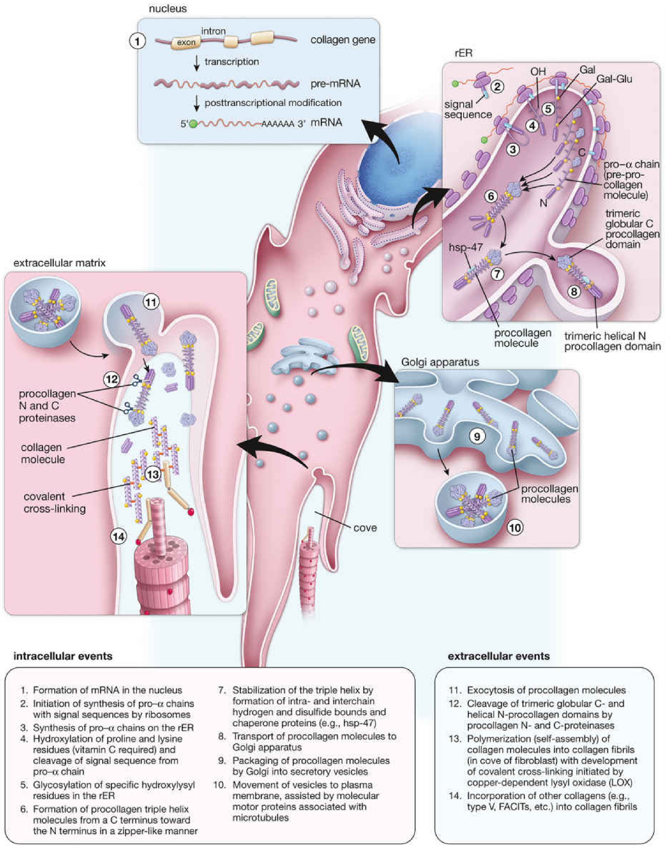
1.5 Collagen Degradation
Normal collagen fibers have half-lives of several years. Degradation is carried out by matrix metalloproteinases (MMPs) (collagenases), which cleave type I collagen at a specific site, generating 3/4 + 1/4 fragments, which are then degraded by other proteinases.
1.6 Clinical Disorders of Collagen (Collagenopathies)
| Disease | Defect | Features |
|---|---|---|
| Scurvy | Vitamin C deficiency → impaired prolyl/lysyl hydroxylation | Perifollicular hemorrhage, poor wound healing, bleeding gums |
| Ehlers-Danlos Syndrome (EDS) | Mutations in collagen types I, III, V or deficiency of processing enzymes (lysyl hydroxylase, N-procollagen peptidase) | Skin hyperextensibility, joint hypermobility; vascular EDS (type III) = arterial rupture risk |
| Osteogenesis Imperfecta | Mutations in type I collagen genes (often a Gly substitution in Gly-X-Y) | Brittle bone disease; dominant-negative effect |
| Lathyrism | Lysyl oxidase inhibited by beta-aminopropionitrile (in Lathyrus odoratus seeds) | No cross-linking → skeletal and vascular weakness |
| Menkes Syndrome | Copper deficiency (X-linked) → lysyl oxidase dysfunction | Kinky hair, connective tissue fragility |
- Lippincott, p. 152-157
PART 2: EXTRACELLULAR MATRIX - Collagen and Proteoglycans
2.1 Components of the ECM
The ECM has two major structural components:
- Fibrous proteins: Collagen (tensile strength) and elastin (recoil)
- Ground substance: Proteoglycans + glycoproteins (fibronectin, laminin)
2.2 Proteoglycans
Proteoglycans are macromolecules consisting of a core protein covalently linked to one or more glycosaminoglycan (GAG) chains. They are found in every tissue, primarily in the ECM.
Structure: The "bottle-brush" model - a central core protein with GAG chains radiating outward like bristles on a brush. Multiple proteoglycan monomers can non-covalently associate with one molecule of hyaluronic acid (via link proteins) to form enormous proteoglycan aggregates.
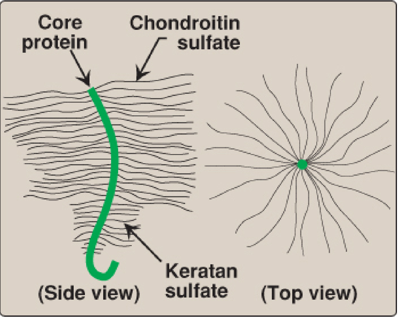
2.3 Glycosaminoglycans (GAGs)
GAGs are long, unbranched polysaccharide chains made of repeating disaccharide units (one amino sugar + one uronic acid or galactose). They are highly negatively charged (sulfate and carboxylate groups), attract Na+ and water, and give the ECM its gel-like, hydrated character.
| GAG | Sugar composition | Key Location | Key Features |
|---|---|---|---|
| Hyaluronic acid | GlcUA + GlcNAc | Cartilage, synovial fluid, skin, vitreous | Only unsulfated GAG; not covalently linked to protein; forms backbone of proteoglycan aggregates |
| Chondroitin 4- and 6-sulfate | GlcUA + GalNAc-sulfate | Cartilage, bone, tendons | Most abundant GAG; important for compressibility of cartilage |
| Keratan sulfate | Gal + GlcNAc-sulfate | Cornea, cartilage, bone | No uronic acid |
| Heparan sulfate | GlcUA + GlcNAc-sulfate | Basement membranes, cell surfaces | Co-receptor for growth factors |
| Heparin | IdUA + GlcNAc-sulfate | Mast cell granules, lung, liver | Anticoagulant |
| Dermatan sulfate | IdUA + GalNAc-sulfate | Skin, heart valves, arteries |
Functions of proteoglycans:
- Structural support - resist compressive forces in cartilage (chondroitin + hyaluronic acid)
- Act as sieves - restrict passage of large macromolecules into ECM while allowing free diffusion of small molecules
- Bind growth factors (e.g., decorin binds TGF-beta) - modulate cell signaling
- Attract water by osmosis - the polyanion GAG chains bind Na+/K+ by counterion attraction, drawing water in and providing turgor
- Lippincott, p. 460-464; Harper's, p. 550
GAG synthesis: Occurs in the Golgi, by sequential addition of alternating sugars donated from their UDP-sugar derivatives, catalyzed by specific glycosyltransferases.
2.4 Proteoglycans in Cartilage and Osteoarthritis
In cartilage, aggrecan (the major cartilage proteoglycan) contains multiple chondroitin sulfate and keratan sulfate chains linked to a core protein, which in turn binds hyaluronic acid via link proteins. This enormous aggregate, combined with type II collagen fibers, gives cartilage its ability to withstand compressive loads.
In osteoarthritis, proteoglycans are degraded and lost from cartilage by MMPs and aggrecanases, destroying its compressibility - resulting in pain, stiffness, and progressive joint destruction.
- Lippincott, p. 462-463 (Clinical Application 14.1)
PART 3: BIOCHEMICAL COMPOSITION OF BONE AND CARTILAGE
3.1 Bone
Bone is a specialized connective tissue that provides mechanical support, serves as a calcium/phosphate reservoir, and houses the bone marrow.
Composition:
| Component | % Dry Weight | Details |
|---|---|---|
| Mineral phase (inorganic) | ~65% | Hydroxyapatite crystals: Ca10(PO4)6(OH)2. Also contains CO32-, Mg2+, F-, Na+ |
| Organic matrix (osteoid) | ~35% | ~90% type I collagen; ~10% non-collagenous proteins |
Organic matrix (Osteoid):
- Type I collagen (90%): Provides tensile strength. Cross-linked fibers arranged at angles to resist mechanical shear from all directions.
- Non-collagenous proteins (10%):
- Osteocalcin (bone Gla-protein): vitamin K-dependent; binds calcium and hydroxyapatite; marker of bone turnover
- Osteopontin: cell attachment; involved in bone remodeling
- Bone sialoprotein: nucleates hydroxyapatite crystal formation
- Osteonectin (SPARC): binds collagen and calcium; involved in mineralization
- Alkaline phosphatase: enzyme of osteoblasts; releases inorganic phosphate to promote mineralization
- Proteoglycans: biglycan, decorin (smaller amounts than in cartilage)
Cells of Bone:
- Osteoblasts: synthesize osteoid (type I collagen + matrix proteins); express alkaline phosphatase
- Osteocytes: embedded osteoblasts; mechanosensing
- Osteoclasts: multinucleated; resorb bone by secreting H+ (acidification) and cathepsin K (protease)
Mineralization: Alkaline phosphatase on osteoblast surfaces cleaves phosphate from pyrophosphate, increasing local [Pi], driving precipitation of hydroxyapatite onto collagen fibers.
3.2 Cartilage
Cartilage is avascular and aneural. It relies on diffusion for nutrition.
| Component | Details |
|---|---|
| Water | 65-80% of wet weight (highest in articular cartilage) |
| Type II collagen | Provides tensile strength; resists swelling pressure of proteoglycans |
| Aggrecan (proteoglycan) | Core protein + chondroitin sulfate + keratan sulfate; binds hyaluronic acid |
| Hyaluronic acid | Backbone for proteoglycan aggregates; attracts water |
| Minor collagens | Types IX, XI (regulate fibril diameter and cross-linking) |
| Non-collagenous proteins | COMP (cartilage oligomeric matrix protein), matrilin |
| Chondrocytes | Only cells; synthesize and maintain all matrix components |
Function of water + proteoglycans: Under compressive load, water is squeezed out; when load is removed, the negative charges of GAGs cause rapid re-hydration (spring-back effect), enabling cartilage to function as a shock absorber.
PART 4: VITAMIN D METABOLISM
4.1 Sources and Structure
Vitamin D is a group of fat-soluble sterol hormones.
- Vitamin D2 (ergocalciferol) - from plants; dietary source
- Vitamin D3 (cholecalciferol) - from animal tissues and endogenous synthesis in skin from 7-dehydrocholesterol via UV-B radiation
Both forms are biologically inactive until activated by two sequential hydroxylations.
- Lippincott, p. 1086
4.2 Metabolic Activation (The Two Hydroxylations)
7-Dehydrocholesterol  →[UV light, skin]→  Cholecalciferol (D3)
                                              ↓
                                  [Liver: 25-hydroxylase]
                                              ↓
                            25-OH-D3 (Calcidiol) — major serum storage form
                                              ↓
                          [Kidney: 1-alpha-hydroxylase]  ←stimulated by PTH, low Pi
                                              ↓
                      1,25-diOH-D3 (Calcitriol) — ACTIVE FORM
                      OR
                          [Kidney: 24-hydroxylase] → 24,25-diOH-D3 (inactive)
Key details:
- 25-hydroxylase (liver) - not regulated; produces calcidiol (circulates bound to vitamin D-binding protein; major storage form)
- 1-alpha-hydroxylase (kidney) - the regulated step; stimulated by low serum Pi, PTH, and low Ca2+; inhibited by calcitriol itself (auto-feedback)
- Both hydroxylases are cytochrome P450 enzymes
- Lippincott, p. 1087
4.3 Actions of Calcitriol (1,25-diOH-D3)
Calcitriol acts like a steroid hormone - it binds to the cytosolic Vitamin D Receptor (VDR), the complex translocates to the nucleus, and interacts with Vitamin D Response Elements (VDREs) on DNA to regulate gene transcription.
Target organs and actions:
| Organ | Action | Mechanism |
|---|---|---|
| Intestine | ↑ Ca2+ and Pi absorption | Induces calbindin (Ca2+-binding protein), TRPV6 channels |
| Kidney | ↑ Ca2+ reabsorption (distal tubule) | Induces calbindin in DCT |
| Bone | ↑ Ca2+ mobilization (when blood Ca2+ is low) | Stimulates osteoclast activity (via RANK-L on osteoblasts) |
| Parathyroid | ↓ PTH synthesis (negative feedback) | Suppresses PTH gene transcription |
| Kidney | ↑ 24-hydroxylase activity | Degrades itself (auto-inactivation) |
Lippincott, p. 1088-1091
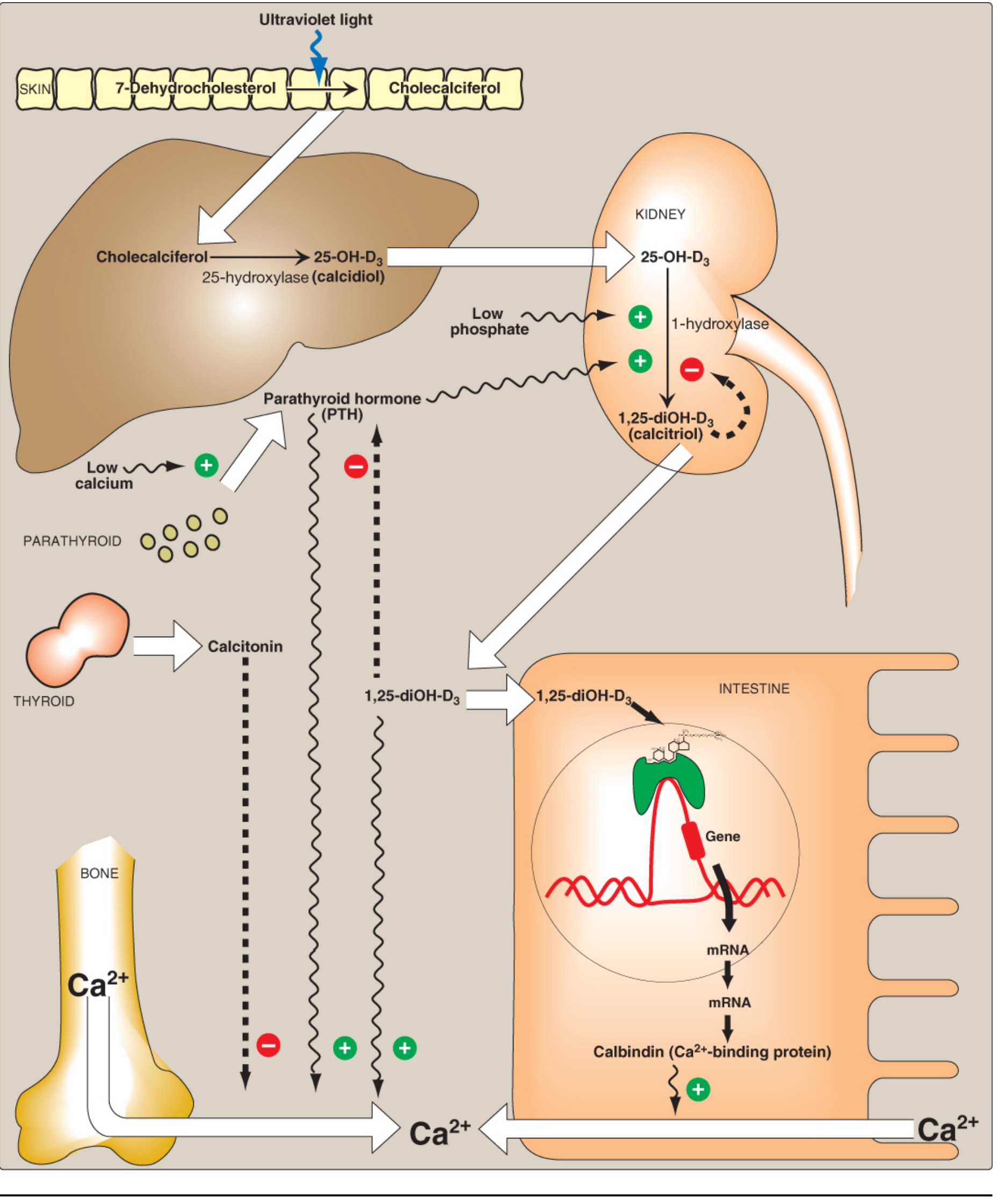
PART 5: REGULATION OF CALCIUM AND PHOSPHATE - Hormonal Interregulation
5.1 Normal Values
| Parameter | Normal Range | Form |
|---|---|---|
| Serum Ca2+ | 8.5-10.4 mg/dL (2.1-2.6 mM) | 50% ionized, 40% protein-bound (albumin), 10% complexed |
| Serum Phosphate | 2.5-4.5 mg/dL | Mostly HPO42- and H2PO4- |
99% of body calcium is in bone and teeth. Only the ionized Ca2+ is physiologically active (muscle contraction, nerve conduction, coagulation, enzyme activation).
5.2 The Three Hormones - Overview
| Hormone | Source | Effect on Ca2+ | Effect on PO4 |
|---|---|---|---|
| PTH | Chief cells, parathyroid | ↑ | ↓ (phosphaturia) |
| Calcitriol (1,25-diOH-D3) | Kidney (activated) | ↑ | ↑ |
| Calcitonin | Parafollicular C-cells, thyroid | ↓ | ↓ |
| FGF-23 | Osteocytes | No direct effect | ↓↓ (phosphaturia) |
5.3 Parathyroid Hormone (PTH)
Stimulus: Low serum ionized Ca2+ sensed by the calcium-sensing receptor (CaSR) on parathyroid chief cells.
PTH is a polypeptide hormone (84 amino acids); its 1-34 N-terminal fragment is biologically active.
Actions on Ca2+ (all raise Ca2+):
- Bone: PTH stimulates osteoclast-mediated bone resorption (indirectly via osteoblasts expressing RANK-L) → releases Ca2+ and PO4 from bone
- Kidney:
- ↑ Ca2+ reabsorption in distal tubule (DCT)
- ↓ Phosphate reabsorption in proximal tubule (PCT) → phosphaturia - this lowers serum PO4
- ↑ 1-alpha-hydroxylase activity → ↑ calcitriol production
- Intestine (indirect): Via increased calcitriol → ↑ Ca2+ and Pi absorption
PTH signal transduction: Binds PTH/PTHrP receptor (PTHR1, a Gs-coupled GPCR) → ↑ cAMP → PKA activation → phosphorylation of target proteins.
5.4 Calcitonin
Stimulus: High serum Ca2+.
Source: Parafollicular C-cells of the thyroid gland.
Actions (all lower Ca2+, oppose PTH):
- Bone: Inhibits osteoclast activity → prevents bone resorption → ↓ Ca2+ release
- Kidney: Inhibits tubular reabsorption of Ca2+ and PO4 → ↑ excretion of both
- Intestine: Inhibits Ca2+ absorption
Calcitonin is physiologically less important than PTH in adult humans but is clinically used to treat hypercalcemia and Paget disease.
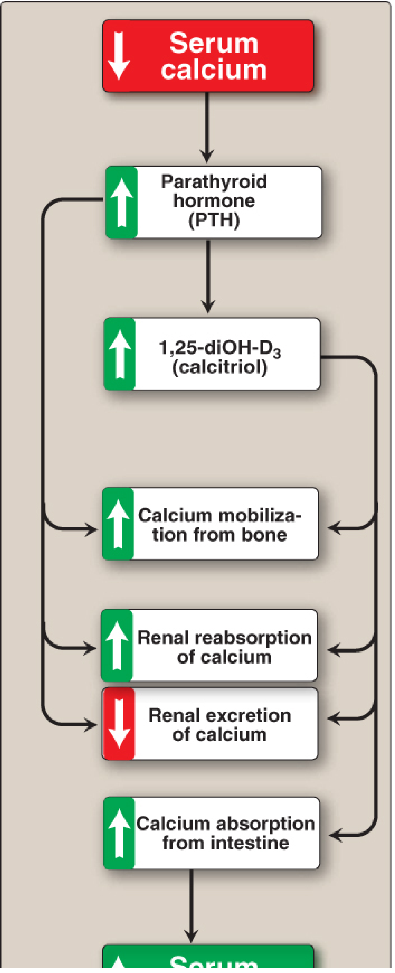
5.5 The Integrated Interregulatory Mechanism
This is the heart of the question - how PTH, calcitriol, and calcitonin interregulate each other and respond to changes in Ca2+ and PO4.
Response to LOW serum Ca2+ (Hypocalcemia):
LOW [Ca2+] serum
      ↓
CaSR on parathyroid chief cells detects ↓Ca2+
      ↓
PTH secretion ↑↑
      ↓
[Bone]              [Kidney]                    [Kidney - indirect]
Osteoclast ↑    DCT: Ca2+ reabsorption ↑      1α-hydroxylase ↑
↑ Ca2+ release  PCT: PO4 excretion ↑ (↓ PO4)       ↓
                                         Calcitriol (1,25-diOH-D3) ↑↑
                                                ↓
                                    [Intestine] Calbindin ↑
                                    Ca2+ absorption ↑ + Pi absorption ↑
                                                ↓
                                    Serum [Ca2+] RESTORED
Negative feedback loop:
- Restored Ca2+ → CaSR activated → PTH secretion ↓
- Calcitriol itself → directly inhibits PTH gene transcription
- Calcitriol activates 24-hydroxylase (inactivating enzyme) and inhibits 1-hydroxylase (self-limiting)
Response to LOW serum Phosphate (Hypophosphatemia):
- Low Pi directly stimulates renal 1-alpha-hydroxylase → ↑ calcitriol → ↑ intestinal Pi absorption and ↑ renal Pi reabsorption
- Low Pi also inhibits FGF-23 secretion (which normally promotes phosphaturia)
Response to HIGH serum Ca2+ (Hypercalcemia):
HIGH [Ca2+]
      ↓
Parafollicular C-cells secrete CALCITONIN
      ↓
Osteoclasts inhibited       Renal excretion of Ca2+ and PO4 ↑
      ↓
[Ca2+] falls back to normal
FGF-23 and phosphate regulation (newer axis):
- Secreted by osteocytes in response to high serum PO4 and high calcitriol
- Acts on kidney: ↓ NaPi cotransporters → ↓ PO4 reabsorption (phosphaturia)
- Also inhibits 1-alpha-hydroxylase (↓ calcitriol) and stimulates 24-hydroxylase (degrades calcitriol)
- Net effect: ↓ serum PO4, ↓ calcitriol
- Goodman & Gilman's, p. based on hormonal regulation section
5.6 Summary: Calcium-Phosphate Regulation at a Glance
| Stimulus | Hormone released | Bone | Kidney (Ca2+) | Kidney (PO4) | Intestine | Net result |
|---|---|---|---|---|---|---|
| ↓ Ca2+ | ↑ PTH | ↑ resorption | ↑ reabsorption | ↓ reabsorption (↑ excretion) | ↑ (via calcitriol) | ↑ Ca2+, ↓ PO4 |
| ↓ Ca2+, ↓ PO4 | ↑ Calcitriol | ↑ mobilization | ↑ Ca2+ reabsorption | ↑ reabsorption | ↑ Ca2+ + Pi absorption | ↑ Ca2+, ↑ PO4 |
| ↑ Ca2+ | ↑ Calcitonin | ↓ resorption | ↑ excretion | ↑ excretion | ↓ absorption | ↓ Ca2+, ↓ PO4 |
| ↑ PO4 | ↑ FGF-23 | - | - | ↑ excretion | ↓ (via ↓ calcitriol) | ↓ PO4 |
PART 6: CLINICAL CORRELATIONS (High-yield for MS-1)
| Condition | Defect | Ca2+ | PO4 | PTH | Calcitriol | Key feature |
|---|---|---|---|---|---|---|
| Hypoparathyroidism | Low/absent PTH | ↓ | ↑ | ↓ | ↓ | Tetany, Chvostek/Trousseau signs |
| Primary Hyperparathyroidism | PTH-secreting adenoma | ↑ | ↓ | ↑ | ↑ | Stones, bones, groans |
| Vitamin D deficiency (Rickets) | Low calcitriol | ↓ | ↓ | ↑ (2° hyperPTH) | ↓ | Soft bones, bowing legs, craniotabes |
| Chronic Kidney Disease | ↓ 1α-hydroxylase, ↑ PO4 retention | ↓ | ↑ | ↑↑ (2° hyperPTH) | ↓ | Renal osteodystrophy |
| Pseudohypoparathyroidism | PTH receptor resistance | ↓ | ↑ | ↑↑ | ↓ | Normal PTH but end-organ resistance |
Quick-Reference Summary
Collagen biosynthesis order (Lippincott mnemonic):
"Pro-chain → Hydroxylation (vit C) → Glycosylation → Triple helix (procollagen) → Secretion → Propeptide cleavage (tropocollagen) → Fibril assembly → Cross-linking (lysyl oxidase, Cu2+)"
Vitamin D activation:
Skin (UV) → Liver (25-OH) → Kidney (1-OH, regulated by PTH/low Pi) → Active calcitriol
Calcium homeostasis hormones:
PTH saves calcium, wastes phosphate | Calcitriol absorbs both | Calcitonin opposes PTH | FGF-23 wastes phosphate
*Sources: Lippincott Illustrated Reviews: Biochemistry, 8th ed. (p. 141-157, 460-465, 1085-1092) | Harper's Illustrated Biochemistry, 32nd ed. (Ch. 44, 50) | Goodman & Gilman's Pharmacological Basis of Therapeutics (Ch. on Mineral Homeostasis) | Histology: A Text and Atlas with Correlated Cell and Molecular Biology (Ross & Pawlina)*The full MS-1 level explanation is complete above. Here's a quick orientation to what was covered:
What Was Covered (All 5 Topics + Interregulation)
1. Collagen (Part 1)
- Structure: Triple helix, Gly-X-Y repeats, hydroxyproline/hydroxylysine, 25+ types with tissue-specific distribution
- Biosynthesis (step-by-step): Pre-pro-alpha chains → RER hydroxylation (Vit C/Fe2+) → glycosylation → triple helix (procollagen) → secretion → propeptide cleavage (tropocollagen) → fibril assembly → cross-linking (lysyl oxidase, Cu2+)
- Collagenopathies: Scurvy, EDS, OI, Lathyrism, Menkes
2. ECM - Collagen and Proteoglycans (Part 2)
- Proteoglycan = core protein + GAG chains (bottle-brush); aggregates via hyaluronic acid backbone
- GAG types: hyaluronic acid, chondroitin sulfate, keratan sulfate, heparan sulfate, heparin, dermatan sulfate - each with unique composition, location, and function
- ECM functions: structural support, molecular sieving, water retention (osmotic), growth factor binding
3. Bone and Cartilage Composition (Part 3)
- Bone: 65% hydroxyapatite (Ca10(PO4)6(OH)2) + 35% organic (90% type I collagen + osteocalcin, osteopontin, alkaline phosphatase)
- Cartilage: Type II collagen + aggrecan + hyaluronic acid + 65-80% water; avascular, reliant on diffusion
4. Vitamin D Metabolism (Part 4)
- Skin (UV) → Cholecalciferol → Liver (25-hydroxylase, unregulated) → Calcidiol (storage form) → Kidney (1-alpha-hydroxylase, regulated) → Calcitriol (active)
- Acts via nuclear VDR → calbindin induction (intestine), renal Ca2+ reabsorption, bone mobilization
5. Hormonal Interregulation of Ca2+ and PO4 (Part 5)
- The integrated PTH-Calcitriol-Calcitonin-FGF23 axis with a detailed flow of signals, target organs, and feedback loops shown in the tables and diagrams
This is a shared conversation. Sign in to Orris to start your own chat.