
Extensor, posterior और dorsal compartment of... Forearm.
posterior compartment forearm extensor muscles anatomy diagram
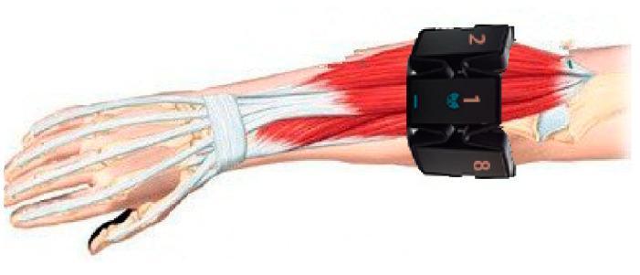
This medical illustration depicts an anatomical diagram of the human forearm and hand with a superimposed wearable surface electromyography (sEMG) device. The underlying anatomy displays the radius and ulna bones, the carpal and metacarpal structures, and the phalanges in white. The musculature of the posterior (extensor) compartment is rendered in red, with tendons highlighted in white crossing the wrist joint beneath the extensor retinaculum. A segmented black armband containing eight dry electrodes is shown positioned on the proximal third of the forearm, approximately one finger-width distal to the elbow joint. The electrodes are numbered, with electrode '1' aligned dorsally with the middle finger, while electrodes '2' and '8' are visible laterally. This visual serves as a technical and anatomical protocol for sensor placement in gesture recognition research, emphasizing the relationship between electrode orientation and underlying superficial muscles and tendons.
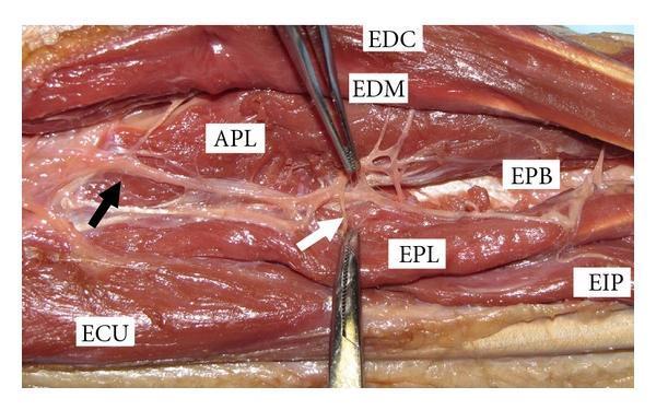
This clinical anatomical photograph displays a cadaveric gross dissection of the human dorsal forearm, highlighting the muscular and nervous anatomy. The image focuses on the extensor compartment, with several muscles clearly labeled: the extensor digitorum communis (EDC), extensor digiti minimi (EDM), abductor pollicis longus (APL), extensor carpi ulnaris (ECU), extensor pollicis brevis (EPB), extensor pollicis longus (EPL), and extensor indicis proprius (EIP). Centrally, the posterior interosseous nerve (PIN) is visible as it exits the arcade of Frohse (indicated by a black arrow). A specific terminal branch of the PIN is shown extending towards the EPL muscle (highlighted by a white arrow). Surgical forceps are positioned to retract tissue, providing a clear view of the neurovascular relationship and the distal innervation pattern of the deep extensor muscles. This visual serves as a critical educational resource for understanding Zone IX forearm anatomy, particularly for neurovascular preservation during surgical procedures or trauma management.
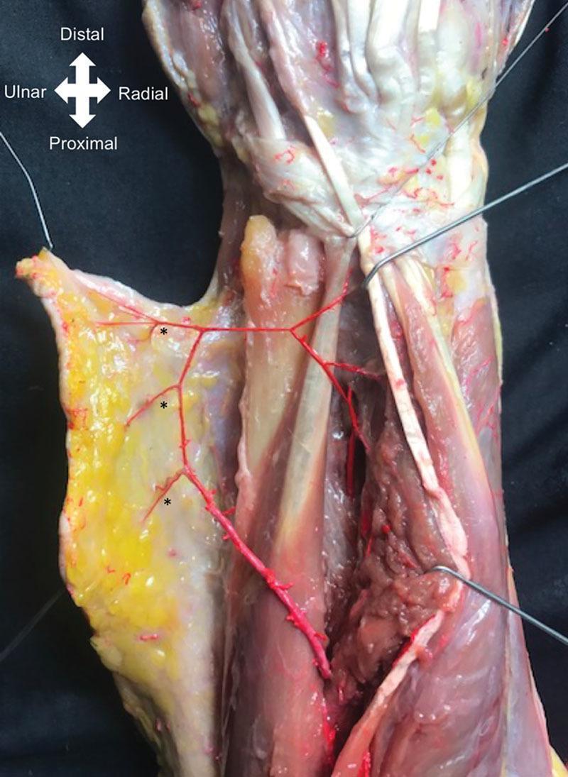
Anatomical dissection of a human cadaver forearm, focusing on the distal third of the posterior compartment. The image demonstrates the surgical anatomy relevant for raising a posterior interosseous artery (PIA) flap. The dissection reveals the Extensor Carpi Ulnaris (ECU) and Extensor Digiti Minimi (EDM) muscles, with a retracted skin and fasciocutaneous paddle reflected ulnarly. Key features include three distinct fasciocutaneous perforators of the PIA, marked with asterisks, emerging through the intermuscular septum between the EDM and ECU. These vessels are highlighted in red to show their branching pattern as they supply the overlying subcutaneous tissue and skin. Directional markers indicate Distal, Proximal, Ulnar, and Radial orientations. This visual is intended for educational purposes regarding reconstructive surgery, specifically illustrating the vascular landmarks and pivot points required for antegrade or retrograde forearm flaps used in hand or elbow defect coverage.
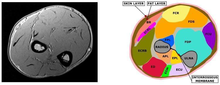
This diagnostic image and anatomical diagram comparison shows a cross-sectional MRI slice of the human forearm (left) alongside its corresponding segmented anatomical map (right). The MRI is a T1-weighted axial scan displaying cortical bone as low-signal (black) voids and muscle bundles as intermediate-signal structures. The segmented diagram identifies key anatomical structures using a color-coding system: the radius and ulna bones are in gray, the interosseous membrane is a black line connecting them, and individual muscles are uniquely colored (e.g., FCR, FDS, FDP, ECU, and ED). The interosseous membrane serves as a landmark dividing the anterior (flexor) and posterior (extensor) muscle compartments. The diagram also illustrates the superficial integumentary layers, with the skin represented in pink and the underlying subcutaneous fat layer in brown. This visual serves as an educational tool for musculoskeletal anatomy, demonstrating the spatial relationships between forearm skeletal structures, deep and superficial muscle groups, and their respective fascial boundaries.
Posterior (Extensor / Dorsal) Compartment of the Forearm
Gray's Anatomy for Students, p. 906-910
Overview
- Wrist joint ko move karna
- Fingers aur thumb ko extend karna
- Supination
Superficial Layer (7 Muscles)
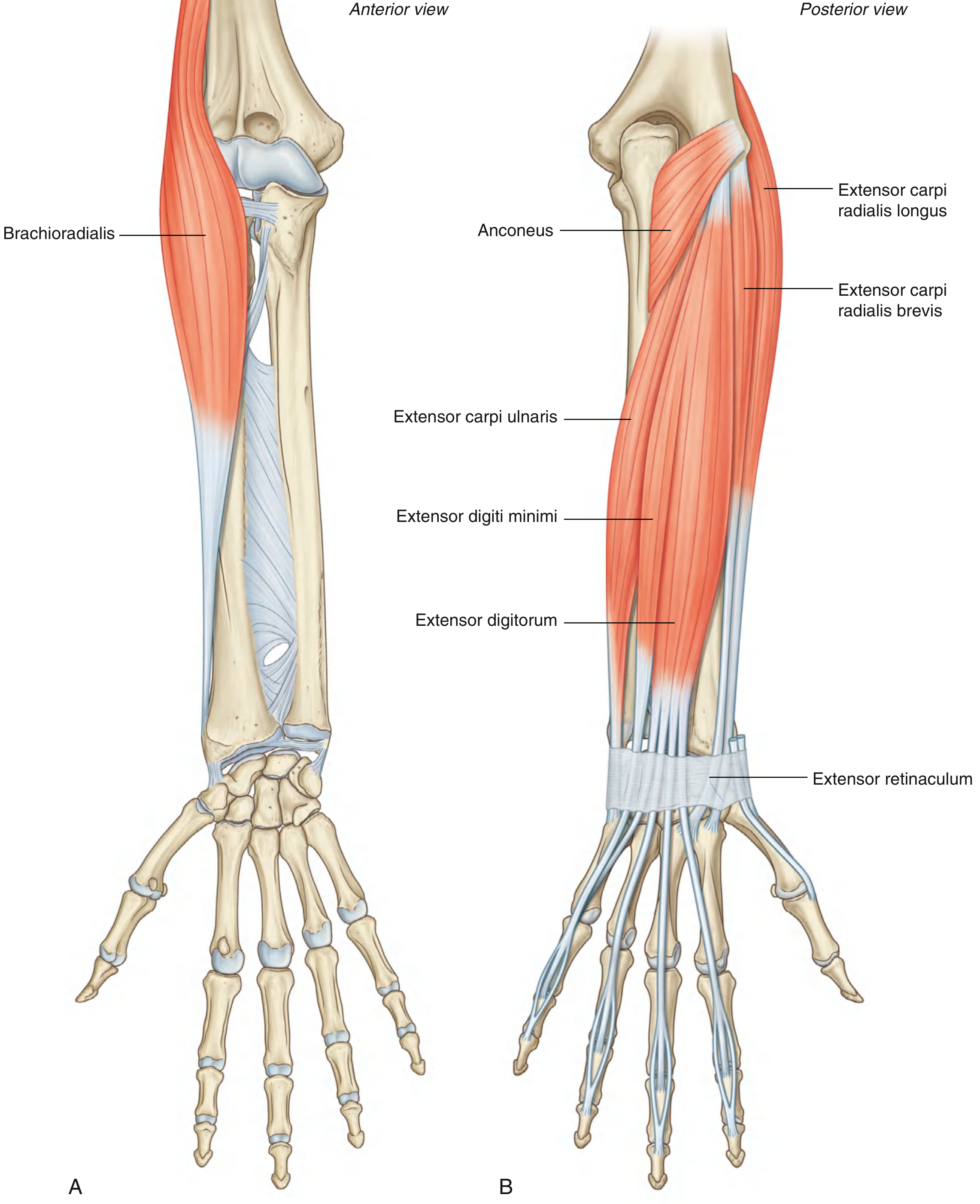
| Muscle | Origin | Insertion | Nerve | Action |
|---|---|---|---|---|
| Brachioradialis | Proximal lateral supraepicondylar ridge | Lateral surface, distal radius (radial styloid proximal) | Radial nerve (C5, C6) - before division | Accessory elbow flexor (midpronation mein) |
| Extensor Carpi Radialis Longus (ECRL) | Distal lateral supraepicondylar ridge | Dorsal base of Metacarpal II | Radial nerve (C6, C7) - before division | Extends + Abducts wrist |
| Extensor Carpi Radialis Brevis (ECRB) | Lateral epicondyle | Dorsal base of Metacarpals II & III | Deep branch of radial nerve (C7, C8) - before supinator | Extends + Abducts wrist |
| Extensor Digitorum | Lateral epicondyle | 4 tendons → extensor hoods → middle & distal phalanges (index to little finger) | Posterior interosseous nerve (C7, C8) | Extends fingers (index, middle, ring, little) |
| Extensor Digiti Minimi | Lateral epicondyle | Extensor hood of little finger | Posterior interosseous nerve (C7, C8) | Extends little finger |
| Extensor Carpi Ulnaris (ECU) | Lateral epicondyle + posterior ulna | Base of Metacarpal V | Posterior interosseous nerve (C7, C8) | Extends + Adducts wrist |
| Anconeus | Lateral epicondyle (posterior surface) | Lateral surface of olecranon + posterior ulna | Radial nerve (C7, C8) | Assists triceps in elbow extension |
Note: Brachioradialis aur Anconeus ke tendons hand mein nahi jate - baaki 5 superficial muscles ke tendons hand tak extend hote hain.
Deep Layer (5 Muscles)
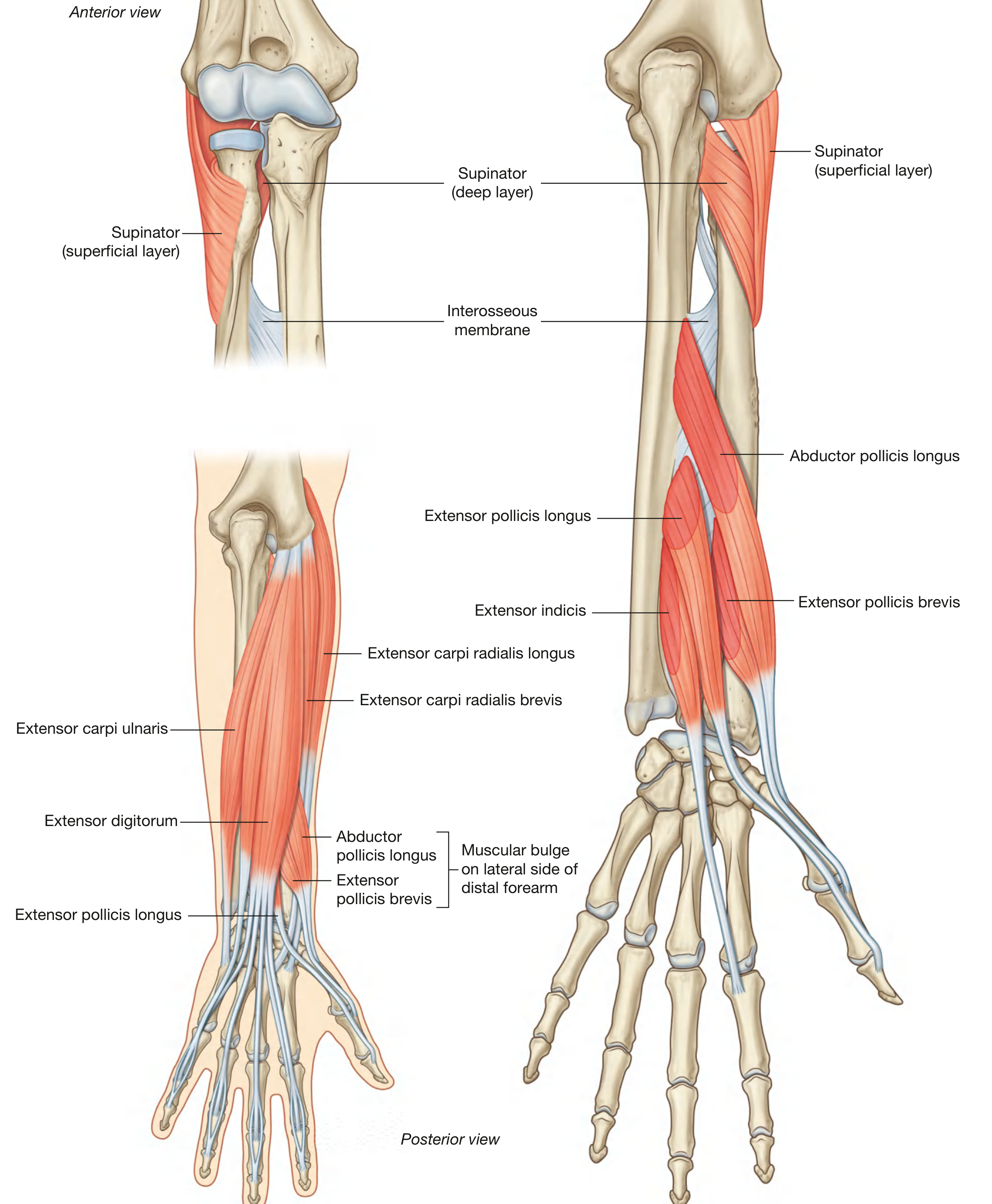
| Muscle | Origin | Insertion | Nerve | Action |
|---|---|---|---|---|
| Supinator | Lateral epicondyle + anular ligament + supinator crest of ulna | Proximal radius (lateral surface, above oblique line) | Deep branch of radial nerve (C6, C7) | Supinates forearm |
| Abductor Pollicis Longus (APL) | Posterior radius + ulna + interosseous membrane | Lateral base of Metacarpal I | Posterior interosseous nerve (C7, C8) | Abducts thumb at CMC joint |
| Extensor Pollicis Brevis (EPB) | Posterior radius + interosseous membrane | Dorsal base of proximal phalanx of thumb | Posterior interosseous nerve (C7, C8) | Extends MCP + CMC of thumb |
| Extensor Pollicis Longus (EPL) | Posterior ulna + interosseous membrane | Dorsal base of distal phalanx of thumb | Posterior interosseous nerve (C7, C8) | Extends all joints of thumb; forms medial border of anatomical snuffbox |
| Extensor Indicis | Posterior ulna (distal to EPL) + interosseous membrane | Extensor hood of index finger | Posterior interosseous nerve (C7, C8) | Extends index finger independently |
Nerve Supply - Summary
Radial Nerve (main trunk)
├── Brachioradialis (C5, C6)
├── ECRL (C6, C7)
│
└── Deep Branch of Radial Nerve
├── ECRB (C7, C8) — before supinator
├── Supinator (C6, C7)
│
└── POSTERIOR INTEROSSEOUS NERVE (after exiting supinator)
├── Extensor Digitorum (C7, C8)
├── Extensor Digiti Minimi (C7, C8)
├── Extensor Carpi Ulnaris (C7, C8)
├── Anconeus (C7, C8)
├── APL (C7, C8)
├── EPB (C7, C8)
├── EPL (C7, C8)
└── Extensor Indicis (C7, C8)
Blood Supply
- Posterior interosseous artery (from common interosseous → ulnar artery) - deep muscles ko
- Anterior interosseous artery - interosseous membrane ke through perforating branches deta hai
- Radial artery - radial side ke extensor muscles ko muscular branches
Clinical Points
- Arcade of Frohse (fibrous arch of supinator) - posterior interosseous nerve yahan compress ho sakta hai → Radial tunnel syndrome / PIN palsy
- Tennis Elbow (Lateral Epicondylitis) - ECRB origin pe common extensor origin pe strain
- Anatomical Snuffbox - EPL medial border, APL + EPB lateral border banaते hain
- APL + EPB distal forearm mein extensor digitorum aur ECRB ke beech se emerge hote hain ek bulge banate hue
Extensor, posterior और dorsal compartment of... Forearm.
posterior extensor compartment forearm muscles superficial deep layer anatomy
This clinical anatomical photograph displays a cadaveric gross dissection of the human dorsal forearm, highlighting the muscular and nervous anatomy. The image focuses on the extensor compartment, with several muscles clearly labeled: the extensor digitorum communis (EDC), extensor digiti minimi (EDM), abductor pollicis longus (APL), extensor carpi ulnaris (ECU), extensor pollicis brevis (EPB), extensor pollicis longus (EPL), and extensor indicis proprius (EIP). Centrally, the posterior interosseous nerve (PIN) is visible as it exits the arcade of Frohse (indicated by a black arrow). A specific terminal branch of the PIN is shown extending towards the EPL muscle (highlighted by a white arrow). Surgical forceps are positioned to retract tissue, providing a clear view of the neurovascular relationship and the distal innervation pattern of the deep extensor muscles. This visual serves as a critical educational resource for understanding Zone IX forearm anatomy, particularly for neurovascular preservation during surgical procedures or trauma management.
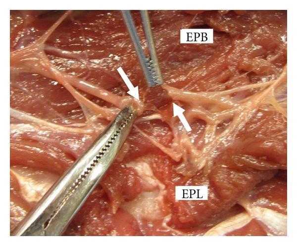
This clinical photograph displays a cadaveric gross dissection of the dorsal forearm, demonstrating a simulated Zone IX laceration injury. The image focuses on the deep muscle layer and neurovascular structures of the posterior compartment. Key anatomical landmarks identified by text labels include the Extensor Pollicis Brevis (EPB) and the Extensor Pollicis Longus (EPL). Two white arrows specifically indicate the severed ends of a motor branch of the posterior interosseous nerve (PIN) as it enters the EPL muscle. Surgical forceps are visible, retracting tissue to provide a clear view of the nerve gap and the surrounding damaged musculotendinous units. This visual emphasizes the clinical significance of identifying nerve involvement in dorsal forearm lacerations, particularly where distal extensor function (thumb and finger extension) is compromised despite intact superficial tendons.
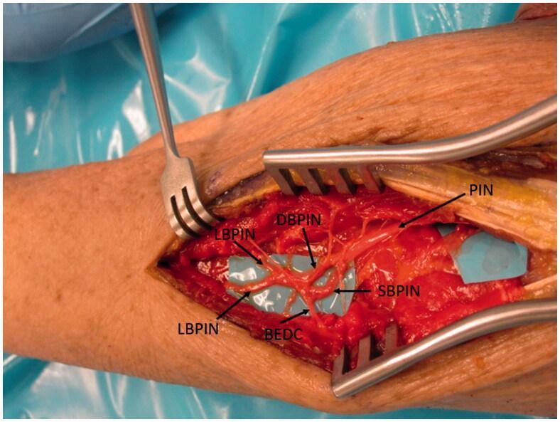
This clinical photograph displays a surgical dissection of the human forearm, focusing on the peripheral nervous system anatomy of the radial nerve. The primary structure shown is the Posterior Interosseous Nerve (PIN) as it courses distally through the extensor compartment. The image highlights a complex branching pattern: the main PIN trunk bifurcates into the Deep Branch (DBPIN) and the Superficial Branch (SBPIN). Further distal arborization is visible, including the lateral branches (LBPIN) and the specific branch to the extensor digitorum communis (BEDC). The nerves are meticulously isolated and placed over blue background material for visual contrast against the red muscular and soft tissue. Surgical retractors are in situ, maintaining exposure of the deep forearm musculature. This anatomical specimen demonstrates the terminal motor innervation of the wrist and finger extensors, critical for understanding PIN palsy, nerve entrapment syndromes, and reconstructive nerve transfer procedures such as AIN-to-PIN transfers.