
Megaloblastic anemia long answer for mbbs pathology
megaloblastic anemia peripheral blood smear hypersegmented neutrophil macrocyte
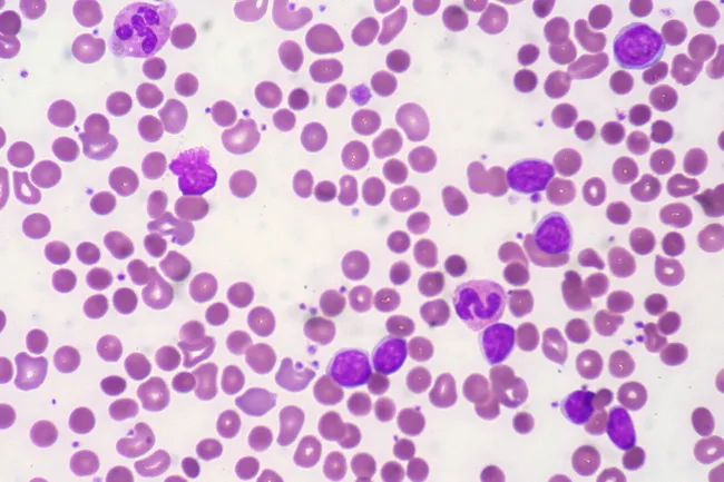
Peripheral blood smear from a patient with known chronic lymphocytic leukemia (CLL) showing autoimmune hemolytic anemia (AIHA). Brightfield microscopy of Wright-Giemsa stained smear at 1000x (oil immersion) reveals two dominant cell populations: numerous small mature lymphocytes typical of CLL and red blood cells showing spherocytic deformation. A smudge cell is evident just left of center, a classical clue for CLL. Red cells display reduced central pallor consistent with spherocytes, indicating intravascular or extravascular hemolysis driven by autoantibodies. The background shows mild polychromasia and rare nucleated RBCs, consistent with a robust reticulocyte response; the corrected reticulocyte count in this case is markedly elevated (>7%). Platelets are not significantly abnormal. These features collectively support autoimmune destruction of erythrocytes in the setting of CLL. Diagnostic significance lies in recognizing AIHA as a common autoimmune complication in CLL, with a clinical picture of sudden Hb decline and compensatory erythropoiesis. Differential considerations include drug-induced hemolysis, other causes of hemolytic anemia, or marrow infiltration by CLL. Clinical correlation includes therapy implications (steroids, rituximab) and close monitoring of hemolysis markers. This image is a valuable teaching resource for hematology morphology, AIHA in CLL, and differential diagnosis in anemia.
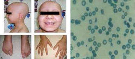
This composite image consists of clinical photographs and a diagnostic peripheral blood smear. Figure 1a (left) shows a pediatric patient with clinical signs including total alopecia (complete absence of scalp hair) and nail pitting affecting both hands and feet, characterized by small punctate depressions on the nail plates. Figure 1b (right) is a microscopic view of a peripheral blood smear demonstrating microangiopathic hemolytic anemia. Visible are numerous schistocytes—fragmented, irregular red blood cells including helmet cells and triangular forms—alongside some normal erythrocytes showing central pallor. The educational focus is on the multi-systemic manifestations of a medical condition, likely a microangiopathic process such as Hemolytic Uremic Syndrome (HUS) or similar systemic pathology, illustrating the correlation between dermatological signs (hair and nail changes) and hematological abnormalities (hemolysis and schistocytosis).
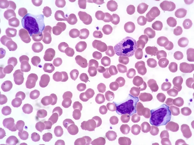
Comprehensive description: This is a brightfield, Wright-Giemsa stained peripheral blood smear examined by light microscopy at high magnification (oil immersion). The specimen shows several activated (atypical) lymphocytes in the peripheral blood, a hallmark of infectious mononucleosis. The lymphocytes have abundant basophilic cytoplasm and irregular, often indented nuclei with clumped chromatin and inconspicuous nucleoli. A normal segmented neutrophil is present for contrast. Erythrocytes exhibit occasional cytoplasmic membrane scalloping around red cells, a feature sometimes observed in EBV-related lymphocytosis. The background cytology is otherwise typical, with uniform erythrocytes and occasional platelets. The activated lymphocytes vary in size and cytoplasmic texture, some appearing plasmacytoid with vesicular basophilia, while others are more indented with coarse chromatin. These morphologic changes reflect an immune response to Epstein-Barr virus infection and are described as reactive lymphocytosis. Clinically, the combination of acute lymphadenitis, fever, a positive Monospot test, and EBV serology supports infectious mononucleosis. The image helps differentiate viral-induced lymphocytosis from bacterial lymphadenitis and from hematologic malignancies when integrated with clinical data. In summary, this smear illustrates the classical hematologic manifestation of EBV-associated mononucleosis. These atypical lymphocytes, sometimes named Downey cells, accompany fever and pharyngitis in EBV infection and aid differentiation from leukemia when combined with serology and clinical data.
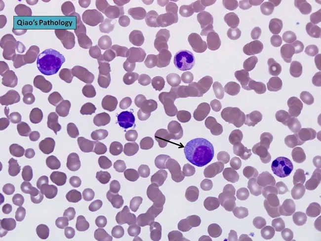
Peripheral blood smear prepared with Wright-Giemsa stain and examined under brightfield illumination using a 100× oil immersion objective reveals a single large plasma cell among a field of mature erythrocytes. The plasma cell displays features typical of clonal plasma cells: basophilic cytoplasm, an eccentrically placed round nucleus, and a conspicuous perinuclear hof; background shows rouleaux formation of red cells. This image exemplifies hematologic involvement by a plasma cell dyscrasia. In plasma cell leukemia, circulating plasma cells constitute more than 20% of leukocytes, a hallmark distinguishing it from conventional multiple myeloma, although both conditions share monoclonal plasma cell proliferation. The presence of circulating plasma cells is associated with younger patient age, higher incidence of anemia, thrombocytopenia, organomegaly, and possible lymphadenopathy; however lytic bone lesions may be less common. Immunophenotypic patterns often include lack of CD56 expression relative to myeloma. Clinically, this morphological finding warrants confirmation with complete blood count, peripheral smear review, quantitative plasma cell percentage, serum protein electrophoresis, and immunofixation to identify monoclonal immunoglobulin isotype (IgD/IgE or light-chain predominance). Early detection of circulating plasma cells supports prognosis assessment and guides therapy decisions in plasma cell dyscrasia management and monitoring, including risk stratification and treatment response evaluation for practice in clinics.
bone marrow megaloblast megaloblastic erythropoiesis nuclear cytoplasmic asynchrony
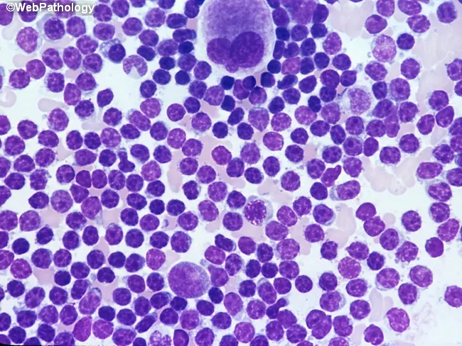
Bone marrow aspirate smear analyzed by light microscopy with high magnification reveals marrow involvement by chronic lymphocytic leukemia (CLL) cells. The predominant population consists of small, dark lymphocytes with scant cytoplasm and condensed, clumped nuclear chromatin, producing high nuclear-to-cytoplasmic ratios. Occasional larger forms and prolymphocytes may be present but are uncommon. A consistent finding is the relative scarcity of cytoplasm and uniform round to slightly irregular nuclei among the bulk of lymphoid cells. The smear may show scattered megakaryocytes, reflecting concurrent hematopoietic activity within the aspirate. Morphology mirrors circulating leukemic cells, consistent with systemic disease. Clinically, such marrow infiltration is associated with cytopenias and advanced disease stage, especially when diffuse, solid infiltration predominates in core biopsy patterns; these patterns correlate with adverse prognosis and with expression of prognostic markers such as ZAP-70. The image corresponds to a high-magnification field intended to document cellular detail rather than architectural marrow patterns. Diagnostic significance lies in confirming lymphoid marrow involvement by CLL, quantifying blast equivalents is not applicable, and differentiating from other small B-cell neoplasms relies on adjunct flow cytometry, cytogenetics, and immunophenotyping. This representation is typical for educational, diagnostic, and research contexts describing CLL marrow morphology.
This histopathology image depicts bone marrow involvement by undifferentiated neuroblastoma. Obtained from a bone marrow biopsy and stained with hematoxylin and eosin, bright-field microscopy at high magnification (approximately 400x). The cellular pattern shows densely packed small round blue neuroblasts with high nuclear-to-cytoplasmic ratio, finely textured hyperchromatic nuclei, inconspicuous nucleoli, and scant cytoplasm. Cells are arranged in diffuse sheets with minimal intervening stroma; occasional Homer Wright–type rosettes may be present, reflecting neural differentiation. Mitotic activity is variable and cytologic features are consistent with embryonal neuroblastic morphology. The marrow is variably replaced by tumor cells, producing a high cellular density and occasional recrudescence of residual hematopoiesis at the periphery. Immunohistochemistry, when performed, typically demonstrates positivity for neuronal markers such as synaptophysin, chromogranin A, and neurofilament protein, supporting neuroblastic lineage; NB84 positivity may be observed in selected cases. Clinically, bone marrow involvement correlates with advanced disease and elevated catecholamines (serum VMA/HVA) in many patients. This image exemplifies a high-grade, poorly differentiated neuroblastoma phenotype, which carries prognostic and therapeutic implications, including risk-stratified chemotherapy, surgical planning, and monitoring for residual disease during follow-up. Recognition of perivascular tumor clusters and subtle stromal response can further refine staging and influence multidisciplinary management decisions in pediatric oncology practice.
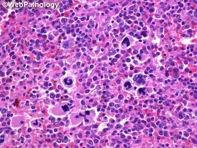
High-magnification brightfield micrograph of a human bone marrow aspirate smear stained with Hematoxylin and Eosin, demonstrating dysplastic megakaryocytes in a hematopoietic background. The megakaryocytes exhibit atypical nuclear morphology, including multinucleation, irregular lobulation, and nuclear fragmentation, with variable cytoplasmic basophilia. Background cellularity shows concurrent dyserythropoiesis and dysplastic myeloid precursors consistent with a myelodysplastic spectrum. Overall architecture may be hypercellular or variably cellular, with scattered normal hematopoietic elements interspersed among abnormal cells. Features are most prominent in the megakaryocytic lineage, where dysplasia is a salient hallmark. The image illustrates morphologic criteria that support a diagnosis in the context of possible progression toward acute leukemia, and should be correlated with peripheral blood counts, marrow blast percentage, cytogenetic and molecular studies. Clinically, such dysplastic megakaryocytes are associated with cytopenias and splenomegaly, and approximately a fifth of patients with related myelodysplastic/myeloproliferative conditions progress to acute myeloid leukemia. This representation is valuable for educational, diagnostic, and research purposes, particularly in studies of megakaryocytic dysplasia, MDS/AML continuum, prognostication, and therapeutic planning focused on palliation and cytopenia management. Careful documentation of features and context enhances reproducibility, supports standardized reporting, and facilitates cross-institutional data sharing for outcome studies and biomarker-driven clinical trials.
Megaloblastic Anemia - Long Answer (MBBS Pathology)
Definition
Etiology / Causes
A. Vitamin B12 Deficiency
| Category | Examples |
|---|---|
| Decreased intake | Strict vegetarianism, macrobiotic diet |
| Intrinsic factor (IF) deficiency | Pernicious anemia (autoimmune), gastrectomy (total/partial) |
| Malabsorption | Ileal resection, Crohn's ileitis, diffuse intestinal disease (lymphoma, systemic sclerosis) |
| Competitive uptake | Diphyllobothrium latum (fish tapeworm), bacterial overgrowth in blind loops/diverticula |
B. Folic Acid Deficiency
| Category | Examples |
|---|---|
| Decreased intake | Alcoholism, indigent diet, infancy, old age |
| Impaired absorption | Malabsorption states, anticonvulsants (phenytoin), oral contraceptives |
| Increased requirement | Pregnancy, infancy, disseminated cancer, hemolytic anemia |
| Increased loss | Hemodialysis |
| Impaired utilization | Methotrexate, trimethoprim (dihydrofolate reductase inhibitors) |
C. Unresponsive to B12 or Folic Acid
- Drugs that inhibit DNA synthesis: methotrexate, hydroxyurea, cytosine arabinoside, 5-fluorouracil
- Erythroleukemia
Biochemistry / Pathogenesis
Vitamin B12 (Cobalamin) Metabolism
- B12 is released from food proteins by pepsin in the stomach
- Binds to haptocorrin (salivary R-protein) in the stomach
- In the duodenum, pancreatic proteases release B12 from haptocorrin
- B12 binds to intrinsic factor (IF) - secreted by gastric parietal cells of the fundic mucosa
- The B12-IF complex travels to the terminal ileum, where it binds the cubilin receptor on ileal enterocytes and is endocytosed
- Inside ileal cells, B12 binds transcobalamin II (TC-II) - its plasma carrier protein
- TC-II delivers B12 to the liver (stores ~3-5 years supply) and bone marrow
Vitamin B12 - Two Key Biochemical Reactions
- Methylcobalamin acts as a cofactor for methionine synthase
- This enzyme converts homocysteine → methionine
- In this reaction, N5-methyl-FH4 (the principal plasma form of folate) donates its methyl group to become FH4 (tetrahydrofolate)
- FH4 is then converted to N5,10-methylene FH4, which is the cofactor for thymidylate synthase in converting dUMP → dTMP (thymidine)
- In B12 deficiency: N5-methyl FH4 cannot be converted to FH4 → "folate trap" → FH4 depleted → dTMP synthesis fails → impaired DNA synthesis
- This is why folic acid partially corrects the anemia of B12 deficiency (but NOT the neurological complications)
- Adenosylcobalamin is a cofactor for methylmalonyl-CoA mutase
- This enzyme converts methylmalonyl-CoA → succinyl-CoA (entering the TCA cycle)
- In B12 deficiency: methylmalonyl-CoA accumulates → methylmalonic acidemia/aciduria
- Accumulated methylmalonyl-CoA leads to abnormal fatty acids being incorporated into myelin → subacute combined degeneration of the spinal cord
- Folic acid does NOT correct this - hence neurological complications persist if only folate is given
Folic Acid Metabolism
- Dietary folates are polyglutamates; intestinal conjugases split them to monoglutamates absorbed in the proximal jejunum
- Converted to 5-methyl-FH4 (transport form) during absorption
- Body stores: only 3-4 months (deficiency develops rapidly)
- Role: FH4 acts as a one-carbon carrier group required for:
- Purine synthesis
- Homocysteine → methionine conversion (requires B12)
- dUMP → dTMP synthesis via thymidylate synthase (most important)
- dTMP is essential for DNA synthesis; its deficiency = the direct cause of megaloblastic change
Pathology (Morphology)
Peripheral Blood Smear
- Macro-ovalocytes (oval macrocytes) - pathognomonic; larger than normal, lack central pallor
- Marked anisocytosis and poikilocytosis - including red cell fragments, dacrocytes (tear-drop cells), microcytes
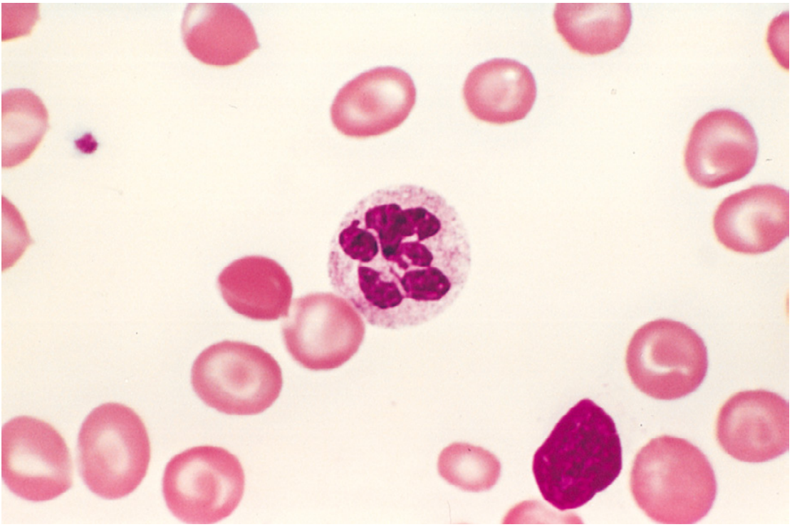
- Hypersegmented neutrophils - 5 or more lobes; any neutrophil with 6+ lobes is abnormal; >5% of neutrophils with ≥5 lobes = hypersegmentation (the single most specific finding)
- Basophilic stippling, Howell-Jolly bodies
- Nucleated RBCs (megaloblasts) may be seen in severe anemia
- Pancytopenia - anemia + leukopenia + thrombocytopenia
- Reticulocyte count is low (ineffective erythropoiesis)
Bone Marrow Aspirate
- Hypercellular marrow (reactive hyperplasia due to elevated EPO)
- Megaloblasts at all stages of erythroid development:
- Promegaloblast: large cell with deeply basophilic cytoplasm, prominent nucleoli, fine ("open") nuclear chromatin
- Basophilic megaloblast: fine chromatin persists
- Polychromatic megaloblast: hemoglobin begins to accumulate but nucleus remains immature (fine chromatin, not condensing)
- Orthochromatic megaloblast (B in Fig. 14.16): cell is hemoglobinized (cytoplasm pink) but the nucleus is not pyknotic - this is the hallmark of nuclear-cytoplasmic asynchrony
- Giant metamyelocytes and band forms in the granulocytic series (also megaloblastic change)
- Megalokaryocytes may be large with bizarre multilobate nuclei
- Increased mitotic figures; cells undergo apoptosis (ineffective hematopoiesis)
- Myeloid:Erythroid (M:E) ratio is reversed - erythroid hyperplasia predominates
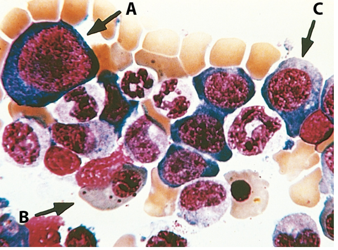
Nuclear-Cytoplasmic Asynchrony (Key Concept)
Pernicious Anemia (Major form of B12 deficiency)
Definition
Pathogenesis (Autoimmune Mechanism)
- Type IV hypersensitivity (cell-mediated) destroys gastric parietal cells
- Three types of autoantibodies are found:
- Type I (Blocking antibody): Binds the B12-binding site of IF → prevents IF binding to B12 (most specific; present in ~75% of cases)
- Type II (Binding antibody): Binds the ileal receptor-binding site of IF → prevents cubilin binding
- Anti-parietal cell antibodies: Against H+/K+ ATPase (proton pump) - present in ~90% of cases but less specific (also found in atrophic gastritis, thyroid disease)
- Autoimmune destruction causes chronic atrophic gastritis with:
- Loss of parietal cells and chief cells
- Mucosal atrophy
- Intestinal metaplasia
- Achlorhydria (reduced stomach acid)
Clinical Features of Pernicious Anemia
- Usually occurs in adults >40 years (peak 60s), slight female predominance
- Associated with other autoimmune diseases: autoimmune thyroiditis (Hashimoto's), Addison's disease, type 1 DM, vitiligo
- Insidious onset (B12 stores last years)
- Triad:
- Anemia - fatigue, pallor, mild jaundice (due to ineffective erythropoiesis with intramedullary hemolysis)
- Glossitis - "beefy red," smooth, sore tongue (Hunter's glossitis)
- Neurological features - subacute combined degeneration of the spinal cord (SACD)
Subacute Combined Degeneration of Spinal Cord (SACD)
- Affects the posterior and lateral columns of the spinal cord
- Posterior column involvement → loss of position sense (proprioception), vibration sense, sensory ataxia
- Lateral column (corticospinal tract) involvement → spastic weakness, exaggerated deep tendon reflexes, Babinski sign
- Also: peripheral neuropathy (glove and stocking pattern), psychiatric manifestations ("megaloblastic madness")
- Key distinction: Neurological complications occur in B12 deficiency only (NOT folate deficiency)
- Folate supplementation alone may mask anemia but will not prevent/treat neurological damage - giving folate to a B12-deficient patient with neurological disease can actually worsen CNS complications
Differences Between B12 and Folate Deficiency
| Feature | Vitamin B12 Deficiency | Folate Deficiency |
|---|---|---|
| Body stores | 3-5 years | 3-4 months |
| Time to manifest | Years | Weeks to months |
| Common cause | Pernicious anemia, ileal disease | Alcoholism, poor diet, pregnancy |
| Neurological complications | Yes (SACD) | No |
| Serum homocysteine | Elevated | Elevated |
| Serum methylmalonic acid | Elevated | Normal |
| Response to folic acid | Anemia improves; SACD does NOT | Full response |
| Schilling test | Abnormal (corrected by IF in pernicious anemia) | Normal |
Laboratory Diagnosis
Hematological Indices
- MCV markedly elevated (>100 fL, often 110-140 fL) - macrocytic anemia
- Hemoglobin reduced; pancytopenia in severe cases
- Low reticulocyte count (ineffective erythropoiesis)
- Elevated LDH and indirect bilirubin (intramedullary hemolysis/ineffective erythropoiesis)
- Elevated serum iron and ferritin (iron utilization decreased)
Specific Tests
| Test | B12 Deficiency | Folate Deficiency |
|---|---|---|
| Serum B12 | Low (<200 pg/mL) | Normal |
| Serum folate | Normal or elevated | Low (<3 ng/mL) |
| RBC folate | Normal | Low (more reliable) |
| Serum homocysteine | Elevated | Elevated |
| Serum methylmalonic acid | Elevated (specific for B12) | Normal |
| Serum anti-IF antibody | Positive (pernicious anemia) | - |
| Serum anti-parietal cell antibody | Positive in PA | - |
Schilling Test (historical, less used now)
- Radioactive B12 given orally → measure urine excretion
- Stage I: Low excretion = impaired absorption
- Stage II: Low excretion corrects with exogenous IF = confirms pernicious anemia (IF deficiency)
- If Stage II still low = ileal disease or bacterial overgrowth
Bone Marrow (if done)
- Hypercellular with megaloblastic changes as described above
Treatment
Vitamin B12 Deficiency
- Pernicious anemia / malabsorption: Intramuscular (IM) cyanocobalamin or hydroxocobalamin
- Initial: 1000 µg IM daily x 7 days, then weekly x 4 weeks, then monthly for life
- High-dose oral B12 (1000-2000 µg/day) also effective due to passive diffusion; useful if injection not feasible
- Dietary deficiency: Oral B12 supplementation + dietary advice (add animal products)
- Response monitoring: Reticulocyte count rises within 5-7 days (reticulocyte crisis); hemoglobin normalizes by 1-2 months; thrombocytopenia and leukopenia correct within 2 weeks
- Neurological disease may not fully reverse - early treatment essential
Folic Acid Deficiency
- Oral folic acid 1-5 mg/day for 3-4 months (or until underlying cause corrected)
- Prophylaxis: 400 µg/day in pregnancy (started pre-conception) - prevents neural tube defects
- Important caution: Always exclude B12 deficiency before giving folic acid alone - folate may correct anemia but mask and worsen neurological complications of B12 deficiency
Complications
- Pancytopenia - susceptibility to infections, bleeding
- Subacute combined degeneration of spinal cord (B12 deficiency only)
- Increased risk of gastric carcinoma - in pernicious anemia due to chronic atrophic gastritis and intestinal metaplasia
- Neural tube defects in fetus (folate deficiency in pregnancy - spina bifida, anencephaly)
- Cardiovascular risk - elevated homocysteine is an independent risk factor for atherosclerosis and thrombosis
- Megaloblastic madness - psychiatric manifestations including dementia, depression, psychosis
Key Exam Points
- The common denominator is impaired DNA synthesis → nuclear-cytoplasmic asynchrony
- Most specific peripheral blood finding: hypersegmented neutrophil (5+ lobes; >5% neutrophils with ≥5 lobes)
- Most specific bone marrow finding: giant metamyelocytes
- Pernicious anemia = autoimmune; blocking anti-IF antibody is most specific
- B12 deficiency: both homocysteine AND methylmalonic acid are elevated
- Folate deficiency: only homocysteine elevated (MMA normal)
- SACD occurs only in B12 deficiency; giving folate alone can precipitate/worsen SACD
- Body stores: B12 = 3-5 years; Folate = 3-4 months
- Ineffective erythropoiesis leads to elevated LDH, elevated indirect bilirubin despite absent hemolysis on peripheral blood smear