
Coumarins derivatives
coumarin derivatives pharmacology classification uses 2024
coumarin warfarin structure anticoagulant mechanism vitamin K
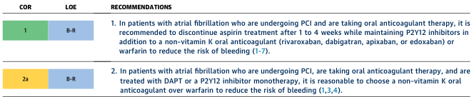
Summary : This figure presents guideline recommendations for managing patients with atrial fibrillation undergoing PCI (percutaneous coronary intervention) who are taking oral anticoagulant therapy, focusing on the discontinuation of aspirin and the choice of anticoagulant to reduce bleeding risk. table: # Columns : • COR (Class of Recommendation): Indicates the strength of the recommendation (1 = strong, 2a = moderate). • LOE (Level of Evidence): Indicates the quality of evidence supporting the recommendation (B-R = moderate quality, randomized). • RECOMMENDATIONS: Detailed clinical guidance for specific patient scenarios. # Rows : • Row 1 (COR 1, LOE B-R, green): – Recommendation: In patients with atrial fibrillation undergoing PCI and taking oral anticoagulant therapy, discontinue aspirin after 1 to 4 weeks while maintaining P2Y12 inhibitors plus a non-vitamin K oral anticoagulant (rivaroxaban, dabigatran, apixaban, edoxaban) or warfarin to reduce bleeding risk. – References: (1-7) • Row 2 (COR 2a, LOE B-R, yellow): – Recommendation: In similar patients treated with DAPT or P2Y12 inhibitor monotherapy, it is reasonable to choose a non-vitamin K oral anticoagulant over warfarin to reduce bleeding risk. – References: (1,3,4) # Design Encodings : • COR 1 is shaded green, COR 2a is shaded yellow. • LOE B-R is shaded blue for both rows. • Recommendations are presented in numbered format for clarity. # Analysis : • The figure emphasizes minimizing bleeding risk in atrial fibrillation patients undergoing PCI by discontinuing aspirin early and preferring non-vitamin K oral anticoagulants over warfarin. • Stronger recommendation (COR 1) is given for discontinuing aspirin, while a reasonable option (COR 2a) is provided for anticoagulant selection. • Both recommendations are supported by moderate-quality randomized evidence.
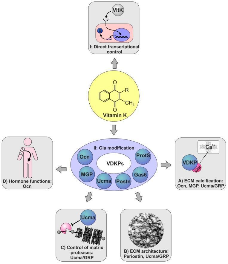
This medical illustration depicts the pathophysiology and signaling pathways of Vitamin K in human skeletal biology. The central element is the chemical structure of Vitamin K, branching into two primary mechanisms. Mechanism I shows 'Direct transcriptional control,' where Vitamin K enters a cell and nucleus to regulate gene expression, likely through receptors like the pregnane X receptor. Mechanism II illustrates 'Gla modification' of Vitamin K-dependent Proteins (VKDPs), including Osteocalcin (Ocn), Matrix Gla protein (MGP), Ucma, Periostin (Postn), Gas6, and Protein S (ProtS). The diagram further details four downstream clinical outcomes of Gla-modified proteins: (A) Extracellular matrix (ECM) calcification, where Gla-residues bind calcium ions (Ca2+); (B) ECM architecture maintenance, specifically involving Periostin and Ucma; (C) Control of matrix proteases, with Ucma/GRP inhibiting protease-mediated degradation of cartilage; and (D) Endocrine or hormone functions, specifically Ocn-mediated crosstalk between bone tissue and systemic glucose metabolism. This flowchart serves as an educational summary of how Vitamin K influences bone development, homeostasis, and pathological calcification.
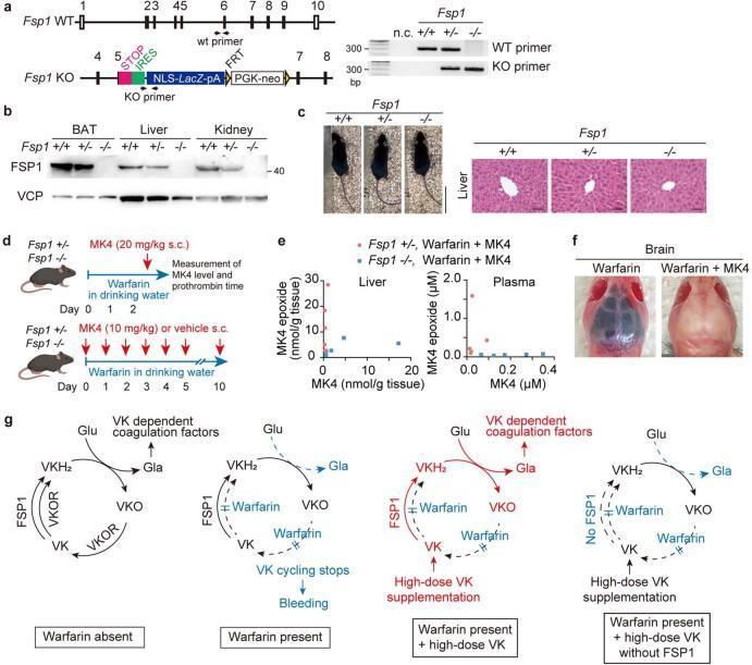
This composite educational graphic illustrates the role of Ferroptosis Suppressor Protein 1 (FSP1) as a warfarin-resistant vitamin K (VK) reductase. Panel (a) shows genomic schematic maps of Fsp1 wild-type and knockout alleles alongside genotyping PCR results. Panel (b) provides an immunoblot for FSP1 expression in adipose tissue, liver, and kidney across genotypes. Panel (c) displays gross morphological and H&E stained liver histology of Fsp1 mice, demonstrating normal architecture. Panel (d) outlines a clinical study design evaluating MK4 (vitamin K2) levels and prothrombin time under high-dose warfarin treatment. Panel (e) uses scatter plots to show the concentrations of MK4 and MK4 epoxide in liver and plasma. Panel (f) presents a clinical photograph comparing mouse brains: the warfarin-only specimen exhibits massive cerebral hemorrhage (dark discoloration), while the warfarin plus MK4 specimen appears normal, illustrating the antidotal effect of MK4. Finally, panel (g) provides pathophysiology flowcharts of the vitamin K cycle, detailing the enzymatic pathways involving VKOR and FSP1, and the mechanism by which high-dose vitamin K bypasses warfarin inhibition to enable carboxylation of coagulation factors.
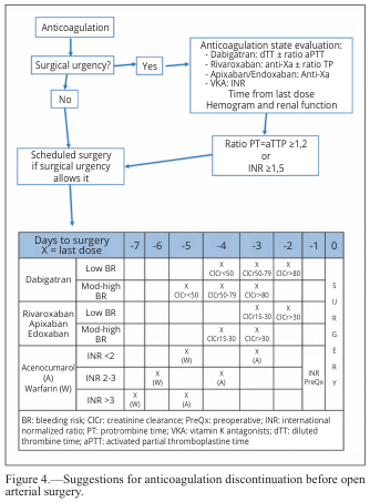
Summary : This figure provides a structured flowchart and reference table for discontinuing anticoagulation therapy before open arterial surgery, including decision points based on surgical urgency, laboratory values, and specific anticoagulant drugs. flowchart: # Nodes : • Anticoagulation (rectangle) • Surgical urgency? (diamond) • No (arrow to "Scheduled surgery if surgical urgency allows it") • Yes (arrow to "Anticoagulation state evaluation: ...") • Anticoagulation state evaluation (rectangle): - Dabigatran: dTT ± ratio aPTT - Rivaroxaban: anti-Xa ± ratio TP - Apixaban/Endoxaban: Anti-Xa - VKA: INR - Time from last dose - Hemogram and renal function • Ratio PT=aTTP ≥1.2 or INR ≥1.5 (rectangle) • Scheduled surgery if surgical urgency allows it (rectangle) # Connectors : • Main flow is top-down, with a decision diamond for "Surgical urgency?" branching to "No" (left) or "Yes" (right). • "No" leads directly to "Scheduled surgery if surgical urgency allows it." • "Yes" leads to "Anticoagulation state evaluation," which then leads to "Ratio PT=aTTP ≥1.2 or INR ≥1.5," and then to "Scheduled surgery if surgical urgency allows it." # Layout : • Flowchart is arranged vertically with a single decision branch. • Supplemented by a reference table below the flowchart. # Reference Table : ## Table Structure : • Title: "Days to surgery X = last dose" • Columns: Days to surgery (-7 to 0), with "SURGERY" spelled vertically in the rightmost column. • Rows: Dabigatran (Low BR, Mod-high BR), Rivaroxaban/Apixaban/Edoxaban (Low BR, Mod-high BR), Acenocoumarol/Warfarin (INR <2, INR >3) • Table cells indicate when to stop each anticoagulant based on bleeding risk and renal function (ClCr thresholds), with "X" marking the last dose day. • Preoperative INR and specific instructions for vitamin K antagonists (VKA) are included. • Abbreviations explained: BR (bleeding risk), ClCr (creatinine clearance), PreOp (preoperative), INR (international normalized ratio), VKA (vitamin K antagonists), dTT (diluted thrombin time), aPTT (activated partial thromboplastine time). # Analysis : • The flowchart provides a clear, stepwise approach for evaluating and discontinuing anticoagulation before surgery, prioritizing urgency and lab values. • The table offers drug-specific timing for cessation based on bleeding risk and renal function, ensuring individualized patient management. • The integration of both flowchart and table supports safe perioperative planning for patients on anticoagulants.
Coumarin Derivatives
1. The Coumarin Scaffold
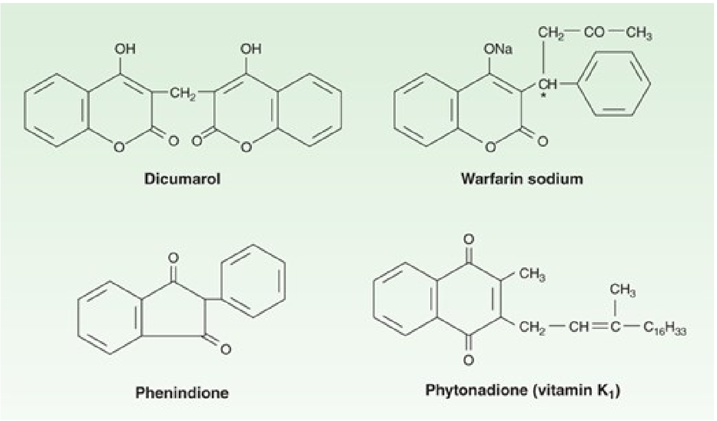
2. Classification of Coumarin Derivatives
A. Simple (Monosubstituted) Coumarins
- Umbelliferone (7-hydroxycoumarin) - antifungal, anti-inflammatory
- Esculetin (6,7-dihydroxycoumarin) - antioxidant
- Osthole - neuroprotective, anti-osteoporotic
- Novobiocin, Coumermycin - antibiotic aminocoumarins
B. Furanocoumarins
- Psoralen, Bergapten, Methoxsalen (8-MOP) - used in PUVA therapy for psoriasis and vitiligo
- Imperatorin - anti-inflammatory
C. Pyranocoumarins
- Linear: Grandivitin, Agasyllin
- Angular: Calanolide A/B (anti-HIV activity), Inophyllum series
D. 4-Hydroxycoumarin Derivatives (Anticoagulants)
- Dicumarol (bishydroxycoumarin) - the first identified, formed in spoiled sweet clover
- Warfarin - the most widely used oral anticoagulant globally
- Acenocoumarol - shorter half-life than warfarin
- Phenprocoumon - longer half-life, used in Europe
3. Mechanism of Action - Anticoagulant Coumarins
- Vitamin K (hydroquinone, KH₂) is a cofactor for gamma-carboxylation of glutamate residues on coagulation factors II (prothrombin), VII, IX, and X - as well as anticoagulant proteins C and S.
- During carboxylation, vitamin K is oxidized to vitamin K epoxide (KO), an inactive form.
- The enzyme vitamin K epoxide reductase (VKORC1) normally reduces KO back to active KH₂.
- Warfarin (and other coumarins) inhibit VKORC1, blocking reactivation of vitamin K.
- Without active vitamin K, the liver synthesizes incomplete, biologically inactive clotting factors (lacking Gla residues, unable to bind Ca²⁺ and phospholipid surfaces).
- Factor VII: 6 h (first to fall)
- Factor IX: 24 h
- Factor X: 40 h
- Factor II (prothrombin): 60 h
4. Warfarin - Key Pharmacokinetics
| Property | Detail |
|---|---|
| Route | Oral (sodium salt) |
| Bioavailability | ~100% |
| Protein binding | >99% (albumin) |
| Volume of distribution | Small (albumin space) |
| Half-life | ~36 hours |
| Stereoisomers | Racemic mixture; S-warfarin is 4x more potent than R-warfarin |
| Metabolism | CYP2C9 (S-form), CYP3A4/1A2 (R-form) |
| Monitoring | INR (target 2-3 for most indications; 2.5-3.5 for mechanical heart valves) |
- Katzung's Basic and Clinical Pharmacology, 16th Ed, p. 961
5. Clinical Indications
- Prevention and treatment of venous thromboembolism (DVT, PE)
- Stroke prevention in atrial fibrillation
- Patients with mechanical prosthetic heart valves
- Secondary prevention after myocardial infarction (selected patients)
6. Toxicity and Adverse Effects
Hemorrhage
Warfarin-induced skin necrosis
- Occurs in the first 1-2 weeks of therapy, especially in patients with inherited protein C deficiency.
- Mechanism: protein C (short half-life) is depleted before the procoagulant factors, causing paradoxical venous thrombosis and necrosis in fatty areas (breast, buttocks, thighs).
Teratogenicity
- Warfarin crosses the placenta freely.
- Causes warfarin embryopathy: abnormal bone formation, nasal hypoplasia, stippled epiphyses - due to inhibition of vitamin K-dependent bone proteins (osteocalcin).
- Also causes fetal hemorrhage.
- Contraindicated in pregnancy - use heparin instead. - Katzung, p. 962
7. Drug Interactions (Warfarin)
| Interaction Type | Examples | Effect on INR |
|---|---|---|
| CYP2C9 inhibitors (↑ S-warfarin) | Fluconazole, amiodarone, metronidazole | Increased (↑ bleeding risk) |
| CYP inducers | Rifampicin, carbamazepine, St John's Wort | Decreased (↑ thrombosis risk) |
| Protein displacement | Aspirin, NSAIDs | Increased; also ↑ GI bleed risk |
| Vitamin K intake (diet) | Green leafy vegetables | Decreased |
| CYP2C9 inhibitors | Vorinostat, romidepsin | Increased (requires close monitoring) - Fitzpatrick's Dermatology |
| Herbal: Dong Quai | Contains coumarin derivatives | Potentiates anticoagulation - Berek & Novak's Gynecology |
8. Reversal of Coumarin Anticoagulation
| Situation | Management |
|---|---|
| Supratherapeutic INR, no bleeding | Hold dose ± oral vitamin K |
| Major hemorrhage | IV vitamin K + Prothrombin Complex Concentrate (PCC) or Fresh Frozen Plasma (FFP) |
| Reversal timeline | Vitamin K requires 12-24 hours for effect (new factor synthesis needed); PCC/FFP works immediately |
9. Other Pharmacological Activities of Coumarin Derivatives
| Activity | Examples | Notes |
|---|---|---|
| Anticancer | Osthole, Scopoletin, synthetic hybrids | Cell cycle arrest, apoptosis induction, inhibition of PI3K/Akt, topoisomerase inhibition; active research area for gynecological and other cancers (Frontiers in Pharmacology, 2024) |
| Antimicrobial | Novobiocin, Coumermycin | Inhibit bacterial DNA gyrase (type II topoisomerase); aminocoumarin antibiotics |
| Anti-inflammatory | Esculetin, Osthole | Inhibit COX, LOX, and NF-κB pathways |
| Antioxidant | Umbelliferone, Fraxetin | Free radical scavenging |
| Neuroprotective | Osthole, Scoparone | MAO inhibition, AChE inhibition (Alzheimer's research) |
| Antiviral | Calanolide A (anti-HIV, NNRTI activity) | Pyranocoumarin with activity against HIV-1 RT |
| Photosensitizers (PUVA) | Psoralen, Methoxsalen | Used in photochemotherapy for psoriasis, vitiligo, CTCL |
| Antifungal | Various 3-substituted derivatives | Disrupts fungal membrane integrity |
| Antitubercular | Coumarin-hydrazide hybrids | Active against Mycobacterium tuberculosis |
10. Newer Oral Anticoagulants vs. Coumarin Derivatives
| Feature | Warfarin (Coumarin) | DOACs (Dabigatran, Rivaroxaban, Apixaban, Edoxaban) |
|---|---|---|
| Target | VKORC1 (indirect, multiple factors) | Thrombin (IIa) or Factor Xa (direct) |
| Monitoring | INR required | Not routinely required |
| Onset | Days | Hours |
| Interactions | Many | Fewer (P-gp/CYP3A4 mainly) |
| Reversal | Vitamin K, PCC | Idarucizumab (dabigatran), Andexanet alfa (Xa inhibitors) |
| Pregnancy | Contraindicated | Contraindicated |
| Mechanical valves | Drug of choice | DOACs NOT recommended |
- Harper's Illustrated Biochemistry, 32nd Ed, p. 697; Katzung's Basic and Clinical Pharmacology, 16th Ed