
Tell me some basics about the lipoproteins LDL and HDL.
LDL HDL lipoprotein structure diagram
![This medical infographic illustrates the structure and pathophysiology of Lipoprotein(a) [Lp(a)] in the context of cardiovascular disease. The diagram shows a central 'LDL-like particle' (yellow) containing an ApoB-100 molecule (blue oval) and associated Apo(a) glycoprotein chains (represented as green hexagonal subunits). The diagram outlines two primary mechanisms of action: 1) Proatherogenic effects, including intimal cholesterol deposition, inflammation, and oxidized phospholipids (OxPL), which lead to Atherosclerosis; and 2) Prothrombotic effects, specifically decreased fibrinolysis, which contribute to Thrombosis. A third clinical outcome, Aortic Valve Calcification, is also highlighted as a downstream effect of elevated Lp(a). The visual summarizes the multifaceted risk factors Lp(a) poses to the vascular and valvular systems, making it an important clinical marker for atherosclerotic cardiovascular disease (ASCVD) and aortic stenosis.](https://cdn.orris.care/cdss_images/pmc_clinical_VQA_7b41b5b4996ac28dd96d6dc75c1abb5b4a22f6b13a2c641d95bd4bd4353c4edc.jpg)
This medical infographic illustrates the structure and pathophysiology of Lipoprotein(a) [Lp(a)] in the context of cardiovascular disease. The diagram shows a central 'LDL-like particle' (yellow) containing an ApoB-100 molecule (blue oval) and associated Apo(a) glycoprotein chains (represented as green hexagonal subunits). The diagram outlines two primary mechanisms of action: 1) Proatherogenic effects, including intimal cholesterol deposition, inflammation, and oxidized phospholipids (OxPL), which lead to Atherosclerosis; and 2) Prothrombotic effects, specifically decreased fibrinolysis, which contribute to Thrombosis. A third clinical outcome, Aortic Valve Calcification, is also highlighted as a downstream effect of elevated Lp(a). The visual summarizes the multifaceted risk factors Lp(a) poses to the vascular and valvular systems, making it an important clinical marker for atherosclerotic cardiovascular disease (ASCVD) and aortic stenosis.
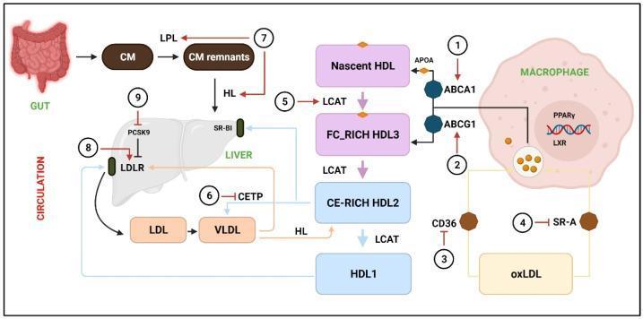
A pathophysiology diagram illustrating cholesterol metabolism, lipoprotein circulation, and Reverse Cholesterol Transport (RCT). The process is divided into exogenous, endogenous, and HDL pathways. In the exogenous pathway, the gut produces chylomicrons (CM), which are hydrolyzed by Lipoprotein Lipase (LPL) into CM remnants for hepatic uptake via SR-BI and Hepatic Lipase (HL) mediation. The endogenous pathway shows the liver secreting VLDL, which matures into LDL; LDL returns to the liver via the LDL receptor (LDLR), a process regulated by PCSK9 and CETP. The RCT/HDL pathway features Nascent HDL (APOA-associated) acquiring cholesterol from macrophages through ABCA1 and ABCG1 transporters. Maturation from FC-rich HDL3 to CE-rich HDL2 and HDL1 is catalyzed by LCAT. Specialized hepatic uptake occurs via SR-BI. On the right, a macrophage is shown accumulating oxidized LDL (oxLDL) via CD36 and SR-A receptors, leading to foam cell formation, regulated by PPARγ and LXR nuclear receptors. Numbered annotations (1-9) indicate potential therapeutic targets for phytochemicals to accelerate RCT and reduce atherosclerosis.
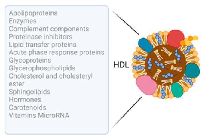
This medical infographic features a schematic diagram illustrating the molecular composition of a High-Density Lipoprotein (HDL) particle. The illustration depicts a cross-sectional view of the HDL, showing a hydrophobic central core containing neutral lipids, represented by intertwined structures. The particle's surface is bounded by a phospholipid monolayer, visualized as small orange globular units, interspersed with larger, multi-colored amorphous shapes representing apolipoproteins and various membrane-associated proteins. A list of cargo molecules associated with HDL is provided, categorized into proteins (Apolipoproteins, Enzymes, Complement components, Proteinase inhibitors, Lipid transfer proteins, Acute phase response proteins, Glycoproteins), lipids (Glycerophospholipids, Cholesterol and cholesteryl ester, Sphingolipids), and other bioactive molecules (Hormones, Carotenoids, Vitamins, MicroRNA). The diagram serves as an educational tool to demonstrate the complexity of HDL as more than just a cholesterol carrier, highlighting its role as a dynamic transport vehicle for a diverse array of proteins and signaling molecules involved in metabolic and immune pathways.
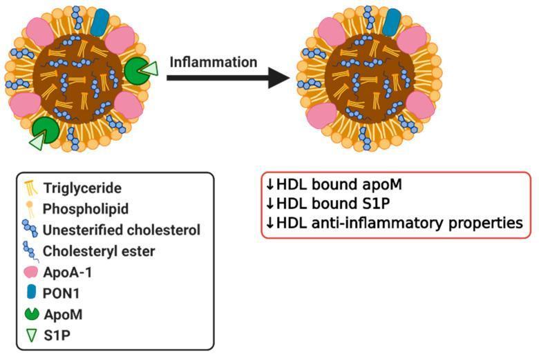
This pathophysiology diagram illustrates the transition of High-Density Lipoprotein (HDL) into a dysfunctional state due to inflammation. The visual displays two spherical HDL particles: one in a healthy state and one post-inflammation, linked by a directional arrow. A detailed legend identifies key components: a hydrophobic core of triglycerides and cholesteryl esters, and a surface monolayer containing phospholipids, unesterified cholesterol, Apolipoprotein A-1 (ApoA-1), Paraoxonase 1 (PON1), Apolipoprotein M (ApoM), and Sphingosine 1-phosphate (S1P). In the healthy state, ApoM (green circles) and its ligand S1P (light green triangles) are bound to the particle surface. Following the 'Inflammation' transition, the second particle shows the loss of ApoM and S1P molecules. An adjacent summary box specifies that inflammation leads to a decrease in HDL-bound ApoM, a decrease in HDL-bound S1P, and a subsequent reduction in the anti-inflammatory properties of the HDL particle. This diagram highlights how systemic inflammatory stimuli alter lipoprotein composition, contributing to increased cardiovascular risk.
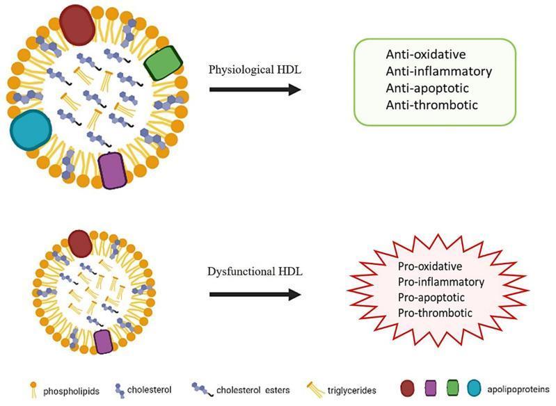
This pathophysiology diagram compares the structural and functional properties of Physiological HDL (High-Density Lipoprotein) versus Dysfunctional HDL. The visual depicts HDL as spherical lipoprotein particles composed of an outer phospholipid monolayer (orange) embedded with free cholesterol and various apolipoproteins (represented by red, purple, green, and teal shapes). The hydrophobic core contains cholesterol esters (blue) and triglycerides (yellow). The top section illustrates Physiological HDL, characterized by a robust complement of apolipoproteins and organized lipid content, leading to anti-oxidative, anti-inflammatory, anti-apoptotic, and anti-thrombotic properties. The bottom section illustrates Dysfunctional HDL, which visually shows a smaller particle size, a relative loss of specific apolipoproteins (green and teal components), and altered internal lipid distribution. These structural modifications result in a functional shift toward pro-oxidative, pro-inflammatory, pro-apoptotic, and pro-thrombotic states. This diagram serves as a medical educational resource for understanding the loss of atheroprotective functions in HDL under pathological conditions like metabolic syndrome or chronic inflammation.
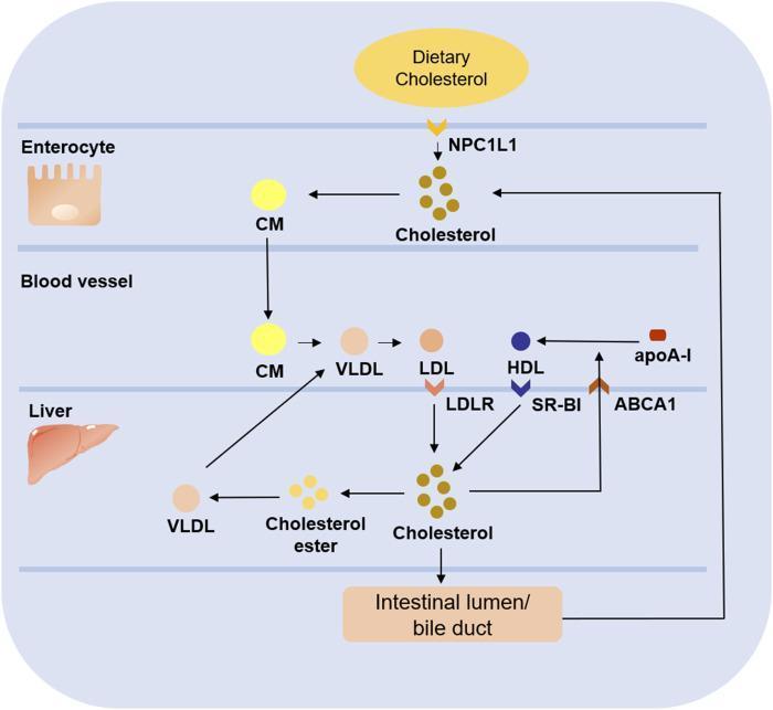
A pathophysiology diagram illustrating the pathways of cholesterol metabolism across four compartments: Enterocyte, Blood Vessel, Liver, and Intestinal Lumen/Bile Duct. Dietary cholesterol enters the enterocyte via the NPC1L1 transporter and is packaged into chylomicrons (CM). CMs enter the blood vessel and undergo conversion into very low-density lipoprotein (VLDL) and then low-density lipoprotein (LDL). LDL is taken up by the liver via the LDL receptor (LDLR). Within the liver, cholesterol can be converted into cholesterol esters to form nascent VLDL for secretion back into the blood vessel, or it can be excreted into the intestinal lumen/bile duct for potential reabsorption via NPC1L1. The diagram also shows reverse cholesterol transport: free liver cholesterol is transferred to the blood via ABCA1, where it combines with apoA-I to form high-density lipoprotein (HDL). HDL then returns cholesterol to the liver through the SR-BI receptor. This diagram highlights key lipoproteins (CM, VLDL, LDL, HDL) and regulatory proteins (NPC1L1, LDLR, ABCA1, SR-BI) involved in systemic lipid homeostasis.
Does low density lipids have a low density and high lipid, or the high lipid and high density?
- Lipoproteins are particles made of lipids + proteins. Protein is denser than lipid.
- The more protein a particle has relative to lipid, the higher its overall density.
- LDL is ~75-80% lipid, ~20-25% protein - lots of lipid, relatively little protein = low density.
- HDL is the opposite - ~50% protein, ~50% lipid - more protein = higher density.
| Particle | Lipid % | Protein % | Density |
|---|---|---|---|
| Chylomicron | ~99% | ~1% | Lowest |
| VLDL | ~92% | ~8% | Very low |
| LDL | ~75-80% | ~20-25% | Low |
| HDL | ~50% | ~50% | High |
HDL is high lipid content or the high density.
- LDL = low density = high lipid, low protein
- HDL = high density = low lipid, high protein