
Acute myeloid leukemia
acute myeloid leukemia blast cells bone marrow smear
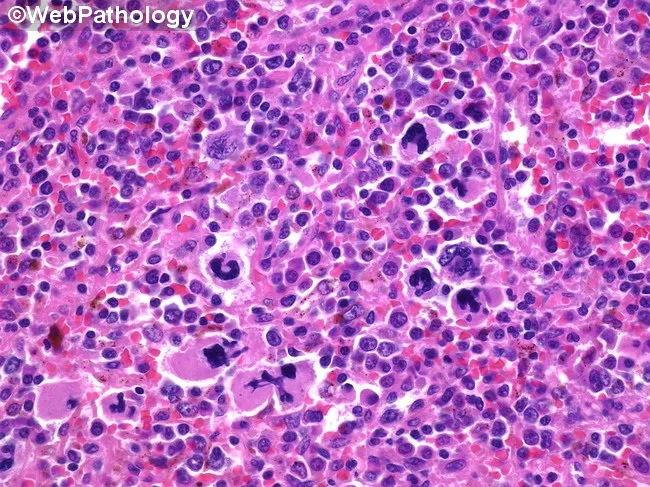
High-magnification brightfield micrograph of a human bone marrow aspirate smear stained with Hematoxylin and Eosin, demonstrating dysplastic megakaryocytes in a hematopoietic background. The megakaryocytes exhibit atypical nuclear morphology, including multinucleation, irregular lobulation, and nuclear fragmentation, with variable cytoplasmic basophilia. Background cellularity shows concurrent dyserythropoiesis and dysplastic myeloid precursors consistent with a myelodysplastic spectrum. Overall architecture may be hypercellular or variably cellular, with scattered normal hematopoietic elements interspersed among abnormal cells. Features are most prominent in the megakaryocytic lineage, where dysplasia is a salient hallmark. The image illustrates morphologic criteria that support a diagnosis in the context of possible progression toward acute leukemia, and should be correlated with peripheral blood counts, marrow blast percentage, cytogenetic and molecular studies. Clinically, such dysplastic megakaryocytes are associated with cytopenias and splenomegaly, and approximately a fifth of patients with related myelodysplastic/myeloproliferative conditions progress to acute myeloid leukemia. This representation is valuable for educational, diagnostic, and research purposes, particularly in studies of megakaryocytic dysplasia, MDS/AML continuum, prognostication, and therapeutic planning focused on palliation and cytopenia management. Careful documentation of features and context enhances reproducibility, supports standardized reporting, and facilitates cross-institutional data sharing for outcome studies and biomarker-driven clinical trials.
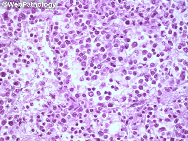
Imaging modality: light microscopy of a hematopathology slide from a bone marrow aspirate smear. Stain: Hematoxylin and Eosin (H&E). Magnification: high-power field (~400x total; 40x objective with 10x ocular). Anatomical location: bone marrow within the medullary cavity (hematopoietic tissue). Visual features: sheets of immature blasts with high nuclear-to-cytoplasmic ratio, round to oval nuclei, fine chromatin, prominent nucleoli, and scant basophilic cytoplasm; increased cellularity with near-complete effacement of normal hematopoiesis; occasional mitotic figures; minimal cytoplasmic granularity; sparse residual neutrophils, erythroid precursors, and megakaryocytes. Pathologic interpretation: diffuse marrow infiltration by blasts, consistent with an acute leukemia until immunophenotyping and genetic studies delineate subtype. Diagnostic significance: morphologic hallmark of an acute hematologic malignancy; requires ancillary tests (flow cytometry, immunohistochemistry, cytogenetics/molecular studies) for lineage classification (myeloid vs lymphoid), prognosis, and treatment planning. Differential considerations: ALL (acute lymphoblastic leukemia), AML (acute myeloid leukemia), lymphoblastic lymphoma with marrow involvement, myelodysplastic syndrome with excess blasts, or nonhematopoietic marrow infiltration. Clinical correlation: commonly presents with cytopenias, fatigue, infections, and bleeding; pediatric ALL vs adult AML guidance; urgent hematology-oncology workup is indicated. Educational use: foundational blast morphology recognition, marrow infiltration patterns, and the need for confirmatory immunophenotyping. This image is valuable for training in blast morphology, differential diagnosis, and education in hematopathology.
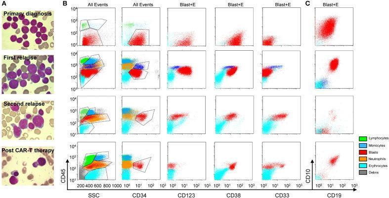
This composite educational graphic illustrates the morphologic and immunophenotypic evolution of a secondary Acute Myeloid Leukemia (AML) case across four clinical stages: primary diagnosis, first relapse, second relapse, and post-anti-CLL1 CAR T-cell therapy. Panel A consists of Wright-Giemsa stained bone marrow aspirate smears. Early stages show dense populations of myeloblasts with high N:C ratios, fine chromatin, and nucleoli. The second relapse reveals increasing dysplasia, while the post-therapy image demonstrates hematopoietic recovery with mature leukocytes and a significant reduction in blast cells. Panels B and C present flow cytometric scatter plots utilizing two gating strategies: 'All Events' and 'Blast+E' (blasts and erythroid cells). Markers analyzed include CD45, Side Scatter (SSC), CD34, CD123, CD38, CD33, CD10, and CD19. Color-coded populations identify blasts (red), lymphocytes (green), monocytes (dark blue), neutrophils (orange), and erythrocytes (light blue). The plots track the lineage switch and immunophenotypic shifts, such as the emergence of myeloid markers (CD33, CD123) in later relapses and the subsequent elimination of the malignant blast population following targeted CAR T-cell therapy.
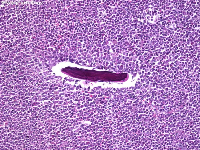
Imaging modality: Histopathology with brightfield microscopy of an H&E stained femoral bone lesion. The specimen shows a dense, cellular marrow‑like infiltrate occupying bone with disruption of normal trabecular architecture. Individual cells are small to medium in size with high nuclear‑to‑cytoplasmic ratio, finely dispersed chromatin, and prominent nucleoli, compatible with myeloblasts. The cell population forms diffuse sheets with scant cytoplasm and frequent mitotic figures. Invasion surrounds a central vascular channel, and occasional residual bone spicules may be seen, consistent with granulocytic sarcoma (myeloid sarcoma), an extramedullary manifestation of acute myeloid leukemia. In the clinical context of chronic myeloid leukemia in blast crisis, this lesion reflects leukemic infiltration of periosteal and marrow compartments, often signaling systemic progression. When integrated with immunophenotype and molecular studies, this tissue supports diagnosis within the AML/acute leukemia spectrum and guides therapy toward AML‑type chemotherapy and consideration of stem cell transplantation. The image is relevant to differential diagnoses such as lymphoma, Ewing sarcoma, and other small round blue cell tumors, especially when cytochemical stains are unavailable. This histopathologic finding emphasizes the importance of evaluating myeloid neoplasms for extramedullary disease in bone and its implications for prognosis, staging, and multidisciplinary management.
"acute myeloid leukemia"[MeSH Terms] AND treatment
"acute myeloid leukemia"[MeSH Terms] AND "targeted therapy"
Acute Myeloid Leukemia (AML)
Definition
- Robbins, Cotran & Kumar Pathologic Basis of Disease
- Henry's Clinical Diagnosis and Management by Laboratory Methods
Epidemiology
- Most common acute leukemia in the first months of life, but accounts for only ~1/3 of childhood acute leukemia
- Becomes the predominant acute leukemia in adults; median age at diagnosis: 60 years
- Incidence: ~10/100,000/year in those over 60; ~13,000 new cases/year in the US
- Risk factors: ionizing radiation, prior cytotoxic chemotherapy, benzene, smoking, Down syndrome; most cases are de novo with no identifiable cause
Pathogenesis
-
Transcription factor mutations - interfere with myeloid differentiation
- t(8;21): RUNX1::RUNX1T1 fusion - disrupts the core binding factor (CBF) transcription complex
- inv(16): CBFB::MYH11 fusion - similarly disrupts CBF
- t(15;17): PML::RARα fusion - blocks retinoic-acid-dependent myeloid maturation (acute promyelocytic leukemia, APL)
-
Signaling mutations - promote proliferation
- FLT3 ITD (internal tandem duplication): ~25-30%, adverse prognosis
- KIT mutations in exon 8/17: present in up to 30% of CBF-AMLs, negates favorable prognosis
- RAS mutations
-
Epigenetic/chromatin modifiers
- DNMT3A, TET2, IDH1/2, EZH2
-
Cohesion/nucleoporin/splicing mutations
- NPM1 mutation: ~30% of AML, favorable prognosis when FLT3-ITD absent
- CEBPA (biallelic): favorable prognosis
WHO Classification (Key Categories)
| Category | Genetic Aberration | Prognosis |
|---|---|---|
| AML with t(8;21); RUNX1::RUNX1T1 | Core binding factor | Favorable |
| AML with inv(16)/t(16;16); CBFB::MYH11 | Core binding factor | Favorable |
| APL with t(15;17); PML::RARα | Very favorable | |
| AML with KMT2A (11q23) rearrangement | >130 partners | Poor |
| AML with mutated NPM1 | Favorable (if FLT3-ITD low) | |
| AML with biallelic CEBPA | Favorable | |
| AML with t(6;9); DEK-NUP214 | Very poor | |
| AML with inv(3)/t(3;3); GATA2/MECOM | Very poor | |
| Therapy-related AML | Prior chemo/XRT | Poor |
| AML, NOS (by differentiation) | Various | Variable |
ELN Risk Stratification (European LeukemiaNet)
| Risk Group | Genetic Features |
|---|---|
| Favorable | t(8;21); inv(16)/t(16;16); NPM1 mutated without FLT3-ITD or with FLT3-ITD^low; biallelic CEBPA |
| Intermediate | NPM1 mutated + FLT3-ITD^high; wild-type NPM1 without FLT3-ITD^low; t(9;11) |
| Adverse | t(6;9); inv(3)/t(3;3); t(v;11q23) KMT2A; monosomy 5/7; complex karyotype; TP53 mutation |
Clinical Features
- Onset resembles acute infection: fever, malaise, prostration
- Signs of granulocytic insufficiency: oral/pharyngeal ulcerations, infections
- Signs of marrow failure: pallor (anemia), bleeding (thrombocytopenia), recurrent infections (neutropenia)
- Lymphadenopathy, splenomegaly, hepatomegaly are usually mild or absent (unlike ALL)
- Gingival hypertrophy in monocytic variants (KMT2A rearrangement)
- DIC risk - especially in APL (t(15;17)) and monocytic subtypes
- Myeloid sarcoma (extramedullary disease) can present as bone lesion or soft tissue mass
Diagnostic Workup
Blood and Bone Marrow Morphology
- Peripheral blood: blasts, cytopenias (anemia, thrombocytopenia, neutropenia)
- Bone marrow aspirate: 500-cell differential; AML = ≥20% blasts
- Myeloblasts: large cells, fine uncondensed chromatin, prominent nucleoli (3-5), variable granules, Auer rods (pathognomonic - fused primary granules appearing as pink needle-like inclusions)
- APL blasts often have bundles of Auer rods ("faggot cells")
Cytochemistry
| Stain | Positive in |
|---|---|
| MPO (myeloperoxidase) | Myeloblasts |
| Sudan black B (SBB) | Myeloblasts |
| Chloroacetate esterase (CAE) | Granulocytic lineage |
| α-naphthyl acetate/butyrate esterase (NaF-inhibited) | Monocytic lineage |
Immunophenotyping (Flow Cytometry)
| Marker | Lineage |
|---|---|
| CD34, CD117, HLA-DR | Immature AML |
| CD13, CD33 | Myeloid |
| CD14, CD15, CD11b | Monocytic |
| CD36, CD71, CD235a (glycophorin A) | Erythroid |
| CD41, CD61 | Megakaryocytic |
| MPO | Myeloid (most specific) |
Cytogenetics and Molecular Testing
- Conventional karyotype (G-banding): essential at diagnosis
- FISH: preferred for detecting specific translocations (e.g., t(15;17) in APL - hematologic emergency)
- Quantitative RT-PCR: MRD monitoring (e.g., PML-RARA, NPM1)
- NGS panels: FLT3, NPM1, IDH1/2, CEBPA, RUNX1, TP53, ASXL1, KMT2A, DNMT3A, TET2 - guides targeted therapy and MRD tracking
Treatment
Induction ("7+3")
- Standard: Cytarabine (ara-C) × 7 days + Anthracycline (daunorubicin or idarubicin) × 3 days
- Achieves CR in 60-80% of younger patients, lower in elderly
APL (AML-M3) - Special Protocol
- ATRA (all-trans retinoic acid) + Arsenic trioxide (ATO): now standard of care for low/intermediate-risk APL
- This combination achieves near-universal CR and cure in most patients
- ATRA syndrome (differentiation syndrome): fever, pulmonary infiltrates - treat with steroids
Consolidation
- CBF-AML (favorable): high-dose cytarabine-based consolidation; responds well, long-term survival achievable
- Intermediate/adverse risk: allogeneic hematopoietic stem cell transplant (allo-HSCT) in first CR
Targeted Therapies (FDA-approved)
| Target | Drug | Indication |
|---|---|---|
| FLT3 | Midostaurin (+ "7+3"), Gilteritinib (relapsed/refractory) | FLT3-mutated AML |
| IDH1 | Ivosidenib | IDH1-mutated AML |
| IDH2 | Enasidenib | IDH2-mutated AML |
| BCL-2 | Venetoclax + azacitidine/low-dose ara-C | Elderly/unfit patients |
| CD33 | Gemtuzumab ozogamicin | CD33+ AML |
Elderly/Unfit Patients
- Venetoclax + azacitidine has become a preferred regimen for older adults not eligible for intensive chemotherapy
- Hypomethylating agents (azacitidine, decitabine) alone as alternative
Prognosis
- Overall 5-year survival: ~30% across all ages; ~50% in patients <60 years with intensive therapy
- APL: highest cure rate (~90%) with ATRA+ATO
- CBF-AML (t(8;21), inv(16)): ~60-70% long-term survival
- Adverse-risk AML (complex karyotype, TP53, monosomy 5/7): <15% 5-year survival
- Minimal residual disease (MRD) negativity after induction is a strong predictor of favorable outcome
Key Histology Images