I now have comprehensive content from four major surgical/gastroenterology textbooks plus clinical images. Let me compose the detailed answer.
Clinical Signs of Inoperability in Gastric Carcinoma
Gastric carcinoma presents with advanced, inoperable disease in a large proportion of patients at the time of diagnosis. The following signs, when found on history, physical examination, or investigations, indicate metastatic/advanced spread that renders the tumor surgically incurable (palliative intent only).
Why These Signs = Inoperability
These signs indicate either distant metastasis (M1 disease) or unresectable locoregional spread, both of which classify the tumor as Stage IV in the AJCC TNM system. Curative (R0) resection is no longer possible once these are present. The goal shifts to palliation, chemotherapy, or targeted therapy.
1. Ascites
What it is: Accumulation of free fluid in the peritoneal cavity.
Mechanism: Gastric carcinoma spreads via the peritoneum (peritoneal carcinomatosis). Tumor cells seed the peritoneal surface, obstruct lymphatic channels, and increase capillary permeability, producing malignant ascites. Peritoneal metastases are a form of M1 disease.
Clinical significance: Malignant ascites from gastric cancer is typically refractory to diuretics - this is a key differentiating feature from hepatic/cardiac ascites. Cytology of ascitic fluid may confirm malignant cells. Peritoneal carcinomatosis is found in up to 40% of advanced gastric cancers, more commonly in the diffuse (signet-ring cell/linitis plastica) subtype. - Goldman-Cecil Medicine, p. 2037
Why inoperable: Peritoneal spread = M1 (distant metastasis) by AJCC staging. Resection is technically futile and confers no survival benefit.
2. Secondaries in the Liver (Hepatic Metastases)
What it is: Palpable hepatomegaly, liver nodularity, jaundice, or liver lesions on imaging.
Mechanism: Gastric cancer spreads hematogenously via the portal vein to the liver. The liver is the most common site of hematogenous metastasis, involved in ~40% of patients who present with metastatic disease. - Sleisenger & Fordtran's Gastrointestinal and Liver Disease, p. [clinical features section]
Clinical features:
- Hepatomegaly with a hard, irregular nodular surface
- Right upper quadrant pain
- Jaundice (from biliary compression or widespread parenchymal replacement)
- Elevated alkaline phosphatase and bilirubin on liver function tests
- Lesions visible on CT/ultrasound
Why inoperable: Liver metastases = M1 disease. While isolated liver metastases have been explored in highly selected cases, this is not standard practice. Widespread hepatic involvement is an absolute contraindication to curative gastrectomy.
3. Para-aortic Lymph Node Involvement
What it is: Involvement of lymph nodes lying along the aorta, outside the standard D2 lymphadenectomy field.
Mechanism: Gastric cancer spreads sequentially through perigastric (N1), regional (N2), and then distant lymph node stations. Para-aortic nodes (Station 16 in the Japanese classification) are considered distant/M1 nodes - they lie beyond the reach of a standard gastrectomy with D2 dissection.
Detection: CT scan, PET-CT, or laparoscopy. Para-aortic nodes are considered bulky/positive when >1 cm on short-axis CT.
Why inoperable: Per AJCC 8th edition, involvement of para-aortic nodes is classified as M1 (distant metastasis), making the tumor Stage IV regardless of T or N1/N2 status. Extended (D3) lymphadenectomy to include para-aortic nodes has been studied but has not demonstrated a survival benefit over D2 in randomized trials, and is not standard.
4. Krukenberg Tumor
What it is: Metastatic deposits in the ovaries from a primary gastric cancer (or other GI malignancy).
Mechanism: Named after Friedrich Krukenberg (1896). Spread occurs via:
- Transcoelomic route (most common): tumor cells shed into the peritoneal cavity and implant on the ovarian surface
- Lymphatic spread: retrograde lymphatic dissemination
- Hematogenous spread: less common
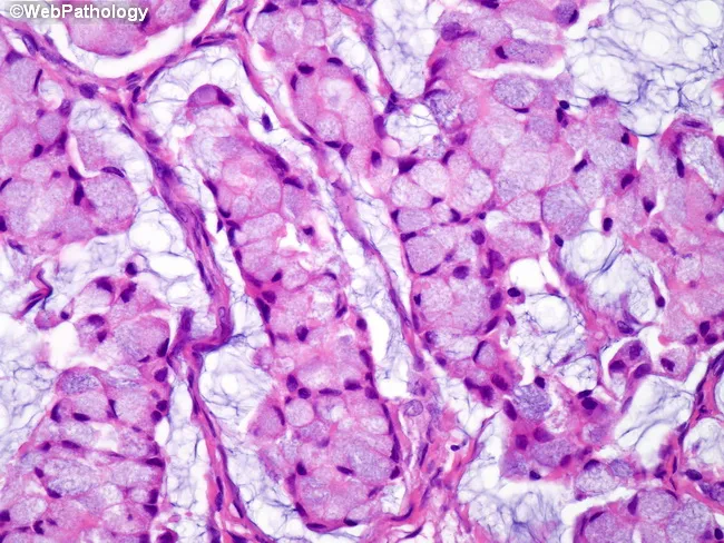
The tumor is characterized histologically by signet-ring cells with intracellular mucin that displaces the nucleus to the periphery, embedded in a fibromyxoid stroma. Gastric diffuse-type adenocarcinoma (linitis plastica, signet ring cell carcinoma) is the most common primary. Krukenberg tumors are classically bilateral (both ovaries involved), which helps distinguish them from primary ovarian malignancy.
Clinical features:
- Palpable pelvic/adnexal mass on bimanual examination
- May cause menstrual irregularities, virilization (rare, from stromal luteinization)
- Can be the presenting feature before gastric primary is identified
Why inoperable: Ovarian metastasis = peritoneal/distant spread = M1 disease, Stage IV.
Krukenberg tumor histology: signet-ring cells with mucin-filled cytoplasm displacing the nucleus, embedded in a fibromyxoid ovarian stroma - metastasis from diffuse-type gastric carcinoma.
5. Sister Mary Joseph Nodule (Umbilical Nodule)
What it is: A palpable, hard nodule at or near the umbilicus, representing metastatic tumor deposit.
Eponym origin: Named after Sister Mary Joseph Dempsey, a surgical assistant to Dr. William Mayo at Mayo Clinic, who first noted that palpating an umbilical nodule in patients with abdominal cancers correlated with advanced inoperable disease.
Mechanism: Tumor cells spread via several routes to the umbilicus:
- Direct peritoneal spread along the falciform ligament
- Lymphatic spread via para-umbilical lymphatics
- Hematogenous spread via para-umbilical veins
- Direct extension via the obliterated umbilical vein or median umbilical ligament
The umbilicus has multiple anatomical connections to the peritoneal cavity that serve as conduits for metastatic spread.
Clinical features:
- Hard, irregular, often painless nodule at the umbilicus
- May be ulcerated or crusted
- Skin color may be reddish or violaceous
- Also called "Sister Joseph's nodule" or periumbilical adenopathy
Why inoperable: Indicates peritoneal carcinomatosis with umbilical seeding = M1 disease. - Sabiston Textbook of Surgery, p. 1800; Maingot's Abdominal Operations, p. 485
6. Blumer Shelf (Rectal Shelf Deposits)
What it is: A firm, shelf-like or mass-like deposit felt on the anterior wall of the rectum on digital rectal examination (DRE), in the rectovesical pouch (men) or pouch of Douglas/rectouterine pouch (women).
Eponym: Named after George Blumer, who described it in the early 20th century.
Mechanism: Peritoneal carcinomatosis results in "drop metastases" - tumor cells suspended in ascitic/peritoneal fluid pool in the most dependent part of the peritoneal cavity by gravity, which is the rectovesical or rectouterine pouch (pouch of Douglas). These deposits accumulate, condense, and form a palpable firm shelf anteriorly on DRE.
Clinical features:
- Felt as a firm, gritty, shelf-like projection on the anterior rectal wall
- Not to be confused with a primary rectal tumor (which arises from the rectal mucosa)
- Usually non-tender
- The term "shelf" refers to its characteristic feel - like a horizontal ledge
Why inoperable: Drop metastases = peritoneal carcinomatosis = M1 disease. This is one of the most clinically significant findings on physical examination, as DRE is simple and rapid. - Maingot's Abdominal Operations, p. 485; Sleisenger & Fordtran's, p. [clinical features]
7. Irish Node (Left Axillary Node)
What it is: An enlarged, palpable lymph node in the left axilla (axillary fossa) from metastatic gastric cancer.
Eponym: Named after Dr. James Britton Irish, who described axillary nodal involvement as a sign of advanced intra-abdominal malignancy.
Mechanism: Tumor emboli spread via the thoracic duct or intercostal lymphatics to reach the axillary nodal basin. Retrograde lymphatic flow can occur when the normal lymphatic pathways are obstructed by tumor.
Clinical significance: The Irish node is analogous to the Virchow node but located in the axilla rather than the supraclavicular fossa. Its presence signifies widespread lymphatic dissemination far beyond the regional nodes. - Sabiston Textbook of Surgery, p. 1800
Why inoperable: Axillary nodal involvement is a distant metastasis = M1 disease.
8. Troisier's Sign: Virchow's Node (Left Supraclavicular Node)
What it is: Enlargement of the left supraclavicular lymph node by metastatic malignancy from an intra-abdominal primary tumor (most classically gastric cancer).
Eponym: Charles Emile Troisier (French physician, 1844-1919) described this sign. Rudolf Virchow (German pathologist) first described the node in a case of gastric cancer.
Mechanism:
- Gastric cancer cells enter the lymphatic system and travel up through the cisterna chyli into the thoracic duct
- The thoracic duct drains into the left subclavian vein at the junction of the left internal jugular vein
- Just before this junction, the left supraclavicular nodes lie along the thoracic duct's terminal segment
- These nodes are the "sentinel nodes" of the thoracic duct, trapping tumor emboli ascending from intra-abdominal organs
Why left side? Because the thoracic duct is on the left side. Right supraclavicular nodes can be involved in right-sided thoracic or pulmonary malignancies (Troisier's sign is conventionally applied to the left).
Clinical features:
- Palpable, hard, rubbery node in the left supraclavicular fossa (behind the clavicular head of the left sternocleidomastoid)
- Often called the "sentinel node" because it is frequently the first distant sign detected on examination
- Other abdominal cancers (pancreas, colon, esophagus) can also cause Virchow node
Why inoperable: Supraclavicular nodal involvement is M1 disease in gastric cancer staging. - Cummings Otolaryngology, p. 2289; Sabiston, p. 1800; Goldman-Cecil Medicine, p. 2037
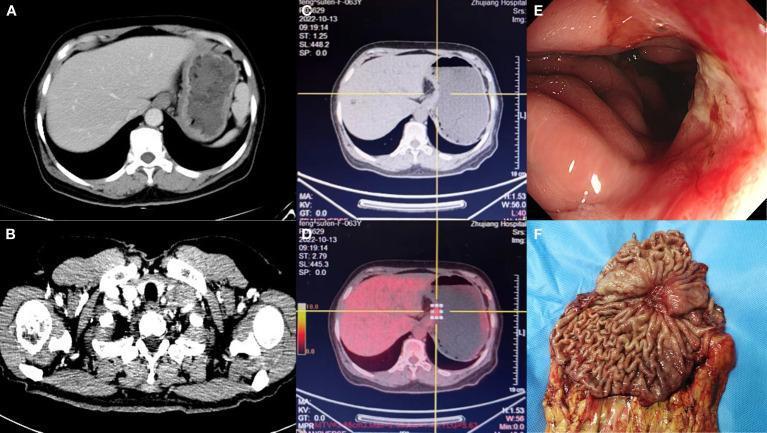
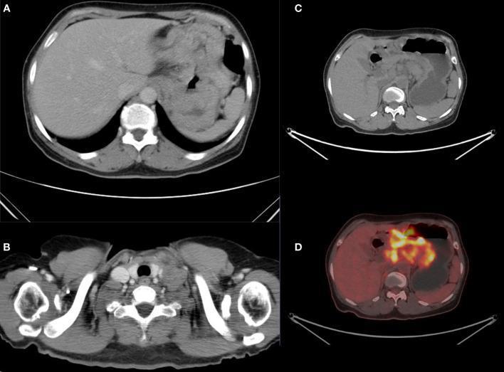
Multi-panel imaging of advanced gastric cancer: panels B shows bilateral enlarged supraclavicular lymph nodes (Virchow's node) on CT; panel A shows thickened gastric wall; panels C-D show PET-CT confirming metabolic activity; panel E shows endoscopic view of the ulcerated mass; panel F shows the gross resection specimen.
9. Paraneoplastic Effects: Trousseau's Syndrome and DVT
Trousseau's Syndrome (Migratory Thrombophlebitis)
What it is: Recurrent, migratory, superficial thrombophlebitis occurring at unusual anatomical sites (arms, chest, abdomen), not limited to the lower limbs. Originally described by Armand Trousseau in 1865 - who later recognized his own migratory phlebitis as a sign of occult visceral malignancy.
Mechanism - cancer-associated hypercoagulability:
Gastric carcinoma (and other mucin-secreting adenocarcinomas) promotes a hypercoagulable state through multiple mechanisms:
| Mechanism | Detail |
|---|
| Tissue factor (TF) expression | Tumor cells overexpress TF, activating the extrinsic coagulation cascade directly |
| Mucin secretion | Mucins (especially MUC1, MUC16) directly activate platelets and L/P-selectin, promoting thrombosis independent of TF |
| Tumor-derived microparticles | Shed from tumor cell membranes, these carry TF and phosphatidylserine, strongly activating coagulation |
| Inflammatory cytokines | IL-1β, TNF-α, VEGF increase endothelial TF expression and reduce anticoagulant mediators (thrombomodulin, protein C) |
| Suppression of fibrinolysis | Tumors produce plasminogen activator inhibitor-1 (PAI-1), suppressing clot dissolution |
Clinical features of Trousseau's syndrome:
- Migratory, recurrent superficial thrombophlebitis - veins become red, tender, cord-like, then resolve and reappear elsewhere
- May precede cancer diagnosis by months to years
- Can involve visceral veins (portal, mesenteric, splenic)
- Associated with non-bacterial thrombotic endocarditis (NBTE/marantic endocarditis) and arterial thromboembolism (cerebral infarcts, limb ischemia)
Deep Vein Thrombosis (DVT)
DVT in gastric cancer patients arises from the same hypercoagulable mechanisms. Key points:
- DVT is the most common thrombotic event in cancer patients overall
- Gastric cancer, pancreatic cancer, and lung cancer are among the highest-risk cancers for VTE
- DVT may present as unilateral leg swelling, pain, and erythema
- Unprovoked DVT in an otherwise well patient should prompt evaluation for occult malignancy, including gastric cancer screening
- Pulmonary embolism (PE) is a major cause of death in advanced gastric cancer patients
Distinction from Trousseau's syndrome:
- Trousseau's syndrome = migratory superficial thrombophlebitis
- DVT = deep venous thrombosis, usually lower limbs
- Both share the same underlying cancer-induced hypercoagulable mechanism
- Together they fall under the umbrella of cancer-associated thrombosis (CAT)
Management context: Low-molecular-weight heparin (LMWH) is preferred over warfarin for cancer-associated VTE, as shown in multiple trials. Direct oral anticoagulants (DOACs) like rivaroxaban and edoxaban are now also guideline-recommended.
Why relevant to inoperability: Trousseau's syndrome and DVT, when occurring in a patient with known gastric cancer, indicate advanced (usually Stage III-IV) disease with aggressive tumor biology. Their presence does not by itself make the tumor inoperable, but they signal:
- Aggressive, mucin-secreting tumor biology
- Systemic pro-coagulant tumor burden
- Significant perioperative thrombotic risk (DVT/PE is a major surgical complication in these patients)
- Goldman-Cecil Medicine, p. 2038; Sleisenger & Fordtran's, p. [paraneoplastic section]
Summary Table: Signs of Inoperability in Gastric Carcinoma
| Sign | Location | What it represents | AJCC implication |
|---|
| Ascites | Abdomen | Peritoneal carcinomatosis | M1 |
| Liver secondaries | RUQ, imaging | Hematogenous metastasis | M1 |
| Para-aortic nodes | CT/imaging | Distant nodal spread (Station 16) | M1 |
| Krukenberg tumor | Ovaries (bilateral) | Transcoelomic/peritoneal spread | M1 |
| Sister Mary Joseph nodule | Umbilicus | Peritoneal spread via umbilical lymphatics | M1 |
| Blumer shelf | Anterior rectum (DRE) | Drop metastases in pouch of Douglas | M1 |
| Irish node | Left axilla | Distant lymphatic spread | M1 |
| Virchow node (Troisier's sign) | Left supraclavicular | Thoracic duct retrograde involvement | M1 |
| Trousseau's syndrome / DVT | Variable | Paraneoplastic hypercoagulability | Indicates advanced disease / aggressive biology |
Memory Aids
- "VIrchow = VIsceral drainage - left supraclavicular" (thoracic duct → left subclavian)
- "Blumer = Bottom of the belly" (gravitational drop metastases to the pouch of Douglas, felt on DRE)
- "Krukenberg = Kitchen (cooking) pot sign" - signet ring cells look like "rings" in a pot - ovarian bilateral
- "Sister Mary Joseph = belly button nodule" (umbilicus = Mary Joseph's landmark)
- "Trousseau's = moving clots" (migratory, not fixed - the hallmark)
Sources:
- Sabiston Textbook of Surgery, 21st ed. - Signs and Symptoms of Gastric Cancer
- Goldman-Cecil Medicine - Physical Examination and Paraneoplastic Syndromes in Gastric Cancer
- Sleisenger & Fordtran's Gastrointestinal and Liver Disease - Clinical Features
- Maingot's Abdominal Operations - Signs and Symptoms of Gastric Cancer