Blood supply of female genital tract
Reading File
Finding Sources
Finding Sources
Finding Sources
Finding Sources
Finding Sources
Reading File
Reading File
Reading File
Reading File
Reading File
Loading Image
Loading Image
Reading File
Finding Sources
Reading File
Reading File
Reading File
Loading Image
I now have all the information needed to compile a thorough, well-illustrated answer. Here is the complete response:
Blood Supply of the Female Genital Tract
The female genital tract receives a rich vascular supply from two main sources: the internal iliac (hypogastric) artery (via its anterior trunk) and the ovarian arteries (direct branches from the abdominal aorta). The two systems are extensively interconnected through anastomoses.
Arterial Supply - Overview
1. Uterus
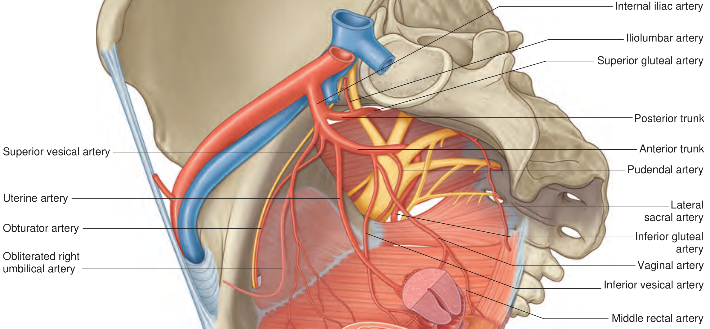
Main artery: Uterine artery (branch of the anterior trunk of the internal iliac artery)
- Course: From the internal iliac artery, the uterine artery runs medially and anteriorly in the base of the broad ligament (cardinal ligament). A surgically critical step occurs here: the uterine artery crosses ABOVE and ANTERIOR to the ureter at the level of the cervix ("water under the bridge"). This relationship makes the ureter vulnerable during hysterectomy when dividing the uterine pedicle.
- At the cervix: the artery reaches the lateral wall of the uterus and divides into:
- Ascending branch - tortuous, spirals up the lateral wall of the uterus supplying the corpus and fundus; at the uterine tube level it curves laterally and anastomoses with the ovarian artery
- Descending branch (vaginal artery) - descends to supply the cervix and upper vagina
- The ascending branch sends an ovarian branch (A5) at the fundus that joins the ovarian artery, and a tubal branch (A7) to the fallopian tube
- A network of valveless veins forms the uterine venous plexus around the body and cervix, draining via uterine veins into the internal iliac veins
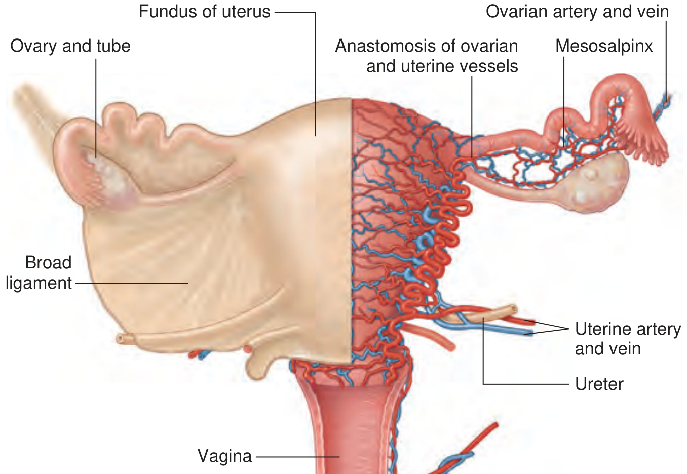
2. Ovaries
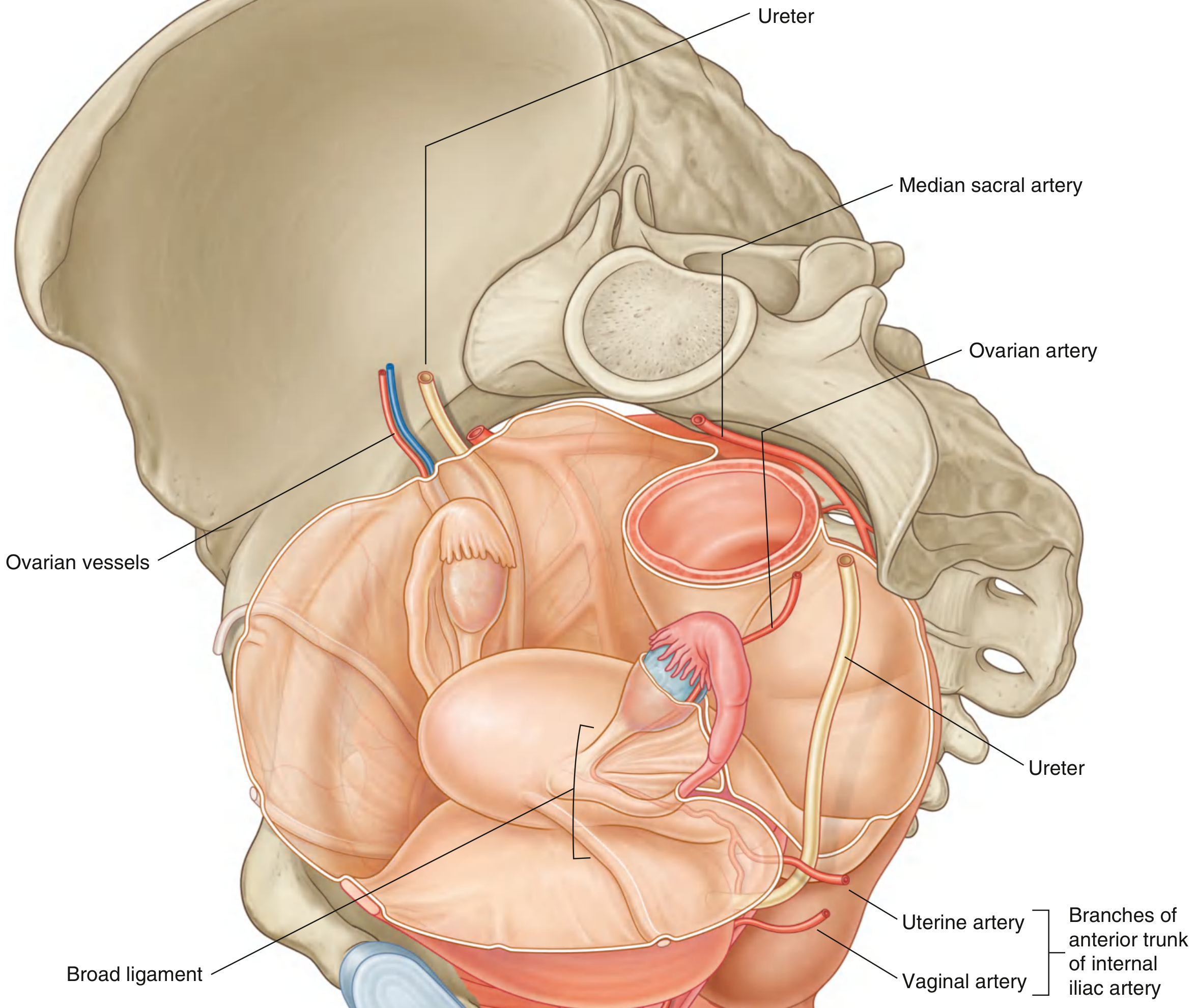
Main artery: Ovarian artery (direct branch of the abdominal aorta, at approximately L2 level)
- The ovarian artery descends retroperitoneally along the lateral abdomen and pelvis
- It crosses the pelvic inlet and travels in the suspensory (infundibulopelvic) ligament to reach the ovary
- Branches pass through the mesovarium to enter the ovary at the hilum
- Anastomoses extensively with the uterine artery through the broad ligament; this dual supply is important - ligation of either vessel alone rarely causes ischemia
- The ovarian arteries enlarge significantly during pregnancy to augment uterine blood supply
Venous drainage: The ovarian veins form from the ovarian plexus:
- Right ovarian vein drains directly into the inferior vena cava
- Left ovarian vein drains into the left renal vein
3. Fallopian Tubes
Dual supply from both uterine and ovarian arteries:
- The tubal branch of the uterine artery (medial supply) and the tubal branch of the ovarian artery (lateral supply) form an anastomotic arcade within the mesosalpinx called the ovarian arcade
- This ensures excellent collateral supply throughout the tube
4. Vagina
Multiple sources:
- Upper vagina (vaginal fornix): supplied by the descending (vaginal) branch of the uterine artery
- Middle vagina: vaginal artery - a direct branch from the anterior trunk of the internal iliac artery (sometimes arising from the uterine artery or inferior vesical)
- Lower vagina and vestibule: branches of the internal pudendal artery (also from the internal iliac, anterior trunk)
- Additional contribution: from the middle rectal artery posteriorly
5. Cervix
- Blood supply arises from a descending branch of the uterine artery, running laterally at the 3 and 9 o'clock positions
- Additional supply from ascending vaginal branches
6. Vulva and External Genitalia (Clitoris)
- Supplied by the internal pudendal artery (branch of the anterior trunk of internal iliac artery)
- The internal pudendal artery is the terminal branch supplying erectile tissue of the clitoris, labia, and perineum
- The clitoral veins drain into the retropubic plexus, which in turn drains via the vesical plexus to the internal iliac vein
Venous Drainage - Summary
Extensive pelvic venous plexuses (bladder, uterine, vaginal, rectal) interconnect and ultimately drain into the internal iliac veins. Key plexuses:
- Uterine (parametrial) plexus - around the uterus and cervix; valveless veins
- Vaginal plexus - interconnects with uterine and vesical plexuses
- Pampiniform plexus of the ovary - forms the ovarian vein (right to IVC; left to left renal vein)
Lymphatic Drainage - Summary
| Structure | Primary Lymph Nodes |
|---|---|
| Uterine fundus & body | Para-aortic nodes (along suspensory ligament); superficial inguinal (via round ligament) |
| Cervix | Internal iliac, obturator, presacral nodes |
| Ovary & tube | Para-aortic (lumbar) nodes at L2 level |
| Vagina (upper 2/3) | Internal and external iliac nodes |
| Vagina (lower 1/3) / Vulva | Superficial inguinal nodes |
Clinical Correlates
- Ureter-uterine artery relationship: The ureter passes 1-2 cm lateral to the cervix and is crossed superiorly by the uterine artery - the most common site of ureteric injury during hysterectomy
- Uterine artery embolization (UAE): Exploits the dominant uterine artery supply to the fibroid; effective for treating fibroids and adenomyosis
- Uterine artery origin variation: Four recognized types of origin from the internal iliac system (Types I-IV); the artery most commonly arises from the anterior trunk but can arise from the inferior gluteal artery
- Ovarian artery-uterine artery anastomosis: This anastomosis is why bilateral uterine artery ligation does not always stop obstetric hemorrhage (ovarian artery can reconstitute flow), and why UAE can reduce ovarian reserve
- Postpartum hemorrhage: B-Lynch sutures and uterine artery/internal iliac artery ligation exploit the segmental supply to control bleeding
Sources: Campbell-Walsh-Wein Urology (2020), p. 3216 | Berek & Novak's Gynecology | Gray's Anatomy for Students | Color Atlas of Human Anatomy Vol. 2 - Internal Organs | Sabiston Textbook of Surgery
This is a shared conversation. Sign in to Orris to start your own chat.