
Important minerals that are asked in biochemistry exams
Reading File
Now let me search the medical library for minerals content, running parallel queries:
Finding Sources
Finding Sources
Excellent! Let me read the most relevant biochemistry and clinical sources in parallel:
Reading File
Reading File
Reading File
Now let me get the detailed biochemistry content on each mineral from Lippincott:
Reading File
Finding Sources
Finding Sources
Reading File
Finding Sources
Finding Sources
Reading File
I now have comprehensive data to compile a thorough answer. Let me also fetch a couple of the key images to include:
Loading Image
Loading Image
I now have all the information needed for a comprehensive response. Here is the full guide on important minerals for biochemistry exams:
Important Minerals in Biochemistry Exams
Sources: Lippincott Illustrated Reviews: Biochemistry 8th ed, Ganong's Review of Medical Physiology 26th ed, Goldman-Cecil Medicine
Overview
Minerals are divided into macrominerals (needed in larger amounts: Ca, P, Mg, Na, K, Cl) and microminerals/trace elements (needed in minute amounts). The 15 trace minerals essential to health are: iron, zinc, copper, chromium, selenium, iodine, fluorine, manganese, molybdenum, cobalt, nickel, tin, silicon, vanadium, and arsenic - but only the first 10 have compelling evidence in humans.
1. Iron (Fe)
Body content: 3-4 g total; ~70% in hemoglobin.
Functions:
- Component of hemoglobin, myoglobin, cytochromes
- Part of Fe-S proteins of the electron transport chain
- Cofactor for hydroxylases (e.g., prolyl hydroxylase in collagen synthesis)
- Note: Free ionic Fe is toxic - generates hydroxyl radicals (ROS)
Absorption & Transport:
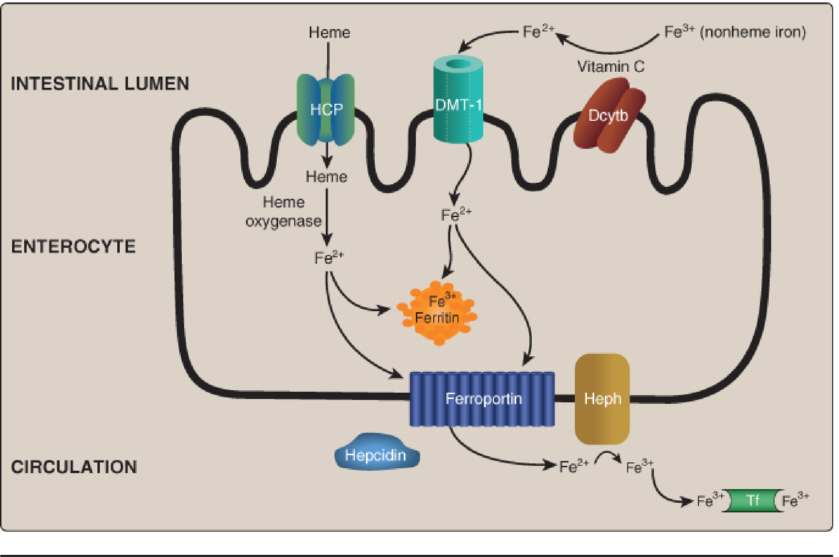
- Heme iron (animal): absorbed via heme carrier protein (HCP) → heme oxygenase releases Fe²⁺
- Non-heme iron (plants, Fe³⁺): reduced to Fe²⁺ by duodenal cytochrome b (Dcytb, requires Vitamin C as coenzyme) → absorbed via DMT-1 (divalent metal transporter-1)
- Storage: Fe³⁺ stored by ferritin (up to 4,500 Fe³⁺/molecule); overflow stored as insoluble hemosiderin
- Export: ferroportin (only known Fe exporter) → oxidized by hephaestin (Cu-containing) → binds transferrin (2 Fe³⁺/molecule, normally 1/3 saturated)
- Recycling: Macrophages recycle Fe from old RBCs via ferroportin + ceruloplasmin (Cu-containing plasma ferroxidase) → meets ~90% of daily need
- Regulation: Hepatic hepcidin is the master regulator - induces degradation of ferroportin, reducing Fe export. Suppressed when Fe is deficient.
| Key protein | Role |
|---|---|
| DMT-1 | Apical uptake of nonheme Fe²⁺ |
| Ferritin | Intracellular Fe³⁺ storage |
| Ferroportin | Only cellular Fe exporter |
| Hephaestin | Oxidizes Fe²⁺ → Fe³⁺ at enterocyte basolateral membrane |
| Transferrin (Tf) | Plasma Fe³⁺ transport (2 Fe³⁺/molecule) |
| Ceruloplasmin | Oxidizes Fe²⁺ → Fe³⁺ in plasma (macrophages) |
| Hepcidin | Master regulator; degrades ferroportin |
Deficiency: Microcytic, hypochromic anemia - most common anemia; serum ferritin low, Tf saturation low, TIBC elevated
Excess / Toxicity:
- Hereditary hemochromatosis (HH): AR disorder (HFE gene mutation) → "bronze diabetes" (hyperpigmentation + hyperglycemia), liver damage, heart disease; tx: phlebotomy / chelation
- Acute poisoning: most common cause of poisoning death in children <6 years; tx: chelation
2. Zinc (Zn)
Functions:
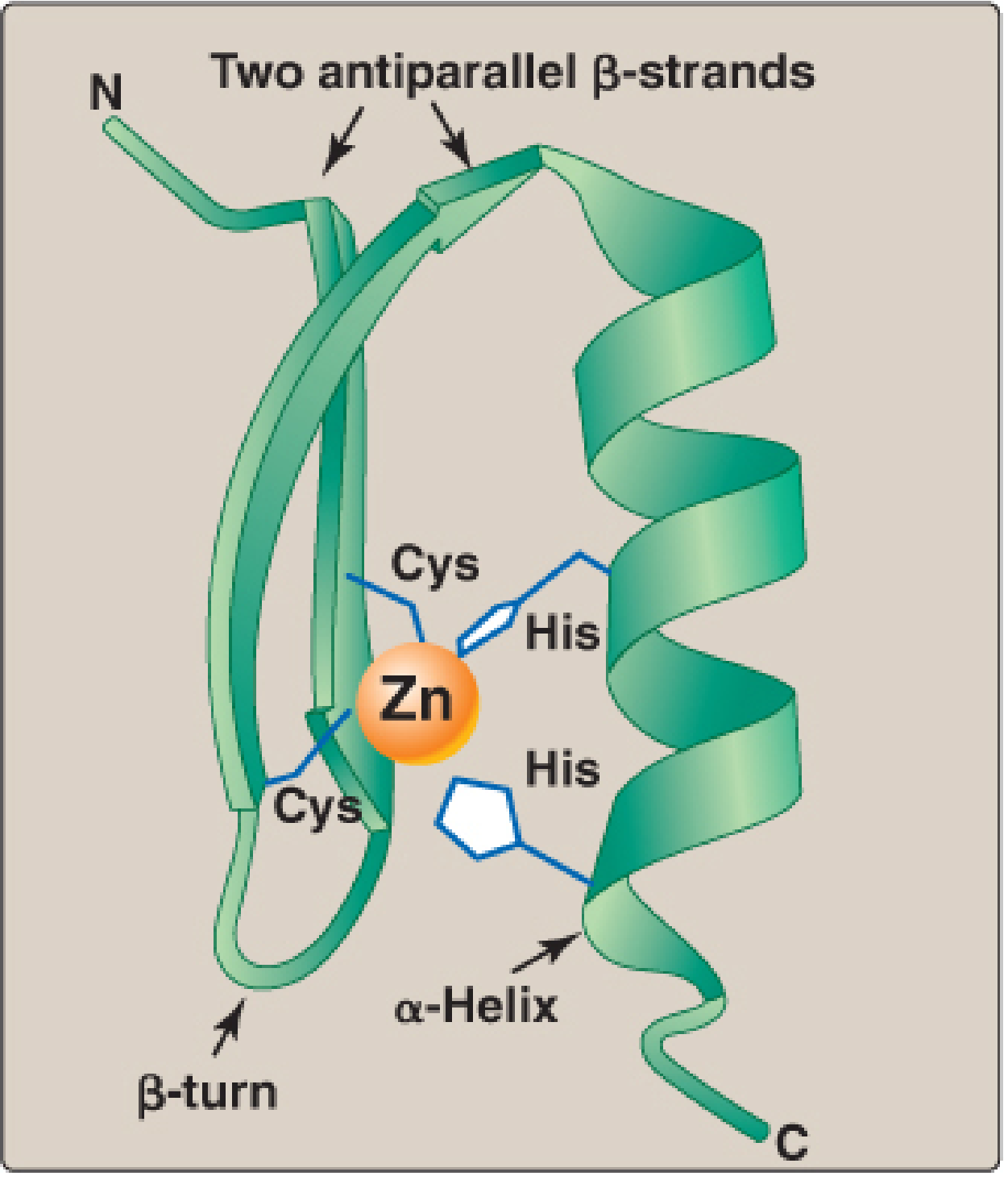
- Zinc fingers - supersecondary structures in transcription factors that bind DNA and regulate gene expression
- Cofactor for hundreds of enzymes:
- Alcohol dehydrogenase (ethanol → acetaldehyde)
- Carbonic anhydrase (bicarbonate buffer system)
- ALA dehydratase (heme synthesis - inhibited by lead, which displaces Zn)
- Superoxide dismutase (Cu/Zn-SOD, cytosolic form)
- Needed in metabolism of Vitamin A (night vision)
Dietary sources: Meat, fish, eggs, dairy. Phytates (in grains, seeds, legumes) irreversibly bind Zn and impair absorption.
Deficiency:
- Skin ulcers, depressed immune responses, hypogonadal dwarfism
- Poor wound healing, growth retardation, alopecia, ageusia (loss of taste), anosmia
- Acrodermatitis enteropathica - AR disorder; defect in intestinal Zn transporter. Triad: periacral/periorificial dermatitis, alopecia, diarrhea
3. Copper (Cu)
Functions:
- Cofactor for cuproenzymes:
- Ceruloplasmin (plasma ferroxidase)
- Hephaestin (intestinal ferroxidase)
- Cytochrome c oxidase (Complex IV of ETC)
- Cu/Zn-SOD (superoxide dismutase)
- Lysyl oxidase (cross-linking of collagen and elastin)
- Dopamine β-hydroxylase (catecholamine synthesis)
- Tyrosinase (melanin synthesis)
Deficiency: Anemia (because Cu needed for Fe mobilization via ceruloplasmin/hephaestin), changes in ossification, neurological problems
Genetic disorders:
| Disease | Gene | Defect | Features |
|---|---|---|---|
| Menkes disease | ATP7A (X-linked recessive) | Defect in Cu absorption/transport; systemic Cu deficiency | Kinky/steely hair, neurodegeneration, seizures, connective tissue defects; low serum Cu and ceruloplasmin |
| Wilson disease | ATP7B (AR) | Defect in Cu incorporation into ceruloplasmin + biliary excretion; Cu accumulates in tissues | Liver damage, Kayser-Fleischer rings in cornea, neuropsychiatric symptoms; low ceruloplasmin, high urinary Cu |
4. Iodine (I)
Functions: Essential component of thyroid hormones (T3 and T4). Incorporated via thyroid peroxidase (TPO).
Dietary sources: Dairy, seafood, meat; iodized salt has greatly reduced deficiency worldwide.
Deficiency:
- Goiter (thyroid enlargement from excessive TSH stimulation)
- Hypothyroidism: fatigue, weight gain, decreased thermogenesis and metabolic rate
- Congenital hypothyroidism (fetal/infant): cretinism - irreversible intellectual disability, hearing loss, spasticity, short stature
Excess: Graves disease (most common hyperthyroidism) - TSH-receptor antibody → nervousness, weight loss, tachycardia, exophthalmos, goiter. Over-ingestion of iodine supplements can also cause hyperthyroidism (UL = 1.1 g/day adults).
5. Selenium (Se)
Functions: Present in ~25 human selenoproteins as selenocysteine (derived from serine):
- Glutathione peroxidase - reduces H₂O₂ to H₂O (antioxidant defense)
- Thioredoxin reductase - reduces thioredoxin (coenzyme of ribonucleotide reductase)
- Deiodinases - remove iodine from thyroid hormones (activation/deactivation of T3/T4)
Dietary sources: Meat, dairy, grains (content depends on soil Se content)
Deficiency: Keshan disease - endemic dilated cardiomyopathy in Se-deficient areas of China
Toxicity (selenosis): Brittle nails, brittle hair, cutaneous and neurological effects (UL = 400 μg/day adults)
6. Chromium (Cr)
Functions: Potentiates the action of insulin (mechanism not fully understood). Assists glucose metabolism.
Sources: Fruits, vegetables, dairy, meat
Deficiency: Insulin resistance (impaired glucose tolerance)
7. Fluorine (F)
Functions:
- As fluoride (F⁻): replaces the hydroxyl group (-OH) in hydroxyapatite → forms fluoroapatite, which is more resistant to acid dissolution by oral bacteria
- Reduces incidence of dental caries
Deficiency: Increased incidence of dental caries
8. Manganese (Mn)
Functions: Cofactor for several enzymes including:
- Pyruvate carboxylase (OAA synthesis)
- Mn-SOD (mitochondrial superoxide dismutase)
- Arginase (urea cycle)
Sources: Whole grains, legumes, nuts, tea
Deficiency: Rare in humans. Toxicity from excess Mn can cause neurological symptoms resembling Parkinsonism.
9. Molybdenum (Mo)
Functions: Cofactor for a small number of oxidases (e.g., xanthine oxidase, sulfite oxidase)
Sources: Legumes
Note: No known dietary deficiency syndrome. Low toxicity (UL = 2 mg/day).
10. Cobalt (Co)
Functions: Essential solely as a component of Vitamin B₁₂ (cobalamin). An isolated cobalt deficiency syndrome has not been described; deficiency manifests as B₁₂ deficiency → megaloblastic anemia.
Calcium (Ca) - Macromineral
Absorption: 30-80% of ingested Ca absorbed; regulated by 1,25-dihydroxycholecalciferol (calcitriol) - increases in Ca deficiency, decreases in excess
Functions: Bone/teeth structure, muscle contraction, nerve conduction, blood clotting, second messenger (calmodulin), enzyme cofactor
Regulation: PTH, calcitonin, Vitamin D
Quick Exam Summary Table
| Mineral | Key Role | Deficiency | Excess/Disease | High-Yield Fact |
|---|---|---|---|---|
| Fe | Hb, ETC, hydroxylases | Microcytic hypochromic anemia | Hemochromatosis (bronze diabetes) | Hepcidin controls ferroportin |
| Zn | Zn fingers, 100s enzymes | Dwarfism, immune deficiency, acrodermatitis enteropathica | Rare | ALA dehydratase inhibited by Pb displacing Zn |
| Cu | Ceruloplasmin, lysyl oxidase, ETC | Anemia, bone changes | Menkes (AR XL, low Cu), Wilson (AR, Cu overload) | Cu needed for Fe export from cells |
| I | Thyroid hormones | Goiter, cretinism | Graves disease | TPO uses I; iodized salt prevents deficiency |
| Se | Glutathione peroxidase, deiodinases | Keshan disease (cardiomyopathy) | Selenosis (brittle nails/hair) | Selenocysteine = 21st amino acid |
| Cr | Potentiates insulin | Insulin resistance | - | Mechanism unknown |
| F | Fluoroapatite in enamel | Dental caries | Dental/skeletal fluorosis | Replaces -OH in hydroxyapatite |
| Mn | Pyruvate carboxylase, Mn-SOD | Rare | Parkinsonism-like | Mitochondrial SOD uses Mn |
| Mo | Xanthine oxidase | None described | Low toxicity | Legumes are main dietary source |
| Co | Vitamin B₁₂ only | Megaloblastic anemia | Cardiomyopathy (beer drinkers) | No isolated Co deficiency - always B₁₂ |
High-yield exam points:
- Hepcidin is the master Fe regulator - suppressed in Fe deficiency, induced by Fe overload/inflammation
- Vitamin C enhances non-heme Fe absorption (Dcytb coenzyme: Fe³⁺ → Fe²⁺)
- Phytates reduce Zn (and Fe) absorption from plant foods
- Lead poisoning inhibits ALA dehydratase by displacing Zn (plus inhibits ferrochelatase) → sideroblastic-type anemia
- Menkes vs Wilson: both Cu disorders; Menkes = low Cu (can't absorb/transport it); Wilson = high tissue Cu (can't excrete it)
- Selenium's key enzyme is glutathione peroxidase - antioxidant defense
- Acrodermatitis enteropathica = Zn deficiency (AR transporter defect): dermatitis + alopecia + diarrhea triad
- Keshan disease = Se deficiency → dilated cardiomyopathy
Sources: Lippincott Illustrated Reviews: Biochemistry 8th ed, pp. 1114-1127; Ganong's Review of Medical Physiology 26th ed; Goldman-Cecil Medicine 26th ed
This is a shared conversation. Sign in to Orris to start your own chat.