
Classic xray chest pattern features of aspiration pneumonia
aspiration pneumonia chest X-ray radiograph
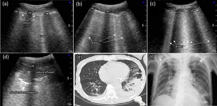
A multi-modal comparison of thoracic imaging findings in aspiration pneumonia, featuring point-of-care ultrasound (POCUS), computed tomography (CT), and chest X-ray (CXR). Panels (a)-(d) demonstrate a spectrum of lung ultrasound findings: (a) shows a single vertical B-line artifact originating from the pleural line; (b) displays multiple (≥3) B-lines; (c) shows confluent (≥5) B-lines creating a 'white lung' appearance; and (d) identifies sub-pleural consolidation (a hypoechoic area) and adjacent pleural effusion. Panel (e) is an axial CT scan of the chest showing significant consolidation and air bronchograms in the left lung, typical of aspiration pneumonia. Panel (f) is a posterior-anterior chest radiograph from the same patient, illustrating a loss of the silhouette of the left diaphragmatic arch behind the heart, indicating left lower lobe pathology. The collection serves as an educational comparison of diagnostic modalities for identifying pulmonary edema, interstitial syndrome, and consolidation in a clinical setting.
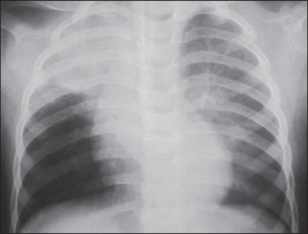
This diagnostic image is an anteroposterior (AP) chest X-ray of an 18-month-old pediatric patient. The radiograph demonstrates focal areas of increased density, specifically consolidative opacities located in the right upper lobe and the upper segment of the left lower lobe. These radiographic findings are highly characteristic of aspiration pneumonia or atelectasis occurring while the patient is in the supine (dorsal decubitus) position. The mediastinal shadow, cardiac silhouette, and diaphragmatic contours appear within normal limits for the patient's age. The skeletal structures, including the ribs and clavicles, show no fractures or deformities. The localized nature of the opacities in dependent lung segments provides significant clinical evidence for aspiration following a vomiting event, especially in the context of recovering from anesthesia. This visual serves as an educational example of typical radiographic patterns in pediatric aspiration-related pulmonary complications.
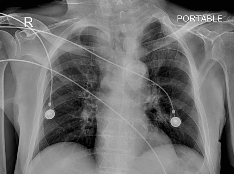
Anterior-posterior (AP) portable chest radiograph. The image depicts the thoracic cage, including the ribs, clavicles, scapulae, and thoracic spine. A right-side marker ('R') and 'PORTABLE' label are visible. The central mediastinal silhouette, including the heart and great vessels, appears within normal limits for this projection. The trachea is centrally located and patent, visible as a radiolucent column extending toward the carina. The lung parenchyma shows bilateral pulmonary vascular markings without clear evidence of consolidation or large pleural effusions. Several external medical monitoring devices are present: multiple radiopaque ECG leads and two circular cutaneous electrode pads are positioned over the chest wall. The radiograph is used clinically to confirm tracheal patency and assess for complications like aspiration pneumonia in patients with chronic dysphagia and instrumentation. The educational focus is on the interpretation of a standard follow-up bedside chest X-ray and identifying normal airway anatomy and medical monitoring hardware.
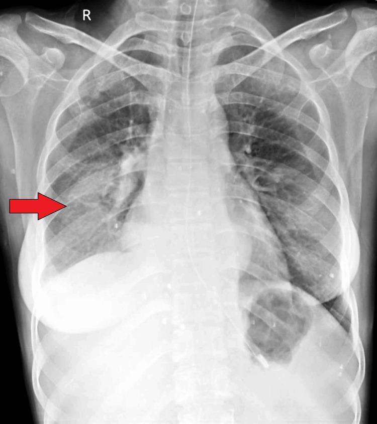
This diagnostic image is a posterior-anterior (PA) chest radiograph illustrating a significant pathological finding in the lower right lung field. A red arrow points to a large, dense, and relatively homogenous opacity localized in the right lower lobe, which partially obscures the right hemidiaphragm and costophrenic angle. This radiographic appearance is consistent with pulmonary consolidation, which can be seen in conditions such as pneumonia, aspiration, or complications arising from a bronchoesophageal fistula (BEF). The cardiac silhouette appears centrally located, and the left lung field remains relatively clear without overt consolidation or effusion. Bony structures, including the clavicles and ribs, appear intact. This image serves as a clinical teaching tool for identifying lobar opacification and its differential diagnoses in patients presenting with respiratory symptoms like cough or fever. The visibility of the gastric bubble and tracheal alignment provide additional anatomical landmarks for standard chest X-ray interpretation.
Classic Chest X-ray Features of Aspiration Pneumonia
1. Location - The Cardinal Feature (Position-Dependent)
| Patient Position | Zones Affected |
|---|---|
| Supine (most common - ICU, obtunded) | Posterior segments of upper lobes + superior segments of lower lobes |
| Upright / Semi-recumbent | Basal segments of lower lobes (especially right) |
| Right lateral decubitus | Right upper lobe |
2. Opacity Type and Pattern
- Patchy consolidation - the most common appearance; ill-defined opacification that does not respect lobar boundaries
- Bronchopneumonic pattern - multifocal, bilateral in severe cases; peribronchial spread
- Air bronchograms may be present within areas of consolidation
- Tree-in-bud pattern - centrilobular nodules connected to branching lines; indicates bronchiolitis and early airway spread
- Volume loss / atelectasis - particularly when a bronchus is plugged by aspirated material; tracheal and mediastinal shift toward the affected side may be seen
3. Distribution
- Usually multilobar in significant aspiration
- More frequently and more extensively right-sided due to airway anatomy
- Can be bilateral in large-volume aspiration or recumbent patients
4. Complications Visible on CXR
| Complication | Radiographic Sign |
|---|---|
| Lung abscess | Thick-walled cavity, often with air-fluid level - classic in the posterior segment RUL or superior segment RLL |
| Pleural effusion | Blunting of costophrenic angle; can be exudative/empyema |
| Pneumothorax | Rare but possible |
| Necrotizing pneumonia | Multiple small cavities within consolidation |
5. Illustrative Images
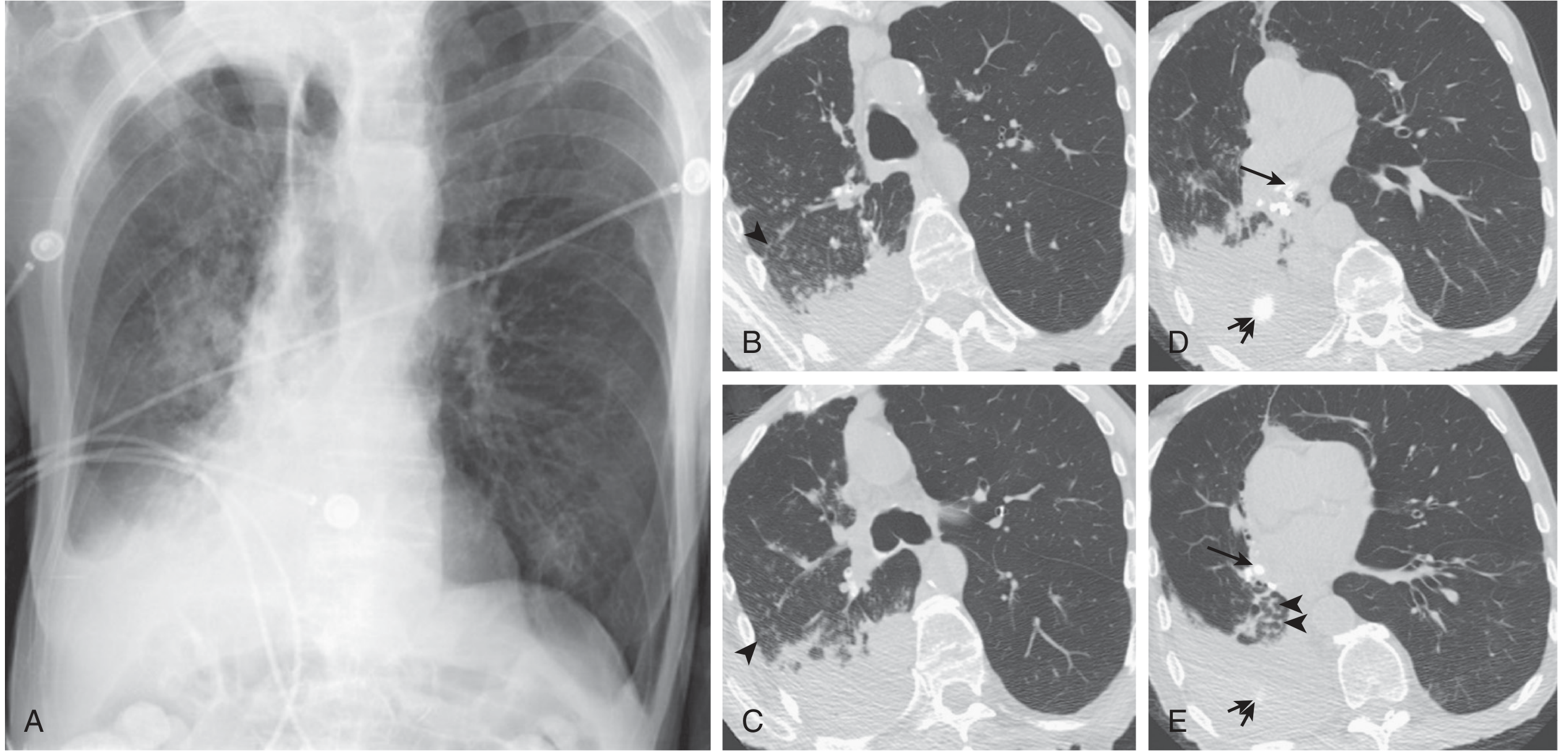
6. Key Differentiating Points from CAP
- Gravity-dependent location rather than a single lobe
- Right > left involvement
- Polymicrobial/anaerobic organisms more likely (putrid sputum, poor dentition)
- Lung abscess formation is more common than in typical CAP
- Pattern ranges from tree-in-bud to patchy consolidation to frank lobar opacity - the radiograph alone cannot reliably distinguish aspiration pneumonia from community-acquired or healthcare-acquired bronchopneumonia without clinical context
Summary Table
| Feature | Classic Finding |
|---|---|
| Location (supine) | Superior segments lower lobes, posterior segments upper lobes |
| Location (upright) | Basilar lower lobes |
| Laterality | Right > Left |
| Opacity type | Patchy consolidation, bronchopneumonic |
| Special patterns | Air bronchograms, tree-in-bud, atelectasis |
| Complications | Lung abscess (air-fluid level), pleural effusion, empyema |
How do you differentiate aspiration pneumonia from aspirated foreign bodies on imaging?
foreign body aspiration chest X-ray air trapping hyperinflation
Differentiating Aspiration Pneumonia vs. Aspirated Foreign Body on Imaging
Head-to-Head Comparison Table
| Feature | Aspiration Pneumonia | Foreign Body Aspiration |
|---|---|---|
| Primary finding | Parenchymal consolidation/infiltrate | Obstructive downstream changes (or direct FB visualization) |
| Location | Dependent zones (posterior upper lobes, superior/basal lower lobes) | Determined by where the FB lodges - right > left bronchus (60:30%) |
| Air trapping | Absent | Key sign - unilateral obstructive emphysema (check-valve mechanism) |
| Volume | Normal or reduced (atelectasis) | Increased on side of FB (hyperinflation) |
| Mediastinal shift | Toward the affected side (if atelectasis dominates) | Away from the affected side (due to air trapping/hyperinflation) |
| Radiopaque FB | Not present | Present in < 25% of cases (coins, batteries, metal) - most organic FBs (nuts, food) are radiolucent |
| Diaphragm | May be elevated ipsilaterally | Contralateral hemidiaphragm elevated; ipsilateral depressed and flattened |
| Bilateral involvement | Common in significant aspiration | Rare - usually unilateral |
| Pattern over time | Progressive infiltrate → may cavitate (abscess) | Persistent collapse/atelectasis + post-obstructive "non-resolving" pneumonia |
Aspiration Pneumonia: Key Imaging Features
- Patchy consolidation in gravity-dependent zones
- Right lung predominance (more vertical right bronchus)
- Bilateral in larger volume aspiration
- May progress to lung abscess (thick-walled cavity + air-fluid level) or empyema
- No obstruction of a central bronchus
Foreign Body: Key Imaging Features
- The FB itself is visible as a radiopaque density in the trachea or bronchus
- Only seen in < 25% of cases - most FBs are organic/food and are radiolucent
- Unilateral obstructive emphysema/hyperinflation - the most important sign; caused by a "ball-valve" mechanism where air enters on inspiration but cannot exit on expiration
- Mediastinal shift away from the FB side - due to air trapping pushing structures contralaterally
- Flattened ipsilateral hemidiaphragm + elevated contralateral hemidiaphragm
- Widened ipsilateral intercostal spaces
- Post-obstructive atelectasis or collapse - when obstruction is complete
- Non-resolving/recurrent pneumonia in the same segment - chronic foreign body
The Expiratory / Decubitus Film - The Critical Maneuver
"In patients with suspected aspiration of a foreign body and a normal examination of the upper airway, inspiratory-expiratory plain chest radiography or decubitus chest radiography should be performed. These may show regional obstructive emphysema resulting from aspirated foreign bodies in the bronchi." - Cummings Otolaryngology
| Technique | What to Look For |
|---|---|
| Inspiratory vs. expiratory films | Normal lung deflates on expiration; FB-obstructed lung stays hyperinflated (remains dark/lucent) |
| Left / right lateral decubitus | The dependent lung normally deflates due to compression; if the FB side is dependent and stays inflated - positive sign |
| Fluoroscopy | "Paradoxical" mediastinal swing toward the normal lung on expiration (Holzknecht sign) |
The Role of CT
| Modality | Aspiration Pneumonia | Foreign Body |
|---|---|---|
| CT chest | Consolidation + air bronchograms + dependent distribution | More sensitive than CXR for FB; can visualize non-radiopaque FB directly, show air trapping with expiratory cuts |
| Virtual bronchoscopy | Not indicated | 3D-reformatted CT generates intraluminal views of the airway - can detect sub-segmental FBs |
| Limitation | - | CT is less sensitive than bronchoscopy; sending an unstable patient to scanner carries risk |
Illustrative Image: Foreign Body with Unilateral Hyperinflation
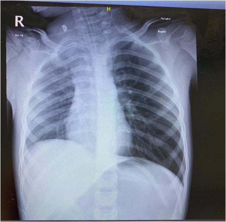
Chronic Retained FB - A Diagnostic Trap
- Post-obstructive pneumonia develops in the distal lung segment
- The consolidation is in the same location repeatedly
- Bronchiectasis may develop
- Granulation tissue can form around organic FBs (especially nuts), which can be mistaken for endobronchial tumor on imaging - Fishman's Pulmonary Diseases
Summary: Decision Algorithm
Consolidation / opacity on CXR
↓
Is it in dependent zones (posterior UL, superior/basal LL)?
→ YES + bilateral / patchy → Aspiration Pneumonia
↓
Is there unilateral hyperinflation or air trapping?
→ YES → Foreign Body until proven otherwise
↓
Get inspiratory-expiratory or decubitus films
→ FB side stays inflated on expiration = positive
↓
CT / Virtual bronchoscopy if still uncertain
↓
Bronchoscopy = Gold Standard
I want to make a presentation on aspiration pneumonia xhest pattern which details should I follow
Who is the target audience? · What format do you want? · Which content sections should be included?
mkdir -p /tmp/workspace/aspiration-pneumonia-pdf/assets && ls /tmp/workspace/
cd /tmp/workspace/aspiration-pneumonia-pdf && python /tmp/skills/shared/scripts/fetch_images.py \ "https://cdn.orris.care/cdss_images/f97f914e6eb5f10ade11a5d80ffbc4d3088ed5b668ac75125607e5420e132c04.png" \ "https://cdn.orris.care/cdss_images/pmc_clinical_VQA_596c35574e51eef345e61ca003d1e6050edc8232db521174c033f93f045d4861.jpg" \ "https://cdn.orris.care/cdss_images/2c59d456830c69adeadd7100a6d09bd9d64c9cb84c4b02bed4d348259dea74e9.jpg" \ "https://cdn.orris.care/cdss_images/69e509e34e98820a75b8f8be6fb1217c6c3ed2d49b4b7c0ee53a26a80143d472.jpg" \ 2>&1 | head -5
Fetch_images.py
Get me the necessary images
What images do you need?
- A specific topic, condition, or concept you want illustrated
- A file or script you had (the error message mentioned a file called
Fetch_images.py— is that related?) - A presentation, document, or report you were working on