
Explain sepsis in detail and septic shock
sepsis pathophysiology diagram
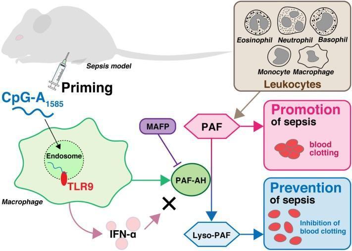
This medical schematic diagram illustrates the pathophysiology and therapeutic priming mechanism in a murine sepsis model. At the cellular level, the diagram depicts a macrophage endocytosing the molecule CpG-A1585, which subsequently binds to Toll-like receptor 9 (TLR9) within an endosome. This signaling pathway triggers the activation of platelet-activating factor acetylhydrolase (PAF-AH). While CpG-A1585 also induces interferon-alpha (IFN-α), a cross mark indicates that IFN-α does not contribute to PAF-AH activation. The diagram further details a leukocyte panel (eosinophils, neutrophils, basophils, monocytes, and macrophages) that produces platelet-activating factor (PAF), a mediator shown to promote sepsis and pathological blood clotting. PAF-AH serves as a critical regulatory enzyme by converting pro-septic PAF into Lyso-PAF, which prevents sepsis through the inhibition of blood clotting. Additionally, the specific inhibitor MAFP is shown antagonizing PAF-AH activity. This illustration highlights the clinical relevance of TLR9-mediated enzymatic pathways in mitigating endotoxin shock and disseminated intravascular coagulation (DIC).
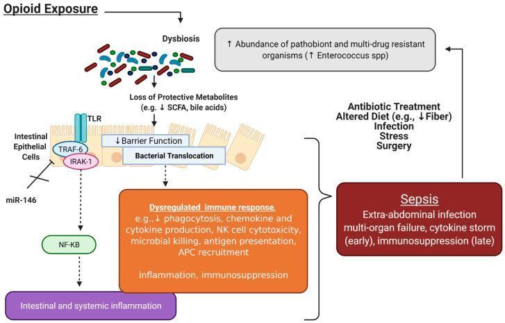
This pathophysiology diagram illustrates the bidirectional relationship between opioid exposure, gut microbial dysbiosis, and the development of sepsis. The process begins with 'Opioid Exposure' triggering dysbiosis, characterized by an increased abundance of pathobionts and multi-drug resistant organisms (notably Enterococcus spp.). This leads to a 'Loss of Protective Metabolites' (decreased SCFA and bile acids), causing 'Decreased Barrier Function' of intestinal epithelial cells and subsequent 'Bacterial Translocation'. The diagram highlights signaling pathways where Toll-like receptors (TLR), TRAF-6, and IRAK-1 activate NF-kB, resulting in 'Intestinal and systemic inflammation'. This inflammation is exacerbated by morphine-induced inhibition of miR-146, which normally regulates IRAK-1 and TRAF-6. These mechanisms culminate in a 'Dysregulated immune response' (e.g., impaired phagocytosis and NK cell cytotoxicity) and ultimately 'Sepsis', characterized by extra-abdominal infection and multi-organ failure. A feedback loop is shown where sepsis and its clinical management (antibiotic treatment, surgery, stress) further perpetuate gut dysbiosis, creating a vicious cycle of immune dysfunction.
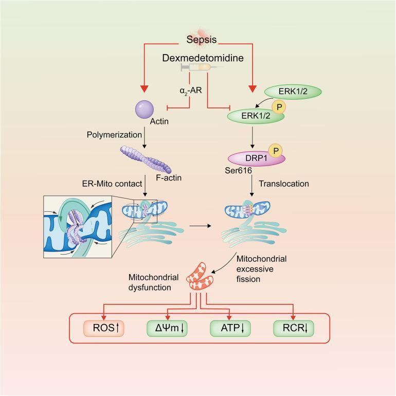
This pathophysiology diagram illustrates the signaling pathways involved in sepsis-induced mitochondrial dysfunction and the therapeutic intervention of dexmedetomidine. The schematic shows sepsis initiating two distinct pathological branches: 1) Actin polymerization into F-actin, which promotes endoplasmic reticulum (ER)-mitochondrial contact and pre-constriction, and 2) Activation of the ERK1/2 pathway, leading to DRP1 Ser616 phosphorylation and its subsequent translocation to the mitochondria. These parallel pathways converge to drive mitochondrial excessive fission and mitochondrial dysfunction. The visual representation highlights the resulting bioenergetic collapse, characterized by increased Reactive Oxygen Species (ROS↑) and decreased mitochondrial membrane potential (ΔΨm↓), Adenosine Triphosphate (ATP↓), and Respiratory Control Rate (RCR↓). Dexmedetomidine, acting via α2-adrenergic receptors (α2-AR), is shown with inhibitory lines suppressing both the actin polymerization branch and the ERK1/2 phosphorylation pathway. This diagram serves as a medical educational resource for understanding the molecular mechanisms of septic vascular endothelial cell injury and the protective role of α2-agonists in maintaining mitochondrial homeostasis.
Sepsis and Septic Shock - A Detailed Overview
1. Definitions (Sepsis-3, 2016)
- Persistent hypotension requiring vasopressors to maintain MAP ≥65 mmHg, AND
- Serum lactate >2 mmol/L (>18 mg/dL) despite adequate volume resuscitation
Historical note: Older definitions relied on the SIRS criteria (≥2 of: temp >38°C or <36°C, HR >90 bpm, RR >20 or PaCO₂ <32 mmHg, WBC >12,000 or <4,000/μL). The Sepsis-3 consensus abandoned "severe sepsis" as a category and incorporated organ dysfunction as the central feature.
2. Epidemiology
- Over 750,000 cases per year in the United States; >48 million cases globally per year
- Sepsis accounts for 6% of adult U.S. hospitalizations and causes as many deaths as acute MI
- Septic shock is the most common cause of death in non-coronary ICUs
- Incidence is rising due to more aggressive surgery, immunosuppressed populations (chemotherapy, HIV, transplant, advanced age), and increasing multidrug-resistant organisms
- Medical costs: ~$20 billion/year in the U.S. alone
- African Americans show higher incidence (6.0 vs. 3.6 per 1000) and higher ICU mortality (32% vs. 29%)
- Goldman-Cecil Medicine, p. 1087
3. Causative Organisms
- Gram-positive bacteria (now the most common in hospitalized patients): MRSA, S. pneumoniae, VRE, enterococci
- Gram-negative bacteria: E. coli, Klebsiella, Pseudomonas, Acinetobacter - their LPS (lipopolysaccharide/endotoxin) is a prototypical trigger
- Fungi: Candida species
- Viruses: SARS-CoV-2 and others
- Blood cultures are positive in only ~1/3 of patients; 20-30% are culture-negative from all sites
4. Pathophysiology
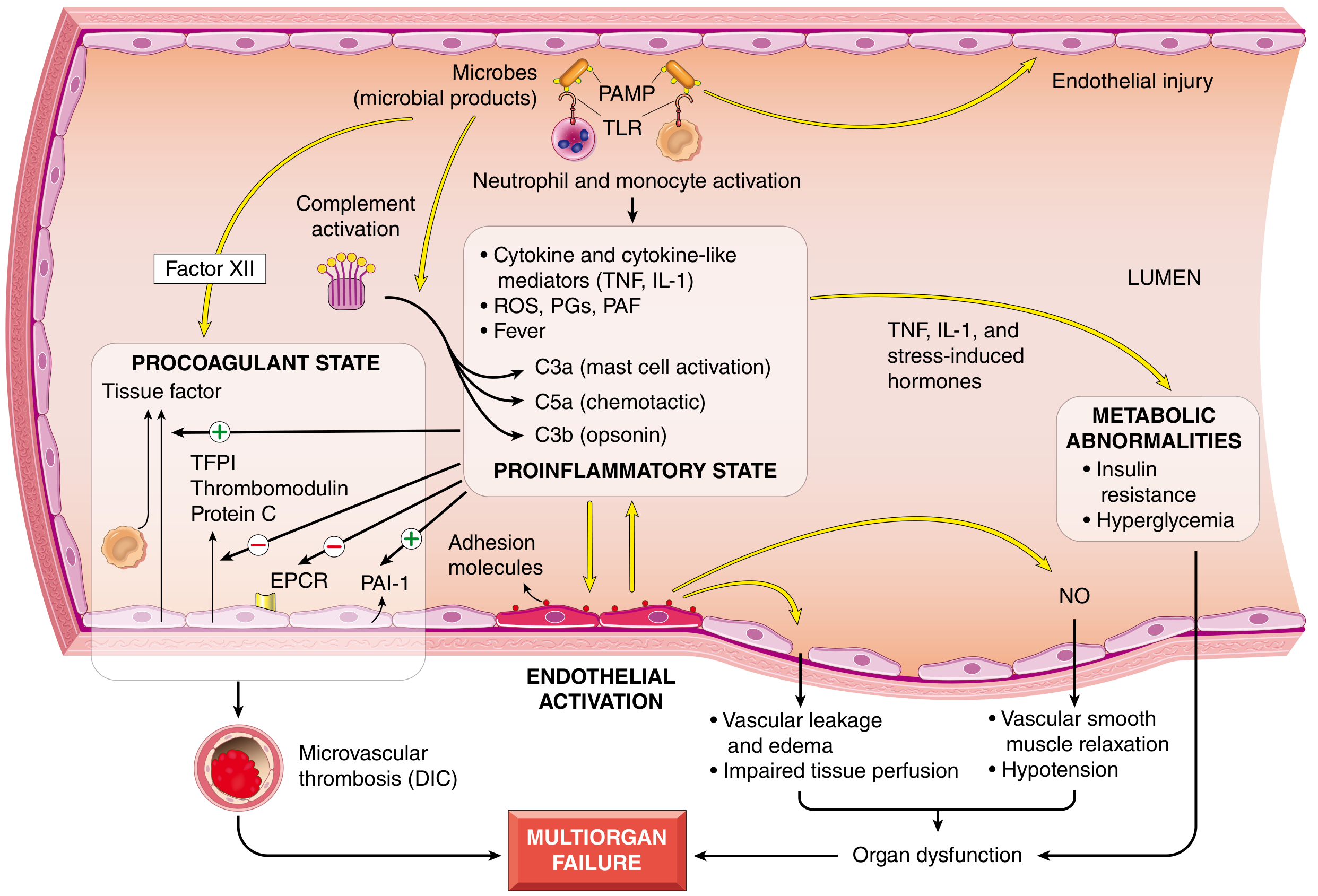
4a. Innate Immune Activation (The Trigger)
- Toll-like receptors (TLRs) on macrophages, neutrophils, dendritic cells, and endothelial cells
- G-protein-coupled receptors (detect bacterial peptides)
- C-type lectin receptors like Dectins (detect fungal antigens)
4b. Proinflammatory State - The Cytokine Storm
| Mediator | Source | Effect |
|---|---|---|
| TNF-α | Macrophages | Fever, hypotension, myocardial depression |
| IL-1β | Macrophages | Fever, endothelial activation |
| IL-12, IL-18, IFN-γ | Macrophages, NK cells | Amplify Th1 response |
| HMGB1 | Many cells (late mediator) | Sustained inflammation |
| ROS | Neutrophils | Tissue damage |
| Prostaglandins & PAF | Multiple | Vasodilation, platelet activation |
| CRP & Procalcitonin | Liver | Markers of acute phase response |
- C3a, C5a (anaphylatoxins) → mast cell activation, vasodilation
- C5a (chemotactic) → neutrophil recruitment
- C3b (opsonin) → phagocytosis
4c. Counter-inflammatory (Immunosuppressive) Response
- Shift from Th1 (proinflammatory) → Th2 (anti-inflammatory) cytokine profile
- Production of IL-10, soluble TNF receptor, IL-1 receptor antagonist
- Lymphocyte apoptosis and T-cell anergy
- Result: septic patients may oscillate between hyperinflammation and immunosuppression during their course, explaining the increased risk of secondary infections
4d. Endothelial Activation and Injury
- Widespread vascular leakage → protein-rich edema throughout the body
- Impaired nutrient delivery and waste removal
- Upregulation of NO (nitric oxide) via iNOS → vascular smooth muscle relaxation → systemic hypotension
- Loss of normal autoregulation of microvascular flow → oxygen delivery/demand mismatch → tissue hypoxia
- Robbins, p. 135
4e. Procoagulant State and DIC
- Increase tissue factor expression on monocytes/endothelium → activates extrinsic coagulation cascade
- Decrease anticoagulants: thrombomodulin, protein C, TFPI (tissue factor pathway inhibitor)
- Increase PAI-1 → dampens fibrinolysis
- Neutrophil extracellular traps (NETs) activate both intrinsic and extrinsic coagulation pathways
- Stasis from vascular leak reduces washout of activated factors
4f. Metabolic Abnormalities
- Insulin resistance and hyperglycemia driven by TNF, IL-1, stress hormones (glucagon, GH, glucocorticoids)
- Lactic acidosis from anaerobic metabolism due to impaired tissue oxygenation - elevated lactate is a key marker of tissue hypoperfusion and mortality predictor
- Mitochondrial dysfunction impairs oxidative phosphorylation at the cellular level
4g. Myocardial Depression (Specific to Septic Shock)
- Circulating TNF-α and IL-1β directly impair cardiomyocyte contractility
- iNOS-derived NO causes negative inotropy
- Impaired mitochondrial oxidative phosphorylation in cardiomyocytes
- This can be detected by advanced echo markers (global longitudinal strain) before overt ejection fraction reduction - Rosen's Emergency Medicine, p. 59
5. The Three Primary Effects of Septic Shock
-
Hypovolemia (both absolute and relative)
- Absolute: from GI losses, tachypnea, sweating, poor intake
- Relative: increased venous capacitance + capillary leak → third-spacing of fluids
-
Cardiovascular depression: direct myocardial dysfunction (see above)
-
Systemic inflammation: capillary leak → ARDS, AKI, hepatic dysfunction
6. Clinical Presentation
Early ("Warm" or Hyperdynamic) Phase
- Fever (>38°C) or hypothermia (<36°C)
- Tachycardia (>90 bpm)
- Tachypnea (>20 breaths/min)
- Flushed, warm skin (vasodilation)
- High cardiac output, low systemic vascular resistance (SVR)
- Leukocytosis or leukopenia; bandemia (≥5-10% bands)
Late ("Cold") Phase
- Cold, clammy skin (peripheral vasoconstriction, decreased CO)
- Hypotension refractory to fluids
- Oliguria/anuria → AKI
- Altered mental status
- Metabolic (lactic) acidosis
- Signs of ARDS, hepatic failure, DIC
7. Organ Dysfunction and the SOFA Score
| System | Parameter | Score 0 | Score 1 | Score 2 | Score 3 | Score 4 |
|---|---|---|---|---|---|---|
| Respiration | PaO₂/FiO₂ (mmHg) | ≥400 | <400 | <300 | <200 + resp. support | <100 + resp. support |
| Coagulation | Platelets (×10³/μL) | ≥150 | <150 | <100 | <50 | <20 |
| Liver | Bilirubin (mg/dL) | <1.2 | 1.2-1.9 | 2.0-5.9 | 6.0-11.9 | >12.0 |
| Cardiovascular | MAP/vasopressors | MAP ≥70 | MAP <70 | Dopamine <5 or Dobutamine | Dopamine 5-15 or Epi/NE <0.1 | Dopamine >15 or Epi/NE >0.1 |
| CNS | Glasgow Coma Scale | 15 | 13-14 | 10-12 | 6-9 | <6 |
| Renal | Creatinine (mg/dL) | <1.2 | 1.2-1.9 | 2.0-3.4 | 3.5-4.9 or UO <500 mL/d | >5.0 or UO <200 mL/d |
- Altered mental status
- Respiratory rate ≥22/min
- Systolic BP ≤100 mmHg
8. Diagnostic Workup
- CBC: leukocytosis/leukopenia, bandemia, thrombocytopenia (DIC)
- Blood cultures (×2 sets, before antibiotics if possible)
- Serum lactate: >2 mmol/L suggests tissue hypoperfusion; >4 mmol/L indicates severe shock
- Procalcitonin (PCT): >0.5 ng/mL suggests bacterial infection; <0.1 ng/mL makes it less likely (but does NOT exclude severe infection)
- CRP: nonspecific inflammatory marker
- Metabolic panel: creatinine, bilirubin, glucose, electrolytes
- Coagulation studies (PT, aPTT, fibrinogen, D-dimer) - screen for DIC
- ABG: respiratory alkalosis early; metabolic acidosis late
- Urine analysis and culture, chest X-ray, imaging for source
- SOFA score calculation
9. Management - The Surviving Sepsis Guidelines
A. Early Recognition and the "Hour-1 Bundle"
- Measure lactate (re-measure if initial >2 mmol/L)
- Obtain blood cultures before antibiotics
- Administer broad-spectrum antibiotics
- Begin 30 mL/kg IV crystalloid for hypotension or lactate ≥4 mmol/L
- Apply vasopressors if hypotensive during/after fluid resuscitation to maintain MAP ≥65 mmHg
B. Volume Resuscitation
- Initial: 30 mL/kg ideal body weight (IBW) IV crystalloid within the first hour
- Balanced crystalloids (e.g., Lactated Ringer's) preferred over normal saline - associated with lower rates of renal dysfunction and potentially improved mortality (based on RCT evidence)
- Multiple trials show no benefit from albumin over crystalloid in septic patients
- Assess volume responsiveness repeatedly (pulse pressure variation, passive leg raise, IVC collapsibility) to avoid volume overload
- Target: lactate normalization (lactate clearance associated with improved mortality) - Washington Manual, p. 296
C. Vasopressors
| Agent | Mechanism | Role |
|---|---|---|
| Norepinephrine | α1 + β1 adrenergic | First-line - shown superior to dopamine (fewer adverse events) |
| Vasopressin | V1 receptor vasoconstriction | Second-line, adjunct to NE; some benefit in less severe shock |
| Epinephrine | α + β adrenergic (dose-dependent) | Third-line; preferred in anaphylaxis |
| Phenylephrine | Selective α1 agonist | Limited evidence in septic shock |
| Angiotensin II | RAAS activation | Investigated in vasodilatory shock |
| Dobutamine | β1 inotrope | Add if myocardial depression/low CO despite adequate filling pressures |
- Washington Manual, p. 296-297
D. Antimicrobials
- Start immediately after blood cultures (ideally within 1 hour of sepsis recognition) - delays increase mortality
- Choose empirical broad-spectrum antibiotics based on suspected source, local resistance patterns, and patient factors (immunocompromised status, recent antibiotics)
- De-escalate once culture and sensitivity results are available
- Use procalcitonin to guide duration and de-escalation
E. Source Control
- Identify and control the anatomical source as soon as possible (drain abscess, debride necrotizing tissue, remove infected device/catheter)
- Especially urgent in necrotizing soft tissue infections and intra-abdominal sepsis
F. Corticosteroids
- Consider hydrocortisone 200 mg/day (IV continuous or intermittent) in patients with refractory septic shock not responding to vasopressors
- Evidence remains mixed; used primarily in vasopressor-refractory cases (relative adrenal insufficiency)
G. Supportive Care
- Lung-protective ventilation for ARDS (TV 6 mL/kg IBW, plateau pressure <30 cmH₂O)
- Glycemic control: target glucose 140-180 mg/dL
- Renal replacement therapy for severe AKI unresponsive to medical management
- DVT prophylaxis, stress ulcer prophylaxis
- Nutritional support (early enteral preferred)
- Avoid unnecessary transfusions (current guidelines do not recommend transfusing to Hct 30% as in early EGDT protocol)
10. Complications (Multi-Organ Dysfunction Syndrome - MODS)
| Organ | Complication |
|---|---|
| Lungs | ARDS (acute respiratory distress syndrome) |
| Kidneys | Acute kidney injury (AKI) - most common organ failure |
| Liver | Septic hepatitis, ischemic hepatopathy, cholestasis |
| Heart | Septic cardiomyopathy, arrhythmias |
| CNS | Sepsis-associated encephalopathy, ICU-acquired weakness |
| Coagulation | DIC (in up to 50% of cases) |
| Adrenals | Relative adrenal insufficiency |
| GI | Ileus, stress ulceration, gut mucosal barrier failure |
11. Prognosis
- Overall sepsis mortality: ~20-30%
- Septic shock mortality: ~40% (rising with number of organ failures)
- Survivors frequently experience post-sepsis syndrome: cognitive impairment, physical disability, psychological effects (PTSD, depression, anxiety), and increased susceptibility to future infections
- Rising SOFA scores correlate directly with increasing mortality
- Robbins, Cotran & Kumar Pathologic Basis of Disease - Pathogenesis of Septic Shock
- Harrison's Principles of Internal Medicine 22e - SOFA Scoring and Sepsis Management
- Goldman-Cecil Medicine - Definition and Epidemiology
- Washington Manual of Medical Therapeutics - Septic Shock Management
- Rosen's Emergency Medicine - Septic Shock Pathophysiology