
Hypoplastic nasal bone at NT scan
Reading File
A hypoplastic (or absent) nasal bone on NT (nuchal translucency) scan is a soft marker for chromosomal abnormalities, most notably trisomy 21 (Down syndrome). It is typically assessed between 11-14 weeks gestation alongside NT measurement.
Searching sources...
Finding Sources
Finding Sources
Searching PubMed
"nasal bone" AND "nuchal translucency" AND "Down syndrome"
Good - found dedicated sections. Let me read the main nasal bone section and the first-trimester nasal bone section.
Reading File
Reading File
Excellent content. Let me get the rest of the imaging and management sections.
Reading File
I now have comprehensive information. Let me also fetch the nasal bone ultrasound image to include visually.
Loading Image
Here is a thorough summary of hypoplastic/absent nasal bone at NT scan:
Hypoplastic/Absent Nasal Bone at NT Scan (11-14 Weeks)
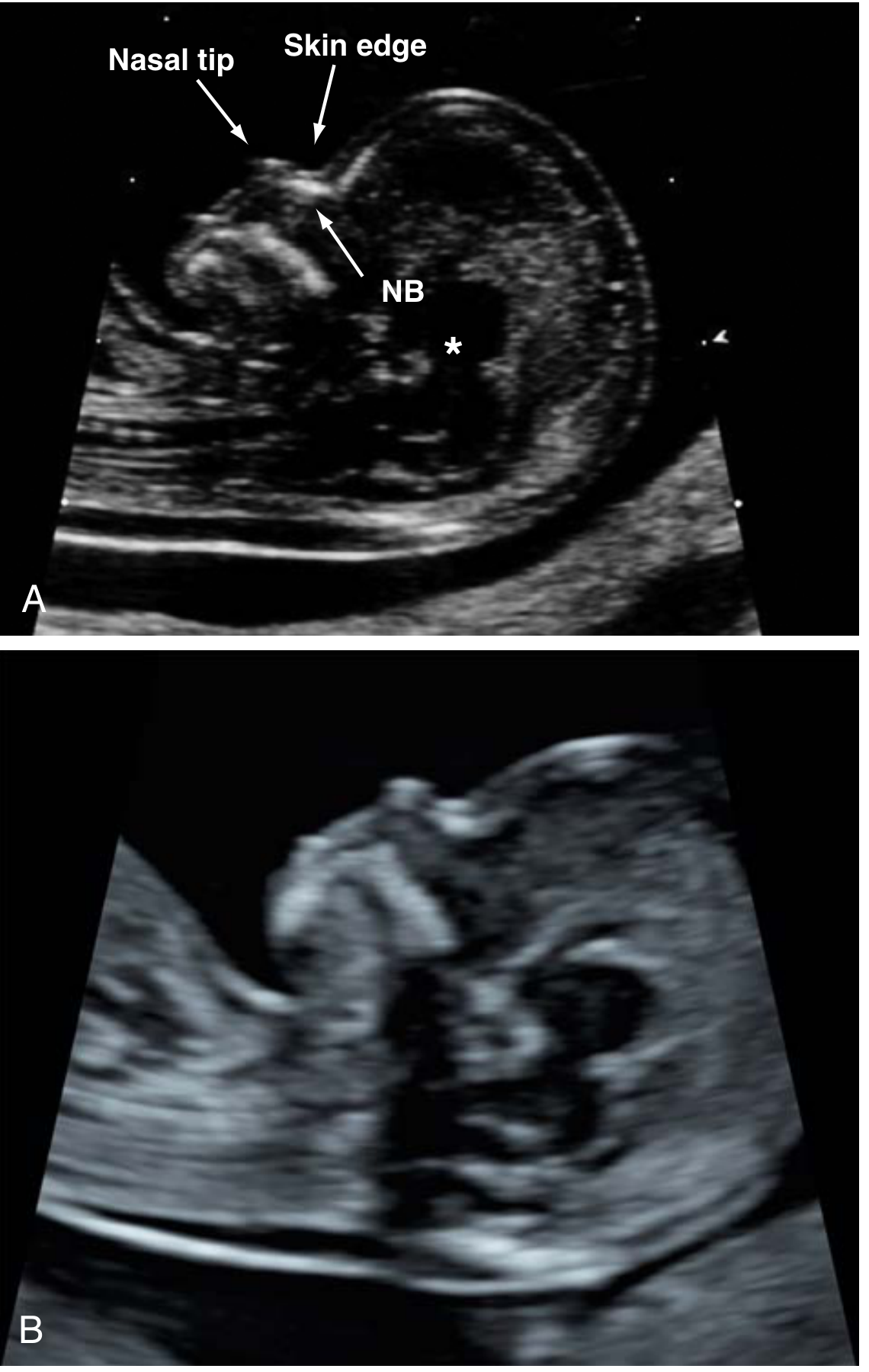
Figure: (A) Normal midsagittal profile - the nasal bone (NB) is the bright linear structure parallel to and below the skin edge, forming an "=" sign with it. (B) Absent nasal bone example.
Definition
Lack of ossification of the fetal nasal bone (NB) results in non-visualization on ultrasound. It is assessed between 11+0 and 13+6 weeks gestation as a marker for aneuploidy. - Creasy & Resnik's Maternal-Fetal Medicine
Incidence and Pathogenesis
In aneuploid fetuses:
| Aneuploidy | Absent NB incidence |
|---|---|
| Trisomy 21 (Down syndrome) | 69% |
| Trisomy 18 (Edwards) | 55% |
| Trisomy 13 (Patau) | 34% |
| Monosomy X (Turner) | 11% |
In euploid (chromosomally normal) fetuses:
- Overall: 0.6-2.6%
- Varies by CRL: 4.7% at CRL 45-54 mm vs. 1% at CRL 75-84 mm
- Varies by race/ethnicity: 2.2% Caucasian, 3.3% East Asian, 5.0% South Asian, 9.0% Afro-Caribbean
- Varies with NT thickness: 1.6% when NT ≤95th percentile, rising to 15% when NT ≥5.5 mm
Differential Diagnosis of Non-Visualization
Pathological causes:
- Aneuploidy (primarily trisomy 21)
- Genetic syndromes
Physiological/technical causes:
- Delayed calcification (normal variant, especially at lower CRL)
- Race (higher false-positive rate in Afro-Caribbean and South Asian)
- Suboptimal fetal position
- Poor imaging conditions (fibroids, high BMI)
How to Correctly Image the Nasal Bone
Correct technique is essential - this assessment should only be performed by a credentialed provider (NTQR or FMF certified):
- Strict midsagittal plane - tip of nose and 3rd/4th ventricles must be visible; fetal orbit and zygoma must NOT be visible
- Angle of insonation ~45 degrees to the profile; transducer parallel to the nose
- Tilt transducer side-to-side to show NB is separate from the overlying skin edge
- Normal NB: echogenicity greater than or equal to overlying skin; parallel and beneath skin edge = the "=" sign appearance
- Absent NB: less echogenic than the skin edge and thinner than overlying skin = not yet ossified
- Average of 80 studies needed to become proficient
Clinical Significance and Screening Performance
| Combination | Down syndrome detection rate | False-positive rate |
|---|---|---|
| NT + serum biochemistry alone | 90% | 5.0% |
| NT + serum + NB assessment | 90% | 2.5% (halved) |
Key statistics for NB alone (Rosen & D'Alton review of 35,312 women):
- Sensitivity: 65% for trisomy 21
- False-positive rate: 0.8%
- If NB absent: likelihood of trisomy 21 increased 87-fold
- Positive predictive value: ~54% (approximately 1 in 2 fetuses with absent NB had trisomy 21)
Associated Findings
- Increased NT
- Other structural anomalies associated with aneuploidy
- Important: An isolated absent NB with NT <95th percentile is not likely to be associated with a chromosomal microarray (CMA) abnormality
Limitations
- Technically challenging; limited availability of skilled providers
- Best used as a contingency marker in intermediate-risk women (risk 1:101 to 1:1000 after NT + biochemistry), not universal primary screening
- Secondary NB evaluation needed in ~10% of women to achieve the 90% detection / 2.5% false-positive target
Clinical Management After Finding
- Combine with other first-trimester data: NT measurement, serum free β-hCG and PAPP-A, maternal age - the absent NB alone is insufficient
- Consider cfDNA (cell-free fetal DNA/NIPT): High sensitivity for trisomy 21 as a next step
- Consider CVS or amniocentesis: For definitive karyotype if risk remains high or patient desires certainty
- Reassess at anomaly scan (18-22 weeks): To check for second-trimester soft markers or structural anomalies
Source: Creasy & Resnik's Maternal-Fetal Medicine: Principles and Practice; The Harriet Lane Handbook, 23rd ed.
This is a shared conversation. Sign in to Orris to start your own chat.