
History of travelling with symptoms Like body pain Headache Fever And red small pimples or bumps on feet and hand
red rash bumps hands feet fever travel
hand foot mouth disease rash vesicles
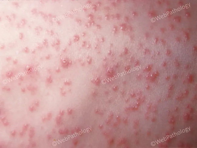
Clinical photography of cutaneous disease. High-resolution macro close-up image of a generalized vesicular rash on the trunk, compatible with Varicella (chickenpox) due to varicella-zoster virus (HHV-3). Anatomical context: integumentary system; skin of the torso (trunk) observed in frontal/anterior view. Visual features: multiple small vesicles (approximately 2-5 mm) containing clear fluid, arranged in crops, at various stages of evolution from vesicles to crusts. Surrounding erythematous halos are common; some lesions appear confluent in areas of diffuse rash. The distribution is trunk-predominant with involvement of the chest and abdomen, often sparing the limbs early in disease; lesions show centripetal dissemination. Classic morphology includes dew drops on a rose petal appearance with vesicles arising on an erythematous base, thin-walled and fragile, possibly eroded by scratching. Pathology: clinical diagnosis supported by history of fever and exposure; virologic confirmation (PCR or DFA) can be used. Diagnostic significance: characteristic vesicular-papular stage lesions in crops establish Varicella diagnosis; differential includes disseminated herpes simplex, hand-foot-mouth disease, disseminated zoster in adults, post-varicella rash. Clinical correlation: consider varicella vaccination status, age, pregnancy status, and risk of bacterial superinfection; contagious during vesicular phase; antiviral therapy indicated for high-risk patients. This image serves as an educational reference for dermatology training and research.
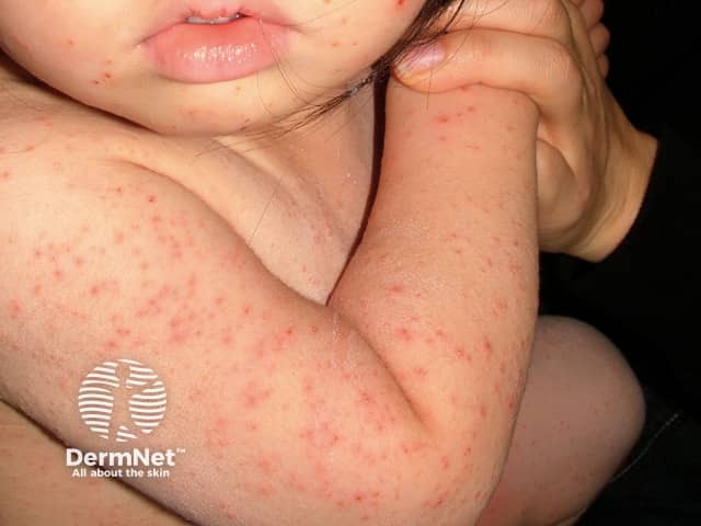
This pediatric dermatology clinical photograph provides a high-resolution, close-up view of a diffuse, non-vesicular erythematous maculopapular eruption on exposed skin surfaces. Lesions are small, round-to-oval papules and flat-topped macules with an erythematous base, distributed across the trunk, proximal limbs, and facial regions (notably the cheek). The pattern includes trunk and limb involvement with milder facial manifestations, and there is no evidence of vesicles, crusts, purpura, or mucosal involvement in the visible fields. The skin shows uniform morphology without scaling or hallmark vesicular halos. Clinically, the image suggests a benign viral exanthem typical of childhood illnesses, such as measles, rubella, roseola, or parvovirus B19, though a non-specific presentation is common. This visualization aids differential diagnosis by contrasting with varicella (vesicles at multiple stages) and hand-foot-mouth disease (palmar/plantar vesicles). Diagnostic significance lies in correlating the rash with febrile or systemic symptoms, exposure history, and vaccination status to guide patient counseling, isolation decisions, and management. Practical educational value includes teaching pediatricians, dermatologists, and infectious disease clinicians about pediatric rash patterns, syndrome-based diagnosis, and telemedicine skin assessments.
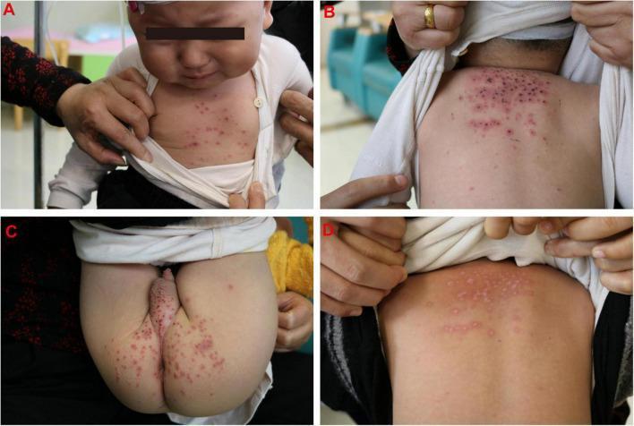
A series of clinical photographs (A-D) demonstrating a pediatric case of varicella-zoster virus (VZV) infection in a 16-month-old male. Panels A-C show the acute phase: (A) demonstrates scattered, erythematous papules on the anterior chest; (B) shows a dense cluster of erythematous papules and vesicles on the upper back and posterior neck, with some lesions exhibiting central umbilication or early crusting; (C) illustrates a similar pleomorphic rash involving the buttocks and genital region. Panel D serves as a comparison, showing the posterior trunk following treatment with acyclovir, where the inflammatory rash has resolved, leaving behind flat, hypopigmented macules and faint scarring. The presentation is characteristic of primary varicella, highlighting a centripetal distribution and the presence of lesions in multiple stages of development. This material is useful for distinguishing VZV from other pediatric exanthems like hand-foot-and-mouth disease (HFMD).
Febrile Traveler with Body Pain, Headache, and Red Papules/Bumps on Hands and Feet
Top Differential Diagnoses
1. Hand, Foot, and Mouth Disease (HFMD) - Most Likely if Lesions are Vesicular
- Fever (low-grade, lasting 2-3 days), malaise, decreased appetite
- Oral lesions - small 4-8 mm vesicles that rapidly ulcerate on buccal mucosa, tongue, soft palate, and gingiva (present in ~90% of cases)
- Skin lesions on hands and feet - asymptomatic red papules that quickly become small gray 3-7 mm vesicles with a red halo. Often oval/linear, running parallel to skin lines on fingers and toes. Distributed on dorsa of fingers/toes and more frequently on palms and soles
- Buttock involvement common in young children
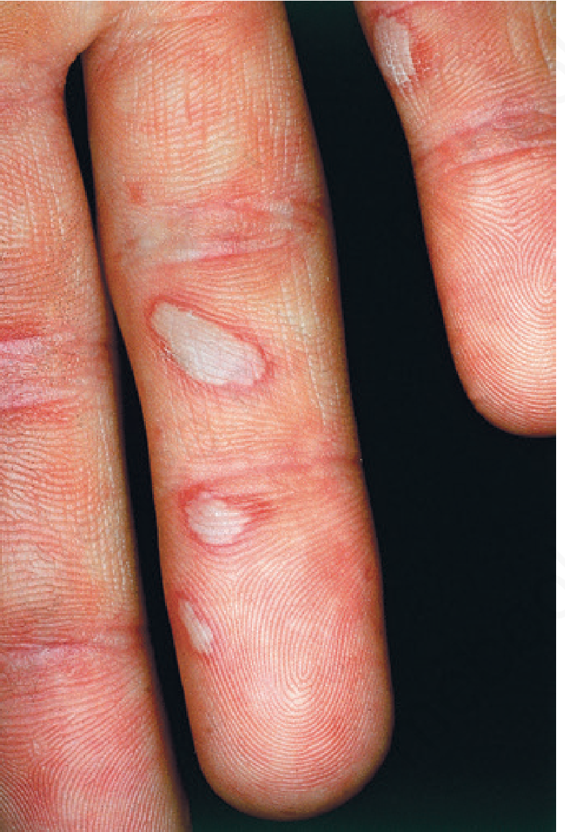
2. Dengue Fever - Must Rule Out in Tropical Travelers
- Sudden high fever, severe headache, retro-orbital pain
- Severe myalgia and bone pain ("breakbone fever")
- Maculopapular rash - typically appears as fever defervesces; generalized or extremity-predominant; confluent with characteristic "islands of white in a sea of red" spared patches
- Petechiae, positive tourniquet test, gingival bleeding
- Facial flushing, leukopenia, thrombocytopenia
3. Rocky Mountain Spotted Fever (RMSF) / Rickettsioses
- Fever, severe headache, myalgia - begins 2-14 days after tick bite
- Rash classically starts on palms and soles, wrists and ankles, then migrates centripetally toward the trunk
- Progresses from macular to petechial/maculopapular
- A traveler who hiked or spent time in wooded areas raises this concern significantly
4. Secondary Syphilis - Cannot Miss
- Fever, headache, malaise, diffuse lymphadenopathy
- Characteristic copper-red papular rash involving the palms and soles - this distribution is nearly pathognomonic
- History of a preceding painless genital ulcer (primary chancre) may be recalled
Differential Summary Table
| Condition | Fever | Headache | Myalgia | Rash Location | Rash Type | Travel Link |
|---|---|---|---|---|---|---|
| HFMD | Low-grade | Mild | Mild | Palms, soles, dorsal fingers/toes | Red papules → vesicles | Childcare, Asia, crowded areas |
| Dengue | High (sudden) | Severe | Severe ("breakbone") | Generalized, extremities | Maculopapular, petechiae | Tropical regions |
| RMSF/Rickettsia | High | Severe | Severe | Starts palms/soles, spreads centrally | Macular → petechial | Tick exposure, outdoors |
| Secondary Syphilis | Low-grade | Present | Present | Palms and soles prominently | Copper-red papules | Sexually transmitted |
| Chikungunya | High | Moderate | Severe arthralgia | Generalized | Maculopapular | Tropical/subtropical |
Red Flags - Seek Emergency Care Immediately If:
- Bleeding from any site (nose, gums, under skin) - suggests dengue hemorrhagic fever
- Confusion, seizure, neck stiffness - meningitis/encephalitis (enterovirus 71, rickettsial)
- Rash that does not blanch (press a glass to it - if it stays red/purple, it is petechial)
- Rapid deterioration or collapse - dengue shock syndrome
What to Do Now
- See a doctor urgently - especially given travel history
- Tell the doctor exactly where you travelled, when symptoms started, and any mosquito/tick exposures
- Blood tests needed: CBC with differential (look for thrombocytopenia, leukopenia), dengue NS1 antigen + IgM, liver function tests, Rickettsia serology if tick exposure
- Do not self-medicate with ibuprofen/NSAIDs if dengue is suspected - it increases bleeding risk. Use paracetamol (acetaminophen) for fever only.
Disclaimer: This is educational information only and does not replace a clinical evaluation. Please see a doctor or go to an emergency department for proper diagnosis and treatment.
Treatment of chickengunia and dangue
Treatment of Chikungunya and Dengue
DENGUE FEVER - Treatment
Phase-Based Management
| Group | Criteria |
|---|---|
| Dengue without warning signs | Fever + 2 of: nausea/vomiting, rash, body aches, leukopenia, positive tourniquet test |
| Dengue with warning signs | Above + abdominal pain, persistent vomiting, fluid accumulation, mucosal bleeding, lethargy, liver enlargement >2 cm |
| Severe dengue | Shock, severe bleeding, severe organ involvement (AST/ALT ≥1000 IU/L, impaired consciousness) |
Treatment by Phase
Febrile Phase (Days 1-3)
- Paracetamol (acetaminophen) - for fever and pain control
- Oral hydration - stay well hydrated, drink fluids liberally
- STRICT AVOIDANCE of:
- Aspirin / salicylate drugs
- NSAIDs (ibuprofen, naproxen, diclofenac)
- These increase bleeding risk significantly
Critical Phase (Days 3-7 - around the time fever breaks)
- Severe abdominal pain
- Persistent vomiting
- Gum or nose bleeding, blood in urine/stool
- Difficulty breathing
- Rapid deterioration, cold/clammy skin, restlessness
- IV fluid therapy (crystalloids first line - normal saline or Ringer's lactate)
- If refractory shock: IV colloids and/or blood products
- Close monitoring of hematocrit, platelet count, blood pressure
- Watch for fluid overload during recovery (convalescent phase)
Early recognition of shock and intensive supportive therapy reduces mortality from ~5-10% down to less than 1%
Convalescent Phase
- Gradual improvement
- Watch for signs of fluid overload as extravascular fluid gets reabsorbed
- Hematocrit may drop (dilutional effect of reabsorbed fluid - this is normal)
Dengue - Key Monitoring Tests
- CBC with differential - look for thrombocytopenia and leukopenia
- Dengue NS1 antigen (detectable days 1-10)
- Dengue IgM antibody (detectable from day 3-5 onward)
- Liver function tests (ALT/AST)
- Hematocrit (rising hematocrit = plasma leakage = danger sign)
CHIKUNGUNYA - Treatment
There is NO specific antiviral treatment.
| Treatment | Details |
|---|---|
| Rest | Essential during acute phase |
| Fluids | Adequate oral hydration |
| Acetaminophen (paracetamol) | Preferred for fever AND joint pain |
| Avoid NSAIDs initially | Until dengue is ruled out - since both diseases co-circulate in the same regions, starting NSAIDs before ruling out dengue can cause dangerous bleeding |
Once Dengue is Ruled Out:
- NSAIDs (ibuprofen, naproxen) - can then be used for joint pain relief
- Corticosteroids - for severe or persistent joint inflammation
- Physiotherapy - for persistent joint stiffness and pain
For Chronic/Persistent Arthritis (weeks to months later):
- Methotrexate - used in severe persistent arthritis
- Hydroxychloroquine - used in some patients with persistent arthritis
Chikungunya - Key Facts to Know
- Fever: Typically >39°C (102°F), lasts several days to 1 week, may be biphasic
- Joint pain: Bilateral, symmetric, most common in hands and feet - can be severely debilitating (the name "chikungunya" means "to become contorted" from the stooped posture due to joint pain)
- Rash: Maculopapular, involves trunk and extremities; palms, soles, and face may be affected
- Acute symptoms: Usually resolve within 7-10 days
- Risk for severe disease: Neonates, elderly (>65 years), people with hypertension, diabetes, cardiovascular disease, or kidney disease
- Chronic arthralgia risk factors: Age >50, severe acute arthritis, prolonged initial infection
Side-by-Side Comparison
| Feature | Dengue | Chikungunya |
|---|---|---|
| Antiviral drug | None | None |
| Fever treatment | Paracetamol only | Paracetamol (until dengue excluded) |
| NSAIDs | Absolutely AVOID | Avoid initially; can use after dengue ruled out |
| IV fluids | Required if warning signs/shock | Rarely needed |
| Hospital admission | Required for severe/warning sign cases | Usually outpatient |
| Duration | 7-10 days acute | 7-10 days acute; arthralgia may persist months-years |
| Biggest danger | Plasma leakage, hemorrhage, shock | Chronic disabling arthritis |
| Vaccine available | Yes (Dengvaxia, for ages 9-45) | No vaccine available |
Prevention (Both Diseases)
- Use insect repellent (DEET-based) consistently
- Wear long-sleeved shirts and long trousers
- Stay in screened or air-conditioned rooms
- Eliminate standing water around the home (mosquito breeding sites)
- Use mosquito nets at night
Important: This is educational information. If you or someone you know has dengue warning signs (bleeding, severe abdominal pain, vomiting that won't stop, signs of shock), go to an emergency department immediately - severe dengue is a medical emergency.