
Serum Hypokalemia is low
Reading File
Hypokalemia means serum potassium (K+) below 3.5 mEq/L. It is one of the most common electrolyte disturbances seen clinically, with effects ranging from mild muscle weakness to life-threatening cardiac arrhythmias.
Searching sources...
Finding Sources
Searching PubMed
"hypokalemia" AND management
Reading File
Reading File
Reading File
Reading File
Reading File
Reading File
Loading Image
Reading File
Here is a thorough overview of serum hypokalemia, drawn from multiple authoritative textbooks:
Hypokalemia (Low Serum Potassium)
Definition
Hypokalemia is defined as a serum potassium (K+) concentration less than 3.5 mEq/L.
- Mild hypokalemia: 3.0 - 3.5 mEq/L
- Moderate hypokalemia: 2.5 - 3.0 mEq/L
- Severe hypokalemia: < 2.5 mEq/L (paralysis and life-threatening arrhythmias can occur at < 2.0 mEq/L)
It is the most common electrolyte abnormality in clinical practice - found in >20% of hospitalized patients and up to 40% of outpatients on thiazide diuretics. - Rosen's Emergency Medicine, p. 2212
Pathophysiology
Potassium is predominantly an intracellular cation (98% intracellular). Two broad mechanisms cause hypokalemia:
- Transcellular (internal) shift - K+ moves from extracellular to intracellular space (acute, transient)
- True external K+ deficit - excess K+ loss via kidneys or GI tract (chronic, more significant)
Each 0.3 mEq/L drop in serum K+ below normal corresponds to approximately 100 mEq total body K+ deficit. - Rosen's Emergency Medicine, p. 2271
Causes
Five Key Categories (Rosen's Emergency Medicine, Box 114.2)
| Category | Examples |
|---|---|
| Renal losses | Diuretics (thiazide > loop), hyperaldosteronism, steroid excess, DKA, renal tubular acidosis, alcohol, aminoglycosides, penicillin |
| Non-renal losses | Diarrhea, vomiting, NG suction, laxative abuse, sweating |
| Decreased intake | Malnutrition, ethanol abuse |
| Intracellular shift | Metabolic alkalosis, hyperventilation, insulin, beta-agonists (e.g., albuterol), decongestants |
| Endocrine | Cushing disease, Bartter syndrome, Gitelman syndrome, insulin therapy |
Key Drug-Specific Notes
- A standard dose of nebulized albuterol reduces serum K+ by 0.2-0.4 mEq/L; a second dose within 1 hour reduces it by nearly 1 mEq/L.
- Vomiting/NG suction: only 5-10 mEq/L K+ is in gastric fluid; hypokalemia is secondary to metabolic alkalosis, chloride depletion, and hyperaldosteronism.
Mechanisms of Diuretic-Induced Hypokalemia
The diagram below shows how diuretics cause hypokalemia through four mechanisms - increased tubular flow, AVP secretion, aldosterone secretion (RAAS activation), and alkalosis, all enhancing K+ secretion via ROMK and BK channels in the collecting duct:
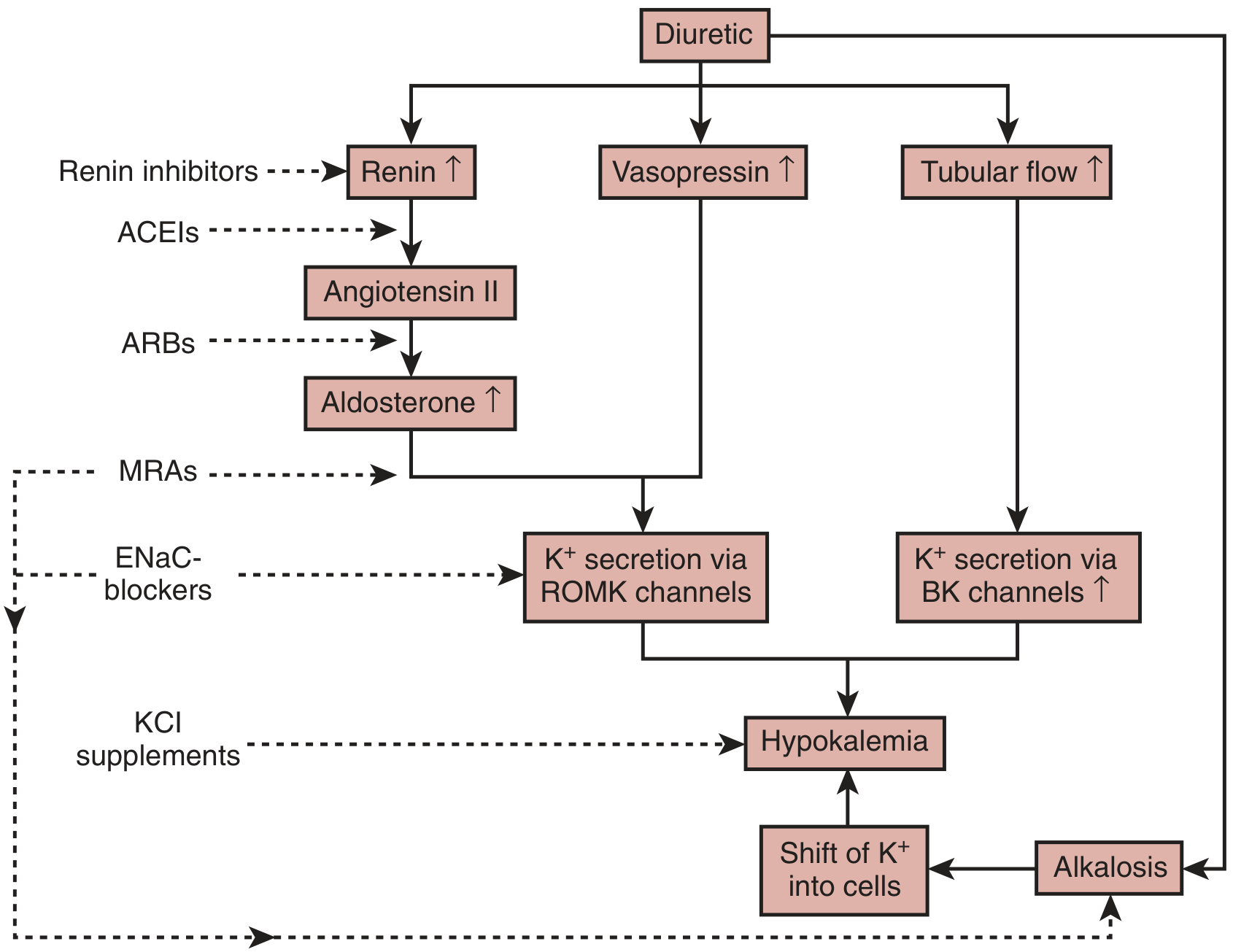
Dashed lines show treatment strategies: renin inhibitors, ACEIs, ARBs, MRAs (spironolactone), ENaC blockers (amiloride, triamterene), KCl supplements. - Brenner & Rector's The Kidney, Fig. 50.20
Clinical Features
Usually asymptomatic in mild cases. Symptoms correlate with the rate of drop (acute drops are worse tolerated than chronic).
| System | Manifestations |
|---|---|
| Neuromuscular | Weakness, muscle pain, fasciculations, depressed deep tendon reflexes, paralysis (K+ < 2.5 mEq/L), rhabdomyolysis, paralytic ileus |
| Cardiac | ST depression, flat/inverted T waves, U waves (pathognomonic), prolonged QT, torsades de pointes (QT > 500 ms), AFib, VFib, asystole |
| Renal | Polyuria/polydipsia, hypokalemic nephropathy |
| Metabolic | Glucose intolerance, metabolic alkalosis |
| CNS | Paresthesias, confusion (at K+ < 2.5 mEq/L) |
In patients without heart disease, cardiac conduction abnormalities are unusual even at K+ < 3.0 mEq/L. But in cardiac ischemia or heart failure, even mild-moderate hypokalemia significantly raises arrhythmia risk. Target K+ > 4.5 mEq/L in acute MI. - Rosen's Emergency Medicine, p. 2219
Diagnostic Approach
- History and physical - usually reveals cause (diuretics, vomiting, etc.)
- ECG - look for U waves, flat T waves, prolonged QT
- Spot urine K+ - if cause is unclear or hypokalemia is severe:
- Urine K+ > 13 mEq/L per gram creatinine → inappropriate renal K+ wasting
- Fractional excretion of K+ > 30% OR TTKG > 6 → renal K+ wasting
- Acid-base status:
- Hypokalemia + alkalosis + hypertension → primary mineralocorticoid excess (hyperaldosteronism, Liddle syndrome, CAH)
- Hypokalemia + alkalosis + normal/low BP → salt-wasting (Bartter, Gitelman)
- Hypokalemia + acidosis → renal tubular acidosis
- Check serum magnesium - always assume concurrent hypomagnesemia (see below)
Treatment
Oral Replacement (mild-moderate, K+ > 2.5 mEq/L, no ECG changes)
- Potassium chloride (KCl) 40-60 mEq orally every 2-4 hours
- Encourage K+/Mg-rich foods: potatoes, avocado, bananas, tomatoes, black beans
- Patients on diuretics: add K+-sparing agent (spironolactone, amiloride) or switch to combination thiazide/K+-sparing diuretic
IV Replacement (severe or unable to take oral)
- KCl at 10-20 mEq/hr via peripheral line (standard)
- > 20 mEq/hr (for K+ < 2.0 mEq/L or QT > 500 ms): requires continuous cardiac monitoring + central line access
- Pediatric dosing: max 0.5 mEq/kg/hr, max concentration 40 mEq/L in saline - Brenner & Rector's The Kidney, p. 3150
Magnesium Replacement
- Always replace magnesium alongside potassium - hypokalemia and hypomagnesemia co-exist frequently; potassium repletion will be refractory if Mg2+ is not corrected. - Rosen's Emergency Medicine, p. 2275
Discharge Criteria
- K+ > 3.0 mEq/L, tolerating oral intake, QT interval < 500 ms
Summary Table
| Feature | Value |
|---|---|
| Normal serum K+ | 3.5 - 5.0 mEq/L |
| Definition of hypokalemia | < 3.5 mEq/L |
| Severe | < 2.5 mEq/L |
| Most common cause | Diuretics (thiazides) |
| Classic ECG finding | U waves, flat T waves |
| Key association | Hypomagnesemia |
| IV replacement rate (standard) | 10-20 mEq/hr |
| IV replacement rate (life-threatening) | > 20 mEq/hr with cardiac monitoring |
Sources: Rosen's Emergency Medicine (9th ed.), Brenner & Rector's The Kidney (10th ed.), National Kidney Foundation Primer on Kidney Diseases (8th ed.)
Does it causes arythmias
Yes, hypokalemia is a well-known cause of cardiac arrhythmias. The risk depends on the severity of the K+ drop and the patient's underlying heart condition.
Searching sources...
Finding Sources
Searching PubMed
"hypokalemia" AND "cardiac arrhythmia"
Reading File
Reading File
Reading File
Yes - absolutely. Hypokalemia is a well-established, direct cause of cardiac arrhythmias, ranging from benign ECG changes to life-threatening rhythms. Here is a detailed breakdown:
Hypokalemia and Cardiac Arrhythmias
Why Does Hypokalemia Cause Arrhythmias? (Mechanisms)
There are three key electrophysiological mechanisms:
1. Paradoxical Depolarization of Cardiac Purkinje Fibers
- Normally, low extracellular K+ hyperpolarizes skeletal muscle (making it harder to fire).
- But in human cardiac Purkinje cells, hypokalemia causes paradoxical depolarization - the opposite effect.
- This is mediated by K2P1 (TWIK-1) channels, which normally carry K+ outward but switch to carrying Na+ inward when K+ falls, destabilizing the resting membrane potential and triggering ectopic firing. - Brenner & Rector's The Kidney, p. 1909-1910
2. HERG Channel Downregulation → Long QT Syndrome
- Hypokalemia accelerates internalization and degradation of HERG (IKr) K+ channels via a clathrin-dependent process.
- HERG encodes the rapidly activating delayed rectifier K+ channel responsible for repolarization (phases 2-3 of the action potential).
- Loss of IKr delays repolarization → prolonged QT interval → torsades de pointes (TdP). - Brenner & Rector's The Kidney, p. 1907
3. Na+/K+-ATPase Downregulation → Intracellular Ca2+ Overload
- Hypokalemia suppresses Na+/K+-ATPase activity.
- Intracellular Na+ rises → impairs the Na+/Ca2+ exchanger → calcium overload inside cardiomyocytes.
- Elevated intracellular Ca2+ activates calmodulin kinase II → activates late Na+ and Ca2+ currents → reduces repolarization reserve → further arrhythmia risk. - Brenner & Rector's The Kidney, p. 1917
Types of Arrhythmias Caused
| Arrhythmia Type | Risk Level |
|---|---|
| Atrial tachycardia (with or without block) | Moderate |
| Atrioventricular (AV) dissociation | Moderate |
| Atrial fibrillation | High (especially post-cardiac surgery) |
| Ventricular tachycardia (VT) | High |
| Ventricular fibrillation (VF) | Life-threatening |
| Torsades de Pointes (TdP) | Life-threatening |
| Asystole | Life-threatening |
Sabiston Textbook of Surgery; Rosen's Emergency Medicine
ECG Changes in Sequence (Severity)
As K+ falls, ECG changes progress:
- Flat or inverted T waves (early)
- Prominent U waves - small deflections after the T wave (pathognomonic)
- ST segment depression
- Prolonged QT interval - most dangerous; once QT > 500 ms, risk of TdP increases 2-3 fold
- Torsades de Pointes - a polymorphic VT that can degenerate to VF and sudden cardiac death
ECG changes are most marked when K+ < 2.7 mmol/L. - Brenner & Rector's The Kidney, p. 1907
Who Is Most at Risk?
Not everyone with hypokalemia will get arrhythmias. Risk is significantly higher in:
| High-Risk Group | Reason |
|---|---|
| Patients on digoxin | Hypokalemia potentiates digoxin toxicity, even at low-normal K+ levels |
| Acute MI / cardiac ischemia | Even mild-moderate hypokalemia significantly increases arrhythmia risk; target K+ > 4.5 mEq/L in AMI |
| Heart failure / dilated or hypertrophied ventricles | Independent risk factor for death; target K+ 4.0-5.0 mEq/L |
| Cardiac surgery patients | K+ < 3.5 mEq/L predicts serious intra- and peri-operative arrhythmias and post-op AFib |
| Patients with LQTS mutations | Hypokalemia + genetic mutation = compounded risk for TdP |
| Athletes post-exercise | Rapid post-exercise K+ drop can trigger TdP and sudden cardiac death |
| Thyrotoxic periodic paralysis | Acute severe hypokalemia + weakness; can be fatal |
"In patients without underlying heart disease, abnormalities in cardiac conduction are extremely unusual, even when serum K+ is below 3.0 mEq/L." - Rosen's Emergency Medicine, p. 2219
Key Clinical Point: Hypomagnesemia Makes It Worse
Hypokalemia almost always coexists with hypomagnesemia. Magnesium is essential for:
- Maintaining intracellular K+ levels
- Stabilizing Na+/K+-ATPase function
- Independently reducing arrhythmia risk
If you only replace potassium without correcting magnesium, hypokalemia (and its arrhythmias) will be refractory. Always replace both together. - Sabiston Textbook of Surgery, p. 705
Bottom Line
| K+ Level | Arrhythmia Risk |
|---|---|
| 3.0 - 3.5 mEq/L (mild) | Low in healthy hearts; significant in cardiac disease |
| 2.5 - 3.0 mEq/L (moderate) | Elevated; ECG monitoring needed |
| < 2.5 mEq/L (severe) | High; VT, VF, TdP, sudden death possible |
| < 2.0 mEq/L | Medical emergency - continuous cardiac monitoring + IV KCl with central access |
Sources: Brenner & Rector's The Kidney (10th ed.), Sabiston Textbook of Surgery, Rosen's Emergency Medicine, DiMaio's Forensic Pathology (3rd ed.)
This is a shared conversation. Sign in to Orris to start your own chat.