
Non surgical management of endodontic leasons 50 marks answer .
endodontic lesion non-surgical treatment
periapical lesion endodontic radiograph root canal
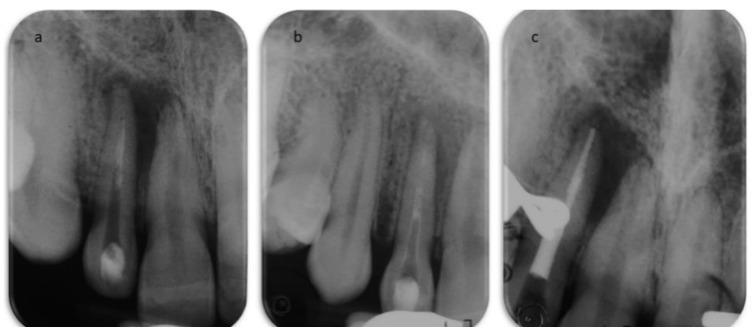
This set of periapical radiographs illustrates the progressive stages of endodontic (root canal) treatment on an upper anterior tooth. (a) Post-access radiograph: The primary canal is largely radiolucent (empty) with minimal radiopaque filling material remaining at the apical third from a prior failed treatment. A large, well-defined periapical radiolucency is visible at the apex, indicating a chronic periapical lesion or abscess. (b) Intermediate stage: The canal is now filled with a moderately radiopaque calcium hydroxide temporary dressing, extending from the coronal access to the apex to facilitate disinfection. (c) Final obturation: The radiograph shows the canal completely sealed with a dense, uniform radiopaque material (gutta-percha and sealer), extending to the radiographic apex. The surrounding alveolar bone structure and the persistent radiolucent lesion are visible for comparison across all stages. These images serve as educational examples of orthograde retreatment and the visual verification of filling density and length in endodontics.
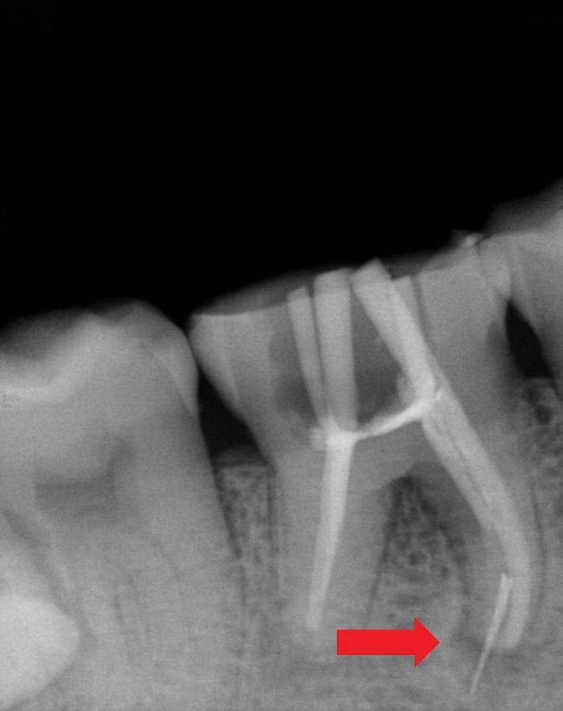
This diagnostic periapical radiograph depicts a mandibular right first molar (tooth 46) undergoing endodontic therapy. The image shows a complex root canal treatment with radiopaque obturation material partially visible within the canal space. A significant endodontic complication is highlighted by a red arrow: a separated NiTi rotary instrument fragment is visible within the mesiolingual root, with approximately 4mm of the fractured file extending beyond the anatomical apex into the periapical bone. A distinct, well-defined radiolucency is observable surrounding the root apices and extending into the furcation area, characteristic of a chronic periapical lesion (periodontitis). The surrounding alveolar bone shows loss of lamina dura and altered trabecular pattern consistent with apical pathology. This clinical photograph is used in dental education to demonstrate procedural accidents in endodontics, specifically instrument separation and its radiographic identification relative to anatomical structures.
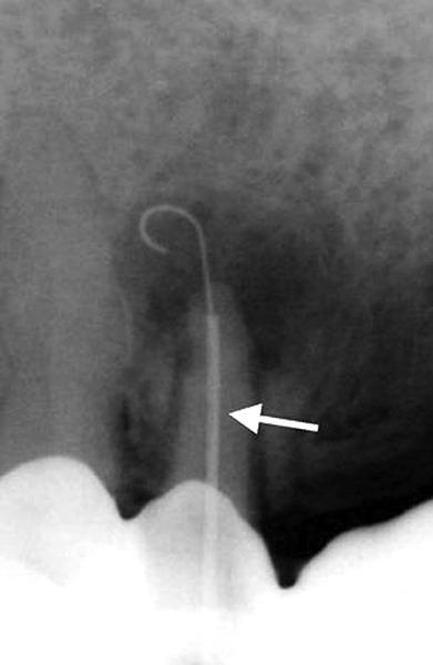
This periapical radiograph demonstrates an endodontic procedure involving a single-rooted tooth with a significant periapical lesion. A radiolucent area, indicating bone resorption and chronic inflammatory or infectious tissue, is clearly visible at the root apex. Within the root canal, a radiopaque Apexum NiTi Ablator is positioned for minimally invasive periapical surgery. The device consists of a straight Nitinol sheath, indicated by a white arrow, which serves as a guide within the canal. Extruded through the apical foramen into the center of the periapical radiolucency is the pre-shaped, curved Nitinol wire (ablator). The curved tip of the ablator is deployed within the lesion, illustrating its mechanical design for grinding and debriding periapical soft tissues through a retrograde approach. This educational visual highlights advanced endodontic techniques for non-surgical management of large periapical pathologies by facilitating tissue removal via the root canal system.
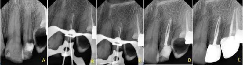
This composite of five periapical radiographs (labeled A-E) documents the step-by-step endodontic management of a maxillary left lateral incisor exhibiting a periapical lesion and complex root canal anatomy. Radiograph A (Preoperative) shows an existing access cavity and a distinct periapical radiolucency suggestive of apical periodontitis. Radiograph B illustrates the working length determination with endodontic files in situ, highlighting the negotiation of two separate root canals in a tooth that typically presents with one. Radiograph C depicts the master cone evaluation using radiopaque gutta-percha points. Radiograph D shows the immediate post-obturation state with homogenous radiopaque filling material extending to the apex. Radiograph E is a six-month follow-up showing a permanent radiopaque coronal restoration and significant reduction (healing) of the periapical radiolucency. The series serves as an educational tool for non-surgical endodontic treatment, anatomical variations in maxillary incisors, and radiographic monitoring of periapical bone regeneration.
non surgical management endodontic lesions root canal treatment steps 2024
Non-Surgical Management of Endodontic Lesions
Introduction
Classification of Endodontic Lesions Relevant to Management
| Type | Features | Management Implication |
|---|---|---|
| Symptomatic irreversible pulpitis | Lingering pain, vital pulp | NSRCT |
| Pulp necrosis | No response to testing, no symptoms | NSRCT |
| Symptomatic apical periodontitis | Percussion sensitivity, radiolucency | NSRCT +/- antibiotics |
| Chronic apical abscess | Sinus tract, chronic drainage | NSRCT |
| Acute apical abscess | Swelling, fluctuance, systemic signs | Drainage + NSRCT +/- antibiotics |
| Previously treated (post-treatment disease) | Persistent/recurrent radiolucency | Non-surgical retreatment |
| Endo-perio lesion (true combined) | Both pulpal + periodontal etiology | NSRCT + periodontal therapy |
I. Diagnosis and Pre-treatment Assessment
A. Clinical Examination
- Thorough history: onset, character, duration, and radiation of pain; response to thermal, mechanical stimuli
- Extraoral: lymphadenopathy, facial swelling, sinus tracts traced with gutta-percha cone
- Intraoral: caries assessment, restorations, probing depths, percussion, palpation
B. Pulp Vitality Testing
- Thermal tests: Cold (ethyl chloride, CO2 snow, refrigerant sprays) and heat (warm gutta-percha, warm water)
- Electric pulp testing (EPT): Screens for pulp vitality; unreliable for calcified canals or immature teeth
- Laser Doppler flowmetry / pulse oximetry: More accurate - assesses blood flow rather than nerve function (especially in traumatized teeth)
C. Radiographic Assessment
- Periapical radiographs: Establish baseline; assess root morphology, number of canals, root length, periapical status (lamina dura integrity, periapical index - PAI score)
- Bitewing radiographs: Assess extent of caries, existing restorations
- Cone Beam CT (CBCT): Indicated for non-healing lesions, complex root morphology, suspected root fractures, calcified canals, resorption; AAE guidelines recommend limited-FOV CBCT as modality of choice for evaluating non-healing post-treatment disease
D. Restorative Assessment
- Tooth restorability must be confirmed before initiating endodontic therapy
- Crown-to-root ratio; periodontal support; strategic value of the tooth
II. Patient Preparation and Infection Control
- Informed consent: discuss alternatives (extraction, implant), treatment sequence, prognosis
- Pre-procedural antimicrobial mouthrinse (0.2% chlorhexidine or 1% povidone-iodine) reduces aerosol contamination
- Rubber dam isolation: Mandatory standard of care - provides a clean operating field, prevents ingestion/aspiration of instruments, and prevents salivary contamination of the canal
III. Access Cavity Preparation
Principles
- Straight-line access to the root canal orifices
- Complete removal of the pulp chamber roof
- Removal of all carious tissue and unsupported tooth structure
- Conservation of tooth structure: "ninja" or conservative access (truss access, contracted endodontic access) advocated to preserve dentinal walls
Steps
- Remove all existing restorations, caries
- Prepare access cavity with high-speed bur (round or Endo-Z bur)
- Identify all canal orifices using DG-16 explorer or endodontic probe
- Use operating microscope (DOM) for calcified or difficult cases
IV. Working Length Determination
- Electronic apex locators (EAL): First-line method (4th generation: Propex Pixi, Raypex 6, Root ZX); measure electrical resistance/impedance; accuracy ~94-96%; not affected by presence of electrolytes
- Radiographic method: Parallel technique with file at estimated WL; adjust for foreshortening or magnification
- Combined method: EAL confirmed with radiograph - gold standard in clinical practice
- Working length = distance from reference point to 0.5-1.0 mm short of radiographic apex (the minor constriction/CDJ)
V. Canal Preparation (Chemo-Mechanical Preparation)
A. Objectives (Schilder's Goals)
- Develop a continuously tapering funnel shape
- Narrow cross-sections at every point apically
- Maintain natural curvature
- Apical foramen should remain in original position
- Apical opening as small as practical
B. Irrigation (Chemical Component)
- Concentration: 0.5-5.25%; most commonly 2.5-3% (balance between efficacy and cytotoxicity)
- Mechanism: dissolves organic matter, necrotic pulp tissue, and has broad-spectrum antimicrobial action via release of free chlorine
- Cannot penetrate dentinal tubules adequately alone
- Volume and agitation more important than concentration alone
- Chelating agent; removes inorganic smear layer
- Used as penultimate rinse before obturation
- Working time: 1 minute; do not use simultaneously with NaOCl (inactivates each other)
- Broad-spectrum, substantivity (~12 weeks on dentinal walls)
- Used as final rinse or intracanal medicament
- CAUTION: Do NOT mix CHX with NaOCl - forms para-chloroaniline precipitate (orange-brown, cytotoxic)
- Passive Ultrasonic Irrigation (PUI): ultrasonic file (passive, not mechanically cutting) placed in canal filled with irrigant; acoustic streaming and cavitation improve irrigant penetration; superior to syringe irrigation alone
- Sonic irrigation (EndoActivator): polymer tip activated at sonic frequencies; safe, no risk of instrument separation
- Laser-activated irrigation: Er:YAG laser; highly effective but requires special equipment
- GentleWave (Multisonic Ultracleaning): proprietary system using broad-spectrum acoustic energy and degassed irrigants; excellent for complex canal anatomy
- 2.5-3% NaOCl throughout preparation (abundant volume, ~20 mL per canal)
- 17% EDTA for 1 minute (smear layer removal)
- Final NaOCl flush (or CHX 2% as final rinse)
- Distilled water flush if using CHX (to prevent PCA precipitate)
C. Mechanical Preparation
- K-files, H-files, reamers (stainless steel)
- Step-back technique (Clem, 1969): canal prepared to small apical diameter, then flare created coronally
- Crown-down technique: begins with larger instruments coronally, progresses apically
- Used in severely curved canals for tactile feedback
- Nickel-titanium alloys: superelastic, shape memory; 2-3x more flexible than stainless steel
- Rotary motion with variable taper (e.g., 0.06, 0.08 taper)
- Systems: ProTaper Universal/Gold/Next, WaveOne Gold (reciprocating), HyFlex EDM, TF Adaptive, Vortex Blue
- Reciprocating motion (WaveOne Gold, Reciproc Blue): single-file systems; CW/CCW motion reduces cyclic fatigue and risk of separation
- Heat-treated alloys (Controlled Memory, Gold wire, Blue wire): increased resistance to cyclic fatigue in curved canals
VI. Management of the Smear Layer
- Smear layer = amorphous layer of organic + inorganic debris created during canal preparation; occludes dentinal tubule orifices
- Removal recommended to expose tubules for irrigant penetration and better adaptation of sealers
- Protocol: 17% EDTA (chelates inorganic component) + NaOCl (dissolves organic component)
- QMix (EDTA + CHX + cetrimide): single-solution smear layer removal with antimicrobial property
VII. Intracanal Medicaments (Between Appointments)
Calcium Hydroxide (Ca(OH)₂) - Gold Standard
- Mechanism: high alkaline pH (~12.5) denatures bacterial enzymes and lipopolysaccharides; stimulates periapical healing; antibacterial (particularly against E. faecalis with long-term exposure)
- Forms: pure powder mixed with saline/water, commercial pastes (Ultracal, Calcipast)
- Placement: placed with lentulo spiral, plugger, or syringe; fill entire canal; verified radiographically
- Duration: minimum 1 week, ideally 2-4 weeks for large lesions; refresh if used >4 weeks (becomes ineffective)
- Note: ineffective against E. faecalis in short-term use; does not eliminate biofilms completely
Antibiotic Pastes
- Triple antibiotic paste (TAP): metronidazole + ciprofloxacin + minocycline; used in regenerative endodontics; minocycline can stain crown - replaced with amoxicillin or clindamycin = modified TAP
- Double antibiotic paste (DAP): metronidazole + ciprofloxacin; avoids staining
- Diluted to 0.1 mg/mL concentration (Hoshino protocol modified) to avoid stem cell toxicity
Formocresol / Ledermix
- Formocresol: devitalizing agent; concerns over cytotoxicity and carcinogenicity; largely replaced by Ca(OH)₂
- Ledermix (triamcinolone + demeclocycline): reduces inflammation and pain; short-term use only
VIII. Obturation
Requirements (Grossman's criteria)
- Easy to manipulate with adequate working time
- Dimensionally stable
- Should seal canal laterally and apically
- Non-irritating to periapical tissues
- Impermeable to moisture
- Resistant to dissolution in tissue fluids
- Bacteriostatic or at least not bacteria-favoring
- Radiopaque
- Easily removable if retreatment needed
Core Material: Gutta-Percha
- Derived from Isonandra gutta tree; gutta-percha + zinc oxide + barium sulfate + heavy metal sulfates
- Alpha (thermoplasticized) and Beta (conventional cones) phases
- Biocompatible, dimensionally stable, radiopaque, easily removed
Sealers
- Zinc oxide eugenol (ZOE) sealers: Grossman's, Tubli-Seal; antimicrobial but sensitizing in some patients
- Resin sealers: AH Plus (bisphenol A-epoxy resin); excellent adaptation, low solubility, excellent flow; gold standard sealer; PMID evidence supports high success rates
- Calcium silicate sealers: BioRoot RCO, iRoot SP, EndoSequence BC Sealer; bioactive, biocompatible, hydrophilic; expand slightly on setting; evidence from systematic review (PMID 38145805) supports good outcomes
- MTA-based sealers: ProRoot Endo Sealer; regenerative potential
- Glass ionomer sealers: Ketac-Endo; water-based but high solubility
Obturation Techniques
- Most widely taught technique
- Master cone (ISO size = working length) + spreader + accessory cones + sealer
- Predictable, controlled; allows retrieval
- Disadvantage: voids possible; does not adapt well to complex anatomy
- Heat carrier (System B plugger) softens gutta-percha
- Wave condensation + back-filling with Obtura or Calamus gun
- Excellent adaptation to complex canal anatomy; fewest voids
- Risk: extrusion of material if not careful
- Single master cone + hydraulic calcium silicate sealer
- Simplified; sealer provides majority of seal
- Well-supported by evidence for bioceramic sealers; PMID 38145805
- Obtura II / Calamus: high-temperature injectable GP
- Thermafil / GuttaCore carriers: pre-heated carrier with GP coating
- Excellent for complex anatomy, lateral canals, fins, isthmuses
IX. Non-Surgical Retreatment (Post-Treatment Endodontic Disease)
Indications
- Persistent or new periapical radiolucency after completed RCT
- Procedural errors (ledging, perforation, short obturation, missed canals)
- Canal contamination due to coronal leakage
- Previously treated tooth requiring retreatment for prosthodontic reasons
Steps of Non-Surgical Retreatment
- Remove crown, post (if present) using ultrasonic instruments, post-pulling devices (Gonon, Eggler extractors), or post-removing sets
- Surgical operating microscope (DOM): mandatory for adequate visualization
- Gutta-percha removal: ProTaper Retreatment files (D1, D2, D3), rotary systems with solvent (chloroform, eucalyptol, halothane); chloroform most effective but cytotoxic with caution
- Paste/sealer removal: ultrasonic irrigation, solvent with hand files
- Carrier-based obturation: plasticized carrier removed with heat (System B), then GP removed as above
- Separated instruments: ultrasonic trephining around fragment, IRS (Instrument Retrieval System), bypass with small file if retrieval not possible; CBCT essential for planning
- Ledges: flexible NiTi files with pre-curved tip to bypass, restore canal patency
- Perforations: mineral trioxide aggregate (MTA) repair; bioceramic cements (Biodentine); prognosis depends on location, size, and time elapsed
- Missed canals: DOM-guided exploration; chlorhexidine or dye staining; CBCT for canal anatomy
- Calcified canals: safe-ended burs (Munce Discovery burs), ultrasonic tips (Spartan, Acteon), CBCT guidance
- Same protocol as initial NSRCT
- Ca(OH)₂ dressing between appointments more strongly indicated
X. Management of Specific Endodontic Lesions
A. Large Periapical Lesions
- Initial NSRCT with Ca(OH)₂ dressing for 3-6 months before obturation
- Biological rationale: alkaline environment promotes periapical healing and stimulates cementogenesis and bone regeneration
- A 2023 meta-analysis (PMID 36890256) confirmed that larger lesion size has a modest negative effect on success but NSRCT remains first line
B. Apical Cysts
- True radicular cysts: self-contained epithelial-lined cavities; may not heal with NSRCT alone
- Bay cysts (periapical pocket cysts): open to root canal, inflammatory cells communicate with canal - NSRCT very likely to resolve
- Clinical approach: attempt NSRCT first; review at 12-24 months; surgical intervention only if no radiographic evidence of healing
C. Endodontic-Periodontal Lesions
- Primary endodontic with secondary periodontal involvement: treat endodontic component first; periodontal component may resolve; then reassess and treat residual periodontal pocketing
- Primary periodontal with secondary endodontic involvement: periodontal therapy first; if pulp becomes non-vital, add NSRCT
- True combined lesion: simultaneous NSRCT + subgingival periodontal instrumentation; two-visit approach with Ca(OH)₂ dressing shown to improve periodontal outcomes by reducing LPS levels (per British Dental Journal 2025)
D. Internal Resorption
- Caused by chronic pulp inflammation activating clastic cells within the canal
- NSRCT is the definitive treatment: eliminates the stimulus; halts progression
- Thorough mechanical + ultrasonic irrigation essential to remove resorptive tissue in irregular lacunae
- Mineral trioxide aggregate (MTA) or bioceramic paste (Biodentine) used to fill resorptive defects
E. External Cervical Resorption
- If pulp remains vital: arrest resorption by surgical exposure + trichloroacetic acid application + restoration; NSRCT only if pulp becomes non-vital
- Non-surgical approach for Class 1 (Heithersay): internal access + MTA/Biodentine filling of defect (minimally invasive)
F. Immature Teeth with Necrotic Pulp (Open Apex)
- Repeated Ca(OH)₂ dressings every 3-6 months until apical barrier formation (calcific bridge)
- Time: 6-24 months; risk of tooth fracture with long-term Ca(OH)₂
- Single visit: place 3-4 mm of MTA at apex, pack with moist cotton for 4 hours, back-fill with GP
- Immediate; predictable; reduces fracture risk
- Disinfect canal with copious NaOCl + EDTA irrigation + antibiotic paste/Ca(OH)₂ (2-4 weeks)
- Second visit: bleed clot evoked by over-instrumenting 2 mm beyond apex; collagen matrix/scaffold placed; MTA or Biodentine coronal plug; composite restoration
- Goal: allow blood clot to act as scaffold for stem cell migration from apical papilla (SCAP)
- Outcomes: continued root development, increased root length and wall thickness, possible pulp-like tissue ingrowth
- Supported by AAE Position Statement (2021); superior for root development in young teeth
XI. Coronal Restoration
- Anterior teeth: composite resin directly post-obturation (same appointment if possible) to prevent coronal leakage
- Posterior teeth: full-coverage crown after NSRCT to prevent cuspal fracture; delay restoration until symptoms resolve
- Studies show coronal leakage can render a technically excellent root canal treatment unsuccessful within months
XII. Prognosis and Review
- Vital pulp tissue at time of treatment
- No periapical pathology pre-operatively
- Absence of symptoms
- Adequate canal preparation and obturation length
- Quality coronal restoration
- Large pre-operative periapical lesion
- Post-treatment disease (retreatment cases)
- Perforations, calcified canals
- Periodontal involvement
- 3 months: clinical review (symptoms, palpation, percussion)
- 6 months: periapical radiograph
- 12, 24 months: further radiographic review
- Healing defined as complete resolution of periapical radiolucency (PAI score 1 or 2)
- Uncertain healing (PAI unchanged at 12 months): continue monitoring to 24 months before declaring failure
Summary Table: Non-Surgical Management Algorithm
Pulpal/Periapical Pathology Detected
           |
    Diagnosis confirmed
           |
   Tooth restorable? → No → Extract
           |
          Yes
           |
    NSRCT (Primary Treatment)
     - Access cavity
     - Working length (EAL + XR)
     - Chemo-mechanical preparation
     - Copious NaOCl irrigation + PUI
     - Smear layer removal (EDTA)
     - Intracanal Ca(OH)₂ if indicated
     - Obturation (GP + sealer)
     - Coronal restoration
           |
    6-24 month review
           |
   Healed? → Yes → Success
           |
          No (Post-treatment disease)
           |
   Non-Surgical Retreatment
     - Disassembly
     - Remove existing obturation
     - Address procedural errors
     - Reinstrument + re-obturate
           |
   Still not healed? → Surgical Endodontics
Recent Evidence Updates
- Periapical lesion size and treatment success (PMID 36890256) - 2023 meta-analysis: larger lesions have modestly lower success but NSRCT remains appropriate first-line treatment
- Non-surgical retreatment vs. endodontic surgery (PMID 35888613) - 2022 meta-analysis: comparable outcomes; non-surgical retreatment preferred as first attempt
- Gutta-percha retreatment outcomes (PMID 38145805) - 2024 meta-analysis: bioceramic sealers show excellent outcomes in retreatment cases
Radiographic Illustration of NSRCT Stages