
heart and vascular system anatomy Full details Deep reserch stape by stape note
Reading File
Now let me search the medical library in parallel across all major cardiovascular anatomy topics.
Finding Sources
Finding Sources
Finding Sources
Finding Sources
Finding Sources
Reading File
Reading File
Finding Sources
Finding Sources
Reading File
Reading File
Reading File
Reading File
Reading File
Reading File
Reading File
Reading File
Reading File
Reading File
I now have comprehensive textbook content. Let me fetch key anatomical diagrams and compile the complete notes.
Loading Image
Loading Image
Loading Image
I now have all the information needed to compile a comprehensive, step-by-step anatomical note. Let me deliver it.
Heart and Vascular System - Complete Anatomy Notes
Sources: Gray's Anatomy for Students | Goldman-Cecil Medicine | Fuster and Hurst's The Heart, 15th Edition
STEP 1 - OVERVIEW AND POSITION OF THE HEART
The heart is a hollow, muscular organ that functions as a double pump enclosed within a fibroserous sac called the pericardium. It lies in the middle mediastinum, between the two lungs, above the diaphragm, and behind the sternum.
Size and weight:
- About the size of a clenched fist
- Weighs approximately 250-350 g in adults
Orientation:
- The base (posterior surface) faces posteriorly and to the right - formed mainly by the left atrium
- The apex points inferiorly, anteriorly, and to the left - formed by the left ventricle
- The apex sits at approximately the 5th intercostal space, midclavicular line
Position in the chest (cardiac borders on PA X-ray):
- Right border: Superior vena cava + right atrium + inferior vena cava
- Left border: Arch of aorta + pulmonary trunk + left auricle + left ventricle
- Inferior border: Right ventricle + left ventricle (at apex)
STEP 2 - THE PERICARDIUM
The pericardium is a fibroserous sac surrounding the heart and roots of the great vessels. It has two components:
2.1 Fibrous Pericardium
- A tough, cone-shaped bag of dense connective tissue
- Base attached to the central tendon of the diaphragm
- Apex is continuous with the adventitia of the great vessels
- Attached anteriorly to the sternum by sternopericardial ligaments
- Limits cardiac distension and holds the heart in position
2.2 Serous Pericardium
Has two layers:
| Layer | Location |
|---|---|
| Parietal layer | Lines the inner surface of the fibrous pericardium |
| Visceral layer (epicardium) | Adheres directly to the surface of the heart |
- The pericardial cavity is the narrow potential space between these two layers, containing a small amount of serous fluid that allows frictionless cardiac movement
- The two layers are continuous at the roots of the great vessels
2.3 Pericardial Sinuses
- Transverse pericardial sinus: Between the arterial vessels (aorta + pulmonary trunk) anteriorly and the venous vessels (SVC + pulmonary veins) posteriorly - important landmark in cardiac surgery
- Oblique pericardial sinus: Behind the left atrium, between the pulmonary veins
2.4 Pericardial Innervation
- The phrenic nerves (C3-C5) supply the fibrous pericardium
- Pain from pericardial disease can be referred to the shoulder and lateral neck (C3-C5 dermatomes)
- Visceral pericardium (epicardium) has no pain sensation
Clinical Note: In cardiac tamponade, rapid accumulation of fluid in the pericardial cavity compresses the heart and causes biventricular failure. Treatment: pericardiocentesis (needle drainage).
STEP 3 - EXTERNAL ANATOMY OF THE HEART
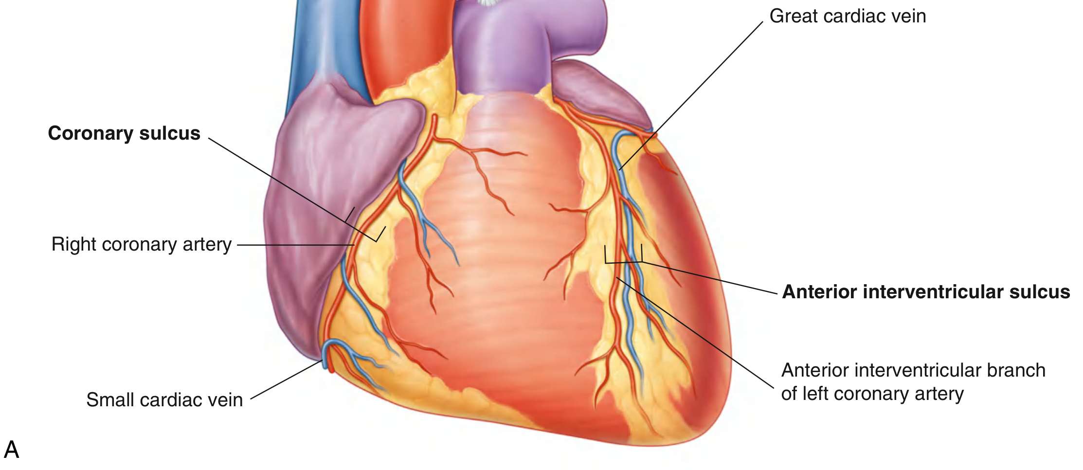
3.1 Surfaces
| Surface | Formed by |
|---|---|
| Anterior (sternocostal) | Mostly right ventricle, part of right atrium and left ventricle |
| Diaphragmatic (inferior) | Left ventricle + small portion of right ventricle |
| Left pulmonary surface | Left ventricle + part of left atrium |
| Right pulmonary surface | Right atrium |
| Base (posterior) | Mainly left atrium; receives pulmonary veins |
3.2 External Grooves (Sulci)
- Coronary sulcus (atrioventricular groove): Circles the heart, separating atria from ventricles; contains the right coronary artery, left circumflex artery, coronary sinus, and small cardiac vein
- Anterior interventricular sulcus: On the anterior surface; contains the anterior interventricular artery (LAD) and great cardiac vein
- Posterior interventricular sulcus: On the diaphragmatic surface; contains the posterior interventricular artery and middle cardiac vein
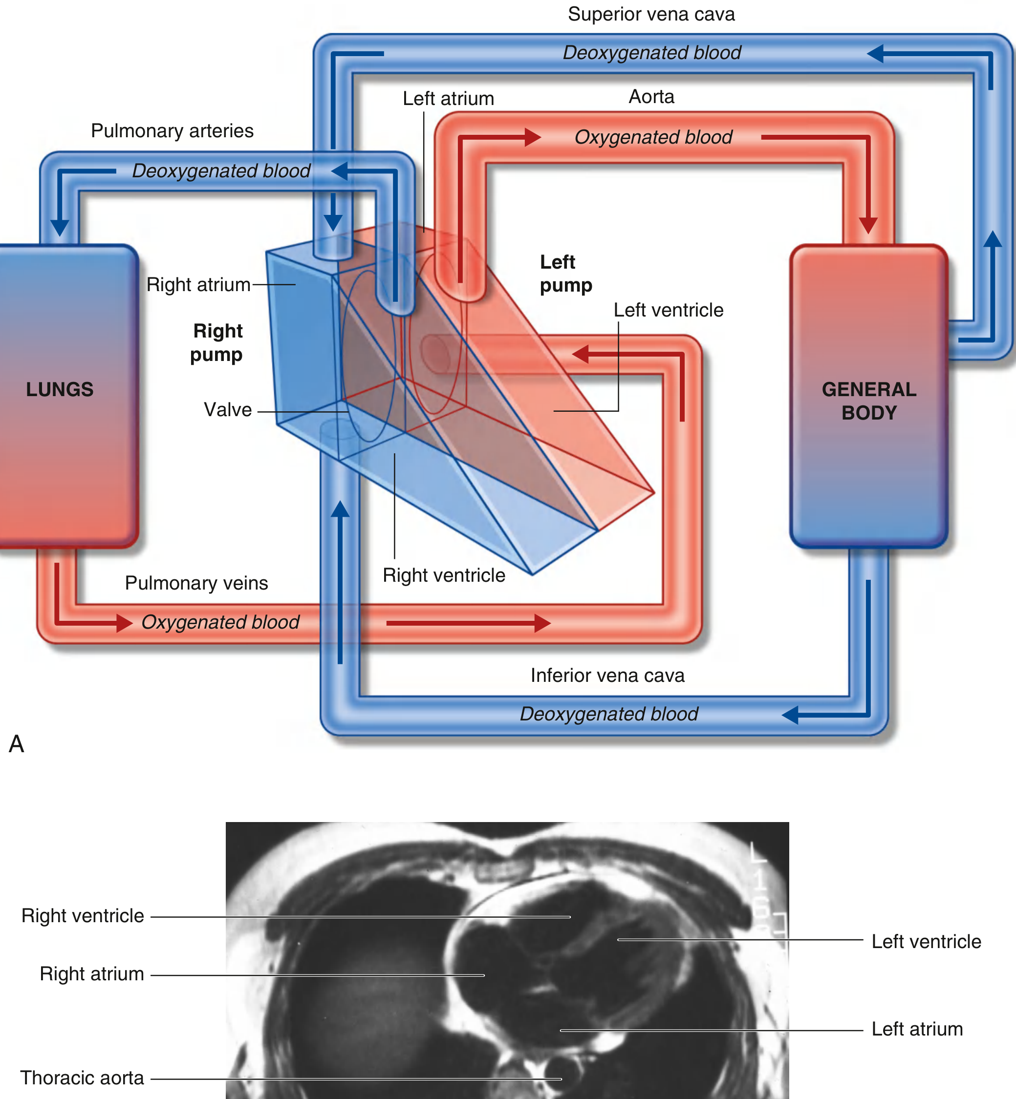
STEP 4 - THE FOUR CARDIAC CHAMBERS
The heart consists of two pumps separated by a central partition:
- Right pump: Receives deoxygenated blood from the body, sends it to the lungs (pulmonary circulation)
- Left pump: Receives oxygenated blood from the lungs, sends it to the body (systemic circulation)
4.1 Right Atrium
- Forms the right border of the heart
- Receives blood from 3 openings: Superior vena cava (upper posterior wall), Inferior vena cava (lower posterior wall), and Coronary sinus (between IVC and tricuspid valve)
- Interior divided by the crista terminalis (muscular ridge) into:
- Sinus of venae cavae (posterior) - smooth-walled; embryological origin: right horn of sinus venosus
- Atrium proper (anterior) - has muscular ridges called musculi pectinati (pectinate muscles)
- Right auricle: Ear-like muscular pouch that overlaps the ascending aorta
- Fossa ovalis: Oval depression on the interatrial septum - remnant of the embryonic foramen ovale
- Tricuspid valve guards the right atrioventricular orifice
4.2 Right Ventricle
- Forms most of the anterior surface of the heart
- Has the thinnest wall of the four chambers (less resistance in pulmonary circulation)
- Interior features:
- Trabeculae carneae - irregular muscular ridges on inner walls
- Papillary muscles (anterior, posterior, septal) - attached to valve cusps via chordae tendineae
- Septomarginal trabecula (moderator band) - muscular band from interventricular septum to anterior papillary muscle; carries the right bundle branch of the conduction system
- Conus arteriosus (infundibulum) - smooth-walled outflow cone that leads to the pulmonary valve
- Pulmonary (semilunar) valve guards the exit into the pulmonary trunk (3 cusps: anterior, left, right)
4.3 Left Atrium
- Forms most of the base (posterior surface) of the heart
- Receives 4 pulmonary veins (2 from each lung) on its posterior surface
- Has left auricle - narrow, hooked pouch
- Smooth posterior wall (derived from the incorporated pulmonary veins)
- Anterior wall has some pectinate muscles (in the auricle)
- Mitral (bicuspid) valve guards the left atrioventricular orifice - 2 cusps (anterior/aortic + posterior/mural)
4.4 Left Ventricle
- Has the thickest wall (~3x the right ventricle wall thickness) to generate high systemic pressure
- Produces most of the left pulmonary surface and the apex
- Interior features:
- Trabeculae carneae (finer than right side)
- 2 papillary muscles (anterior and posterior) with chordae tendineae to mitral valve cusps
- Smooth outflow tract (aortic vestibule) leading to the aortic valve
- Aortic (semilunar) valve guards the aortic orifice - 3 cusps: right, left (coronary cusps), and posterior (non-coronary)
- Above the aortic cusps are 3 aortic sinuses (sinuses of Valsalva) - the right and left coronary arteries arise from the right and left sinuses respectively
STEP 5 - CARDIAC VALVES IN DETAIL
| Valve | Type | Cusps | Location | Prevents |
|---|---|---|---|---|
| Tricuspid (right AV) | Atrioventricular | 3 (anterior, posterior, septal) | Between right atrium and right ventricle | Backflow during RV contraction |
| Mitral/Bicuspid (left AV) | Atrioventricular | 2 (anterior, posterior) | Between left atrium and left ventricle | Backflow during LV contraction |
| Pulmonary | Semilunar | 3 | Between RV and pulmonary trunk | Backflow from pulmonary trunk into RV |
| Aortic | Semilunar | 3 | Between LV and aorta | Backflow from aorta into LV |
Atrioventricular valves depend on the chordae tendineae + papillary muscles to prevent prolapse/regurgitation during ventricular systole. Semilunar valves have no such support.
Cardiac Skeleton
- Dense fibrous connective tissue rings (annuli fibrosi) surrounding all four valve orifices
- Provides attachment for valve cusps and myocardium
- Electrically isolates atria from ventricles - the AV bundle is the only muscular connection through this insulating layer
- Includes: right and left fibrous trigones, and four annuli fibrosi
STEP 6 - THE CARDIAC CONDUCTION SYSTEM
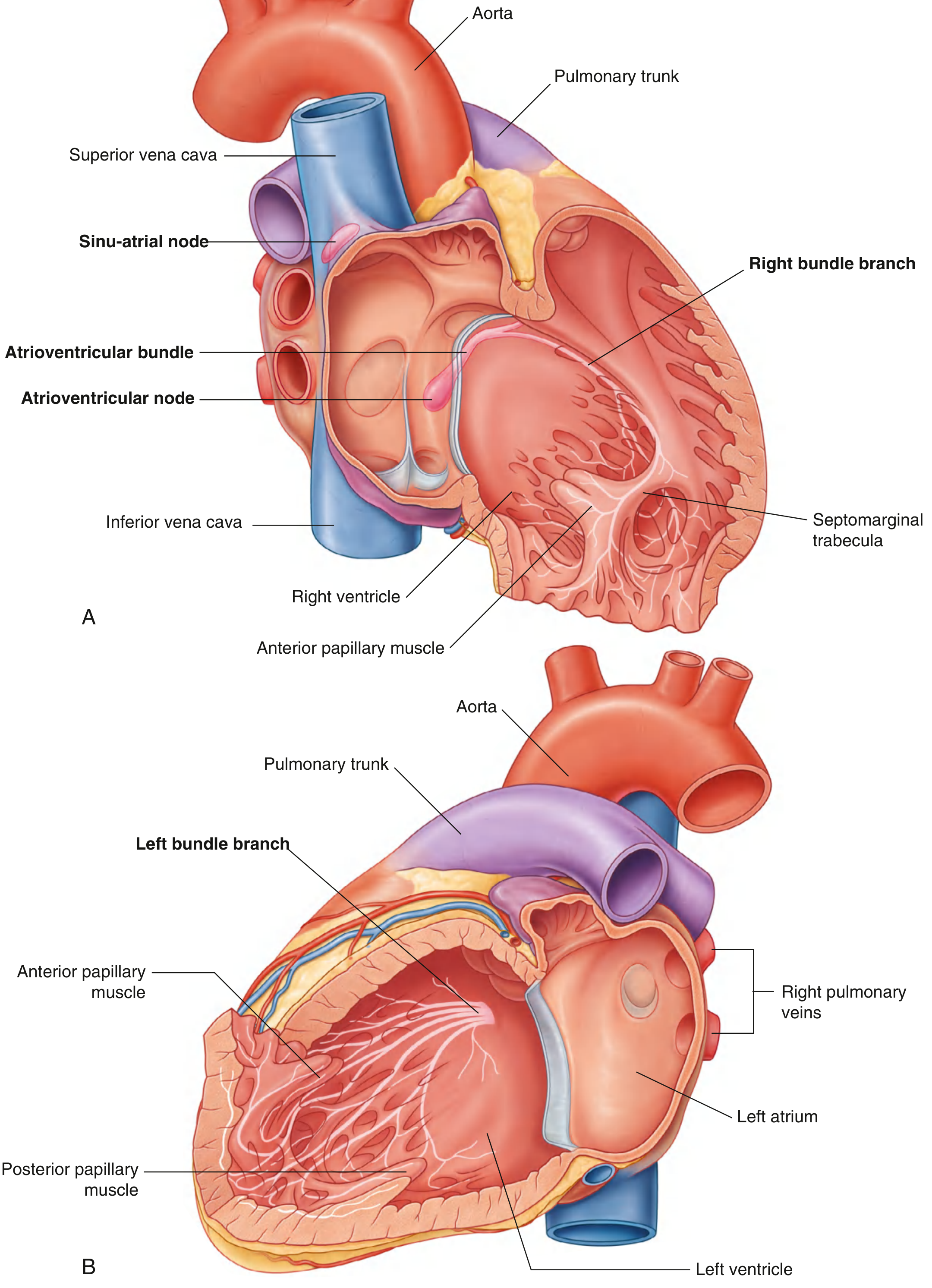
The conduction system initiates and coordinates the heartbeat. It is composed of specialized cardiac muscle cells (NOT neurons) organized into four components:
6.1 Sinoatrial (SA) Node - "Pacemaker"
- Located at the superior end of the crista terminalis, at the junction of the superior vena cava and right atrium
- Sets the rate of the heartbeat (~60-100 bpm at rest)
- Excitation spreads across both atria causing atrial contraction
- Blood supply: SA nodal branch of the right coronary artery (in ~55-60% of people) or left circumflex artery
6.2 Atrioventricular (AV) Node
- Located near the opening of the coronary sinus, close to the attachment of the septal cusp of the tricuspid valve, within the AV septum
- Receives the atrial excitation wave and delays it (allows atrial contraction to complete before ventricular contraction begins - the "PR interval" on ECG)
- Blood supply: AV nodal branch of the right coronary artery (~90% of cases)
6.3 Atrioventricular Bundle (Bundle of His)
- Direct continuation of the AV node
- Runs along the lower border of the membranous interventricular septum
- Then splits into right bundle branch and left bundle branch
- The right bundle branch: passes down the right side of the septum, enters the septomarginal trabecula, reaches the anterior papillary muscle, then spreads as Purkinje fibers
- The left bundle branch: passes to the left side of the muscular septum, descends to the apex, gives off branches, becomes Purkinje fibers
6.4 Subendocardial Plexus (Purkinje Fibers)
- Network of specialized cells spreading throughout both ventricles
- Contacts increase greatly at the subendocardial level - enables synchronized ventricular contraction from apex upward toward the outflow tracts
- Creates a unidirectional wave of excitation from papillary muscles/apex toward the arterial outflow tracts
Important principle: Large branches of the conduction system are insulated from the myocardium by connective tissue - prevents inappropriate stimulation.
STEP 7 - CORONARY VASCULATURE
7.1 Coronary Arteries
Both coronary arteries arise from the aortic sinuses just above the aortic valve.
Right Coronary Artery (RCA):
- Originates from the right aortic sinus
- Descends vertically in the coronary sulcus between the right atrium and right ventricle
- Turns posteriorly at the inferior margin to the diaphragmatic surface
- Key branches:
- SA nodal branch - to the sinoatrial node (runs posterior to SVC)
- Right marginal branch - runs along the acute (inferior) margin toward the apex
- Posterior interventricular (descending) artery - in the posterior interventricular sulcus (in "right-dominant" hearts, ~70% of people)
- AV nodal branch
Left Coronary Artery (LCA):
- Originates from the left aortic sinus
- Short main stem (left main coronary artery) passes between the pulmonary trunk and left auricle
- Divides into 2 major branches:
- Anterior interventricular (LAD - Left Anterior Descending) artery: Descends in the anterior interventricular sulcus to the apex; supplies the anterior wall of both ventricles and the anterior 2/3 of the interventricular septum; most commonly occluded artery in MI
- Circumflex artery: Continues in the coronary sulcus, wraps around the left side to the posterior surface; supplies the left atrium and posterior left ventricle; in "left-dominant" hearts, gives off the posterior interventricular artery
Coronary Dominance:
- ~70% right-dominant (RCA gives posterior interventricular artery)
- ~10% left-dominant (circumflex gives posterior interventricular artery)
- ~20% co-dominant
Coronary blood flow timing:
- Flows to the epicardium during both systole and diastole
- Flows to the endocardium predominantly during diastole (endocardium is compressed by LV pressure during systole)
- Flow is regulated by metabolic signals: nitric oxide, adenosine, bradykinins, prostaglandins, CO2
7.2 Cardiac Veins
Most cardiac venous drainage returns via the coronary sinus (in the coronary sulcus on the posterior surface between LA and LV), which empties into the right atrium.
| Vein | Follows | Drains |
|---|---|---|
| Great cardiac vein | Anterior interventricular sulcus, then coronary sulcus | Anterior surfaces of both ventricles and LV lateral wall |
| Middle cardiac vein | Posterior interventricular sulcus | Posterior ventricular walls |
| Small cardiac vein | Coronary sulcus on right side | Right atrium and right ventricle |
| Posterior cardiac vein | Posterior surface of LV | Left ventricle |
| Anterior cardiac veins | Anterior RV surface | Drain directly into right atrium (bypass coronary sinus) |
| Venae cordis minimae (Thebesian veins) | Within walls | Drain directly into cardiac chambers (mainly RA, RV) |
7.3 Coronary Lymphatics
- Lymphatics follow the coronary arteries
- Drain mainly to brachiocephalic nodes (anterior to brachiocephalic veins) and tracheobronchial nodes (at the inferior end of the trachea)
STEP 8 - CARDIAC INNERVATION
The autonomic nervous system regulates heart rate, force of contraction, and cardiac output via the cardiac plexus.
The cardiac plexus has two parts:
- Superficial part: Inferior to the aortic arch, between the aortic arch and pulmonary trunk
- Deep part: Between the aortic arch and the tracheal bifurcation
8.1 Parasympathetic Supply (Vagus Nerve - CN X)
- Preganglionic fibers: cardiac branches of right and left vagus nerves
- Synapse in ganglia within the plexus or walls of the atria
- Effects:
- Decreases heart rate
- Reduces force of contraction
- Constricts coronary arteries
8.2 Sympathetic Supply
- Preganglionic fibers: upper 4-5 thoracic spinal cord segments (T1-T5)
- Synapse in cervical and upper thoracic sympathetic ganglia
- Postganglionic fibers: bilateral cardiac nerves to cardiac plexus
- Effects:
- Increases heart rate
- Increases force of contraction
- Dilates coronary arteries
8.3 Visceral Afferents (Pain Fibers)
- Travel with sympathetic trunk fibers
- Enter spinal cord at T1-T5
- Referred pain: Cardiac ischemia pain is referred to the chest, left arm, jaw, and neck because these share the same spinal cord levels (T1-T5)
- Vagal afferents sense blood pressure and chemistry (reflex functions, not pain)
STEP 9 - GREAT VESSELS
9.1 Pulmonary Trunk
- Arises from the conus arteriosus of the right ventricle
- Contained within the pericardial sac
- Associated with the ascending aorta in a common sheath
- Bifurcates at the level of the T4/T5 intervertebral disc into:
- Right pulmonary artery: Passes right, posterior to ascending aorta and SVC
- Left pulmonary artery: Passes inferior to the aortic arch, anterior to descending aorta
9.2 Ascending Aorta
- Contained within the pericardial sac
- Above the aortic valve are 3 aortic sinuses (left, right, posterior)
- Right and left coronary arteries originate from the right and left sinuses
9.3 Arch of Aorta
- Gives off 3 branches (left to right in the superior mediastinum):
- Brachiocephalic trunk - divides into right subclavian + right common carotid
- Left common carotid artery
- Left subclavian artery
9.4 Superior Vena Cava
- Formed by union of left and right brachiocephalic veins
- Inferior half lies within the pericardial sac
- Enters right atrium at approximately the 3rd right costal cartilage
9.5 Inferior Vena Cava
- Passes through the diaphragm at vertebra T8
- Enters the fibrous pericardium; short segment within pericardial sac before entering right atrium
9.6 Pulmonary Veins
- Usually 2 from each lung (4 total)
- Enter the posterior surface of the left atrium
- Short segment of each within the pericardial sac
STEP 10 - BLOOD FLOW THROUGH THE HEART (Complete Circuit)
BODY (deoxygenated blood)
     |
     v
Superior Vena Cava / Inferior Vena Cava / Coronary Sinus
     |
     v
RIGHT ATRIUM
     |  (tricuspid valve opens)
     v
RIGHT VENTRICLE
     |  (pulmonary valve opens)
     v
PULMONARY TRUNK --> Right & Left Pulmonary Arteries
     |
     v
LUNGS (gas exchange - CO2 released, O2 absorbed)
     |
     v
4 Pulmonary Veins (oxygenated blood)
     |
     v
LEFT ATRIUM
     |  (mitral valve opens)
     v
LEFT VENTRICLE
     |  (aortic valve opens)
     v
AORTA --> SYSTEMIC CIRCULATION (body)
STEP 11 - VASCULAR SYSTEM (Systemic Vessels)
11.1 Vessel Wall Layers (Tunica Structure)
All blood vessels except capillaries have three coats:
| Layer | Contents | Function |
|---|---|---|
| Tunica intima | Endothelium + basement membrane + subendothelial layer | Non-thrombogenic surface; gas/nutrient exchange |
| Tunica media | Smooth muscle + elastic tissue | Regulates vessel diameter (vasoconstriction/dilation) |
| Tunica adventitia | Collagen + vasa vasorum (vessels of large vessels) | Structural support; anchoring |
11.2 Types of Blood Vessels
| Type | Characteristics | Function |
|---|---|---|
| Elastic arteries (aorta, pulmonary trunk, carotid) | Thick media with many elastic laminae | Conduct blood; buffer systolic pressure (Windkessel effect) |
| Muscular arteries (coronary, femoral, radial) | More smooth muscle; less elastic tissue | Distribute blood to organs |
| Arterioles | Mostly smooth muscle | Regulate resistance; control blood pressure |
| Capillaries | Single endothelial layer only | Site of gas/nutrient/waste exchange |
| Venules | Thin walls; postcapillary venules are where WBCs migrate | Collect blood from capillaries |
| Veins | Thin walls; large lumens; venous valves prevent backflow | Return blood to heart |
11.3 Major Systemic Arteries
Aorta divisions:
- Ascending aorta - coronary arteries
- Aortic arch - brachiocephalic, left common carotid, left subclavian
- Descending thoracic aorta - bronchial, esophageal, posterior intercostal arteries
- Abdominal aorta - celiac trunk, SMA, suprarenal, renal, gonadal, IMA, common iliac arteries
- Bifurcates into common iliac arteries at L4
11.4 Major Systemic Veins
Venous drainage returns via:
- Superior vena cava (SVC): Drains head, neck, upper limbs, and upper thorax
- Inferior vena cava (IVC): Drains lower limbs, pelvis, abdomen
- Azygos system: Drains thoracic wall, posterior mediastinum
- Portal vein: Drains GI tract and spleen to liver before returning to IVC (via hepatic veins)
STEP 12 - WALL OF THE HEART (Histology)
The heart wall has three layers:
| Layer | Structure | Details |
|---|---|---|
| Epicardium | Visceral layer of serous pericardium | Outermost; contains coronary arteries, veins, nerves, and fat |
| Myocardium | Cardiac muscle mass | Bulk of heart wall; thickest in left ventricle |
| Endocardium | Endothelium + subendocardial layer | Lines all chambers; continuous with vessel endothelium |
Cardiac muscle cells (cardiomyocytes):
- Striated, branching, uninucleate cells
- Connected end-to-end by intercalated discs (containing gap junctions - allow electrical coupling, and desmosomes - mechanical coupling)
- ~60-70% of heart cells are actually cardiac fibroblasts (not muscle), which produce the extracellular collagen matrix
Non-muscle cells:
- Cardiac fibroblasts produce the collagen scaffold
- In pathological states (hypertension, MI, heart failure), fibroblasts produce excess extracellular matrix leading to cardiac fibrosis
- Therapies including statins, ACE inhibitors, beta-blockers, and ARBs reduce fibrosis (favorable "reverse remodeling")
QUICK REFERENCE SUMMARY TABLE
| Structure | Key Fact |
|---|---|
| SA node | Right atrium/SVC junction; pacemaker; RCA supply |
| AV node | Near coronary sinus opening; RCA supply in 90% |
| Tricuspid valve | 3 cusps; right AV orifice |
| Mitral valve | 2 cusps; left AV orifice; most common site of rheumatic valve disease |
| Fossa ovalis | Remnant of foramen ovale in interatrial septum |
| LAD artery | "Widow maker"; supplies anterior wall + anterior 2/3 of septum |
| Coronary sinus | Main venous channel; opens into right atrium |
| Moderator band | Carries right bundle branch across RV |
| Cardiac skeleton | Electrically isolates atria from ventricles |
| Referred cardiac pain | Felt at T1-T5 dermatomes (chest, left arm, jaw) |
Sources: Gray's Anatomy for Students | Goldman-Cecil Medicine, 2-Vol Set | Fuster and Hurst's The Heart, 15th Edition
This is a shared conversation. Sign in to Orris to start your own chat.