
Pathogenesis and lab investigations in DIC md pathology exam question and robbins based answer
Reading File
Reading File
Finding Sources
Finding Sources
Searching PubMed
"disseminated intravascular coagulation" AND laboratory
Searching the Web
Merck Manual disseminated intravascular coagulation diagnosis laboratory findings PT PTT fibrinogen D-dimer
Finding Sources
Finding Sources
Finding Sources
Reading File
Reading File
Reading File
Finding Sources
Reading File
Reading File
Finding Sources
Reading File
Reading File
Disseminated Intravascular Coagulation (DIC)
Definition: DIC is an acquired, secondary thrombohemorrhagic syndrome caused by systemic activation of coagulation. It produces widespread fibrin thrombi in the microcirculation, consumption of platelets and coagulation factors, and secondary activation of fibrinolysis. Thus, it causes both thrombosis/organ ischemia and bleeding.
Robbins, Cotran & Kumar Pathologic Basis of Disease, pp. 623-625.
Robbins, Cotran & Kumar Pathologic Basis of Disease, pp. 623-625.
Pathogenesis of DIC
Initiating mechanisms
Robbins describes two major mechanisms:
-
Release of tissue factor or other procoagulants into the circulation
- Obstetric complications: placental tissue, amniotic fluid, retained dead fetus
- Major trauma, burns, extensive surgery: damaged tissue releases tissue factor
- Malignancy: especially mucin-producing adenocarcinomas, in which mucin may activate factor X; acute promyelocytic leukemia is also a classic association.
-
Widespread endothelial injury or activation
- Especially important in sepsis
- Endothelial damage exposes subendothelial matrix, activates platelets and coagulation cascade.
- In sepsis, endotoxin and cytokines, particularly TNF, induce endothelial tissue-factor expression and reduce thrombomodulin expression.
- Reduced thrombomodulin means reduced activation of protein C, so inhibition of factors Va and VIIIa is impaired, favoring coagulation.
Robbins, Cotran & Kumar Pathologic Basis of Disease, pp. 623-625.
Sequence of events
Underlying disorder
(sepsis / obstetric catastrophe / trauma / malignancy)
                    ↓
Tissue factor release and/or diffuse endothelial injury
                    ↓
Widespread thrombin generation
                    ↓
Fibrin deposition in small vessels + platelet activation
                    ↓
Microvascular thrombi
                    ↓
Tissue ischemia, infarction, multiorgan dysfunction
                    ↓
Consumption of platelets, fibrinogen, and clotting factors
                    ↓
Secondary fibrinolysis with plasmin formation
                    ↓
Fibrin degradation products + depletion of hemostatic factors
                    ↓
Bleeding tendency
Consequences
1. Microvascular thrombosis
- Fibrin thrombi form in small vessels, particularly in the brain, lungs, heart, kidneys, adrenals, spleen, and liver.
- Causes tissue hypoxia, ischemia, infarction, and organ dysfunction.
- Red cells passing through fibrin strands are fragmented, producing microangiopathic hemolytic anemia with schistocytes.
2. Hemorrhagic diathesis
Bleeding occurs because of:
- Consumption of platelets
- Consumption of coagulation factors, including fibrinogen
- Plasmin-mediated degradation of fibrin and factors V and VIII
- Fibrin degradation products that inhibit platelet aggregation, fibrin polymerization, and thrombin activity.
Hence, DIC is also called consumption coagulopathy.
Robbins, Cotran & Kumar Pathologic Basis of Disease, pp. 624-625.
Robbins, Cotran & Kumar Pathologic Basis of Disease, pp. 624-625.
Important causes of DIC
Remember: "Sepsis, Surgery/trauma, Obstetric complications, and Malignancy."
| Category | Examples |
|---|---|
| Sepsis | Gram-negative or gram-positive septicemia, meningococcemia |
| Obstetric causes | Abruptio placentae, amniotic fluid embolism, retained dead fetus, severe preeclampsia/eclampsia |
| Malignancy | Acute promyelocytic leukemia; mucinous adenocarcinoma of pancreas, lung, stomach, colon |
| Tissue injury | Major trauma, burns, extensive surgery, shock |
| Others | Severe pancreatitis, hemolytic transfusion reaction, snake bite, giant hemangioma |
Laboratory Investigations in DIC
DIC is diagnosed by a combination of an appropriate clinical setting and serial coagulation tests. A single normal result does not exclude early or chronic DIC.
| Investigation | Finding in overt DIC | Basis |
|---|---|---|
| Platelet count | Decreased, often progressive thrombocytopenia | Platelet consumption in microthrombi |
| PT/INR | Prolonged | Consumption of extrinsic/common pathway factors |
| aPTT | Usually prolonged | Consumption of intrinsic/common pathway factors |
| Thrombin time (TT) | Prolonged | Hypofibrinogenemia and effect of FDPs |
| Plasma fibrinogen | Decreased | Consumption in fibrin formation |
| D-dimer | Markedly increased | Fibrin formation and degradation |
| Fibrin degradation products (FDPs) | Increased | Secondary fibrinolysis |
| Peripheral smear | Schistocytes/helmet cells may be present | Mechanical fragmentation of RBCs in microthrombi |
| Hemoglobin | May be decreased | Bleeding and microangiopathic hemolysis |
| Factor V and VIII | May be reduced | Consumption and plasmin-mediated degradation |
| Antithrombin | Often reduced | Consumption during excess thrombin generation |
The typical laboratory profile is: thrombocytopenia + prolonged PT/aPTT/TT + low fibrinogen + high D-dimer/FDP.
Tietz Textbook of Laboratory Medicine, p. 3128.
Tietz Textbook of Laboratory Medicine, p. 3128.
Peripheral smear
- May show microangiopathic hemolytic anemia
- Schistocytes and helmet cells support the diagnosis
- Their absence does not exclude DIC.
Important qualification
- In early DIC, aPTT may be normal or even shortened due to circulating activated clotting factors and increased factor VIII as an acute-phase reactant.
- Fibrinogen can be normal in sepsis or chronic DIC because it is an acute-phase protein. Therefore, a falling trend is often more useful than one isolated value.
ISTH scoring system for overt DIC
Use only in a patient with a disorder known to be associated with DIC.
| Variable | Score |
|---|---|
| Platelets >100,000/µL | 0 |
| Platelets <100,000/µL | 1 |
| Platelets <50,000/µL | 2 |
| No increase in fibrin markers | 0 |
| Moderate increase in D-dimer/FDP | 2 |
| Strong increase in D-dimer/FDP | 3 |
| PT prolongation <3 seconds | 0 |
| PT prolongation 3-6 seconds | 1 |
| PT prolongation >6 seconds | 2 |
| Fibrinogen >100 mg/dL | 0 |
| Fibrinogen <100 mg/dL | 1 |
Score ≥5: compatible with overt DIC.
Score <5: possible non-overt DIC; repeat tests after 1-2 days.
Henry's Clinical Diagnosis and Management by Laboratory Methods, p. 954.
Score <5: possible non-overt DIC; repeat tests after 1-2 days.
Henry's Clinical Diagnosis and Management by Laboratory Methods, p. 954.
Exam conclusion
DIC is an acquired systemic coagulopathy in which excessive thrombin formation causes widespread microvascular fibrin thrombi. This leads simultaneously to organ ischemia and consumption of platelets and clotting factors, followed by secondary fibrinolysis and bleeding. The key laboratory findings are falling platelet count, prolonged PT and aPTT, low fibrinogen, and raised D-dimer/FDPs, with schistocytes sometimes seen on smear.
Create a one-page DIC exam revision sheet
Reading File
Finding Sources
Searching PubMed
"disseminated intravascular coagulation" AND diagnosis
Searching the Web
disseminated intravascular coagulation laboratory findings D-dimer fibrinogen PT platelet count
DIC: One-Page Exam Revision Sheet
Definition
Disseminated intravascular coagulation (DIC) is an acquired secondary thrombohemorrhagic syndrome with systemic activation of coagulation, causing widespread microvascular fibrin thrombi, consumption of platelets and clotting factors, and secondary fibrinolysis.
Result: simultaneous thrombosis, organ dysfunction, and bleeding.
Robbins, Cotran & Kumar Pathologic Basis of Disease, pp. 623-625.
Robbins, Cotran & Kumar Pathologic Basis of Disease, pp. 623-625.
Causes: “SOMT”
- Sepsis, especially severe bacterial sepsis
- Obstetric catastrophes: abruptio placentae, retained dead fetus, amniotic-fluid embolism
- Malignancy: acute promyelocytic leukemia; mucinous adenocarcinoma
- Trauma/tissue injury: burns, shock, major surgery
Other: acute pancreatitis, incompatible blood transfusion, snakebite, giant hemangioma.
Pathogenesis
Two major initiating mechanisms
-
Tissue-factor/procoagulant release
- Trauma/burns/surgery
- Placental or amniotic-fluid material
- Tumor mucin can directly activate factor X
-
Diffuse endothelial injury/activation
- Sepsis: endotoxin and TNF
- TNF causes:
- ↑ endothelial tissue factor
- ↓ thrombomodulin expression
- ↓ protein C anticoagulant pathway
- leukocyte adhesion and endothelial damage
Flowchart
Trigger (sepsis / obstetric event / trauma / cancer)
                       ↓
Tissue factor release + endothelial injury
                       ↓
Systemic thrombin generation
                       ↓
Widespread fibrin microthrombi + platelet consumption
                       ↓
Ischemia / infarction / multiorgan dysfunction
                       ↓
Consumption of platelets + fibrinogen + clotting factors
                       ↓
Secondary plasmin activation and fibrinolysis
                       ↓
FDPs increase + factors V and VIII degraded
                       ↓
BLEEDING
Why both clotting and bleeding?
| Thrombosis | Bleeding |
|---|---|
| Fibrin thrombi occlude microcirculation | Platelets and clotting factors are consumed |
| Tissue ischemia and organ failure | Plasmin causes fibrinolysis |
| RBC fragmentation causes schistocytes | FDPs inhibit platelet aggregation, fibrin polymerization, and thrombin |
Robbins, Cotran & Kumar Pathologic Basis of Disease, pp. 623-625.
Clinical Features
- Oozing from venepuncture, wound, catheter, or surgical sites
- Petechiae, ecchymoses, mucosal bleeding
- Hematuria, GI bleeding, postpartum hemorrhage
- Shock
- Renal failure, respiratory distress, altered sensorium
- Microthrombi may cause ischemia in brain, lungs, heart, kidneys, adrenals, liver, and spleen
Laboratory Investigations
| Test | Typical finding | Reason |
|---|---|---|
| Platelet count | ↓ | Consumption in thrombi |
| PT/INR | ↑ / prolonged | Consumption of clotting factors |
| aPTT | ↑ / prolonged | Consumption of intrinsic/common pathway factors |
| Thrombin time | Prolonged | Low fibrinogen and FDP effect |
| Fibrinogen | ↓ | Used up in fibrin formation |
| D-dimer | Markedly ↑ | Fibrin formation and breakdown |
| FDPs | ↑ | Secondary fibrinolysis |
| Peripheral smear | Schistocytes, helmet cells | MAHA due to fibrin strands |
| Hb | May be ↓ | Bleeding and hemolysis |
| Factor V, VIII | ↓ | Consumption and plasmin-mediated degradation |
Classic laboratory pattern
Low platelets + prolonged PT/aPTT + low fibrinogen + high D-dimer/FDPs
Important exam point: In early DIC, PT/aPTT or fibrinogen can initially be normal. In sepsis, fibrinogen may remain normal or high as an acute-phase reactant. Therefore, use serial trends, especially a falling platelet count and fibrinogen.
Tietz Textbook of Laboratory Medicine, p. 3128.
Tietz Textbook of Laboratory Medicine, p. 3128.
ISTH Overt DIC Score
Use in a patient with a known DIC-associated condition.
- Platelets: low count gives 1-2 points
- Elevated fibrin markers/D-dimer: 2-3 points
- Prolonged PT: 1-2 points
- Fibrinogen <100 mg/dL: 1 point
Interpretation
- Score ≥5: overt DIC
- Score <5: possible non-overt DIC, repeat testing in 1-2 days
Henry's Clinical Diagnosis and Management by Laboratory Methods, p. 954.
One-Line Answer for Viva
DIC is a secondary consumption coagulopathy in which systemic thrombin generation causes widespread microvascular fibrin thrombi, followed by consumption of platelets and coagulation factors plus secondary fibrinolysis, producing both thrombosis and bleeding.
Bring robbins based image and prepare notes with it
Reading File
Finding Sources
query_images_db
Searching Images
Robbins Pathology disseminated intravascular coagulation pathogenesis diagram microvascular fibrin thrombi
Searching PubMed
"disseminated intravascular coagulation" AND diagnosis
Searching the Web
Robbins pathology disseminated intravascular coagulation figure 14.26 pathophysiology
Reading File
query_images_db
Reading File
Disseminated Intravascular Coagulation (DIC)
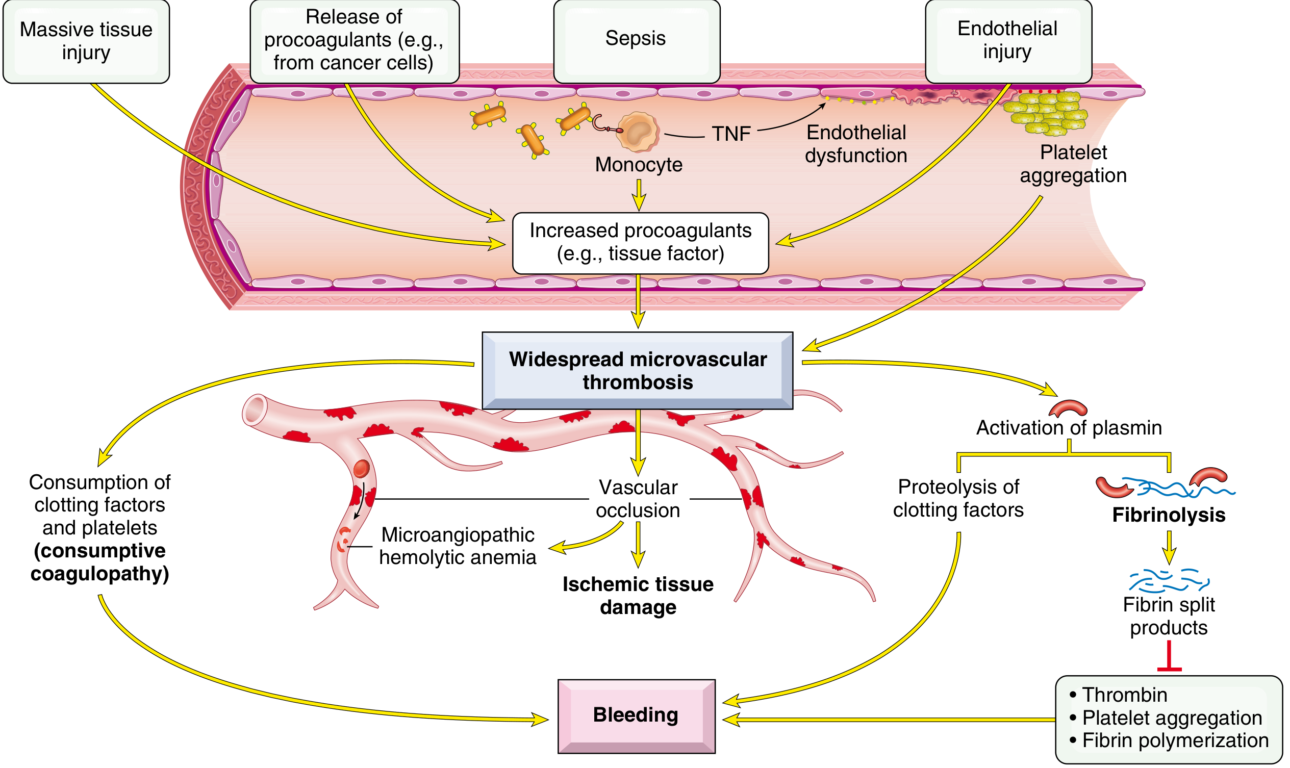
Fig. 14.26. Pathophysiology of DIC. TNF = tumor necrosis factor. Source: Robbins, Cotran & Kumar Pathologic Basis of Disease, p. 626.
Definition
DIC is an acquired, secondary thrombohemorrhagic disorder caused by excessive systemic activation of coagulation. It results in:
- Widespread fibrin thrombi in the microcirculation
- Consumption of platelets, fibrinogen, and clotting factors
- Secondary activation of fibrinolysis
Therefore, DIC produces both microvascular thrombosis with organ ischemia and bleeding due to consumption coagulopathy.
Robbins, Cotran & Kumar Pathologic Basis of Disease, pp. 623-625.
Etiology
Main settings
-
Sepsis
- Especially severe bacterial sepsis
- Endotoxin and cytokines, particularly TNF, activate endothelium and coagulation.
-
Obstetric complications
- Abruptio placentae
- Amniotic fluid embolism
- Retained dead fetus
- Placental tissue entering circulation
-
Malignancy
- Acute promyelocytic leukemia
- Mucin-secreting adenocarcinomas, especially pancreas, lung, stomach, and colon
-
Major tissue injury
- Trauma
- Burns
- Extensive surgery
- Shock
Pathogenesis
Two initiating mechanisms
| Mechanism | Examples |
|---|---|
| Release of tissue factor or procoagulants | Trauma, burns, surgery, placental/amniotic-fluid material, tumor mucin |
| Widespread endothelial injury/activation | Sepsis, immune complexes, burns, heat stroke, infections |
In sepsis
TNF causes:
- Increased endothelial expression of tissue factor
- Reduced expression of thrombomodulin
- Less thrombin-thrombomodulin complex formation
- Reduced activation of protein C, which normally inhibits factors Va and VIIIa
- Endothelial activation and leukocyte-mediated damage
Thus, the normal anticoagulant balance shifts toward uncontrolled coagulation.
Flowchart
Sepsis / trauma / obstetric event / malignancy
                         ↓
Tissue factor release and/or endothelial injury
                         ↓
Systemic activation of coagulation
                         ↓
Excess thrombin generation
                         ↓
Widespread fibrin deposition in microcirculation
                         ↓
Microthrombi → ischemia, infarction, organ dysfunction
                         ↓
Consumption of platelets + fibrinogen + coagulation factors
                         ↓
Secondary fibrinolysis with plasmin activation
                         ↓
FDPs/D-dimer rise + factors V and VIII reduced
                         ↓
BLEEDING
Consequences
1. Thrombotic consequences
- Microvascular fibrin thrombi impair tissue perfusion.
- May cause ischemia and dysfunction of:
- Brain
- Lungs
- Heart
- Kidneys
- Adrenals
- Liver and spleen
- RBC fragmentation in narrowed fibrin-lined vessels causes microangiopathic hemolytic anemia.
2. Hemorrhagic consequences
Bleeding occurs due to:
- Consumption of platelets
- Consumption of fibrinogen and clotting factors
- Plasmin-mediated cleavage of fibrin and factors V and VIII
- Fibrin degradation products inhibiting:
- platelet aggregation
- fibrin polymerization
- thrombin activity
Morphology
- Fibrin thrombi occur mainly in small vessels.
- Kidneys: glomerular thrombi, microinfarcts, sometimes bilateral cortical necrosis.
- Lungs: fibrin thrombi in alveolar capillaries; may be associated with pulmonary edema and hyaline membranes.
- Brain: microinfarcts with possible hemorrhage.
- Adrenals: adrenal hemorrhage in meningococcemia, as in Waterhouse-Friderichsen syndrome.
Robbins, Cotran & Kumar Pathologic Basis of Disease, p. 625.
Clinical Features
- Bleeding from venepuncture, wound, catheter, or surgical sites
- Petechiae, ecchymoses, mucosal bleeding
- Hematuria and gastrointestinal bleeding
- Dyspnea, cyanosis, respiratory failure
- Oliguria and acute renal failure
- Confusion, seizures, coma
- Shock and multiorgan dysfunction
Acute DIC due to sepsis, trauma, or obstetric complications usually presents with bleeding.
Chronic DIC, such as that associated with malignancy, may predominantly present with thrombosis.
Chronic DIC, such as that associated with malignancy, may predominantly present with thrombosis.
Laboratory Diagnosis
| Test | Finding |
|---|---|
| Platelet count | Decreased |
| PT/INR | Prolonged |
| aPTT | Prolonged |
| Plasma fibrinogen | Decreased |
| D-dimer | Markedly increased |
| FDPs | Increased |
| Peripheral smear | Schistocytes/helmet cells may be seen |
| Hemoglobin | May be reduced due to bleeding and MAHA |
Classic DIC pattern
↓ Platelets + ↑ PT/aPTT + ↓ fibrinogen + ↑ D-dimer/FDPs
Exam point: Serial testing is useful. Early DIC may have normal PT/aPTT, and fibrinogen may not be low in sepsis because it is an acute-phase reactant. A downward trend supports ongoing consumption.
Robbins, Cotran & Kumar Pathologic Basis of Disease, p. 625; Tietz Textbook of Laboratory Medicine, p. 3128.
High-Yield Viva Answer
DIC is an acquired consumption coagulopathy due to systemic activation of coagulation. Tissue factor release and diffuse endothelial injury cause excess thrombin generation and microvascular fibrin thrombi. This produces organ ischemia, while consumption of platelets and coagulation factors, together with secondary fibrinolysis, causes bleeding.
Recent evidence does not alter this Robbins-based core mechanism. A 2025 systematic review on DIC diagnosis and outcomes is indexed as PMID 40383152.
This is a shared conversation. Sign in to Orris to start your own chat.