
Best radiology findings for ligament tera
Reading File
The query mentions "ligament tear" - this is a broad radiology/musculoskeletal topic. Before I clarify, let me note this is likely about MRI/imaging findings for ligament tears in general, or possibly a specific joint. Let me ask briefly to give the best answer.
Asking for Preferences
Which ligament/joint are you asking about?
Knee ligament tear radiology - MRI is the gold standard. Let me pull textbook content on this.
Searching sources...
Finding Sources
Finding Sources
Finding Sources
Finding Sources
Good - I have key sources. Let me read the radiology textbook (Grainger & Allison's) and Campbell's content, plus the imaging anatomy atlas for ligament tear findings.
Reading File
Finding Sources
Excellent content found. Let me read more of this section and also get the MCL/PCL/LCL sections from Grainger & Allison's.
Reading File
Reading File
Excellent - I have very rich content. Let me also read the MCL/LCL/posterolateral corner section that was partially cut off, and fetch key images.
Reading File
Loading Image
I now have comprehensive content. Let me also fetch the MCL tear image and PCL image for visual completeness.
Loading Image
Loading Image
I now have everything needed for a thorough, well-illustrated answer. Here it is:
Radiology Findings in Knee Ligament Tears
Modality of choice: MRI - it is the gold standard for evaluating knee ligament injuries, providing excellent soft tissue contrast for all four major ligaments.
ACL (Anterior Cruciate Ligament)
Normal ACL appears as fan-shaped bundles of low-to-mixed signal running parallel to Blumensaat's line (the roof of the intercondylar notch). Two bundles - the anteromedial and posterolateral - can sometimes be differentiated on coronal and axial sequences.
Direct MRI signs of ACL tear:
- Fibre discontinuity - gap or interruption in the ligament substance
- Increased signal within the ligament (T2/PD fat-sat)
- Laxity - abnormal orientation, often with a more horizontal course than the expected 45° angle
- Retraction of torn fibres (complete tears)
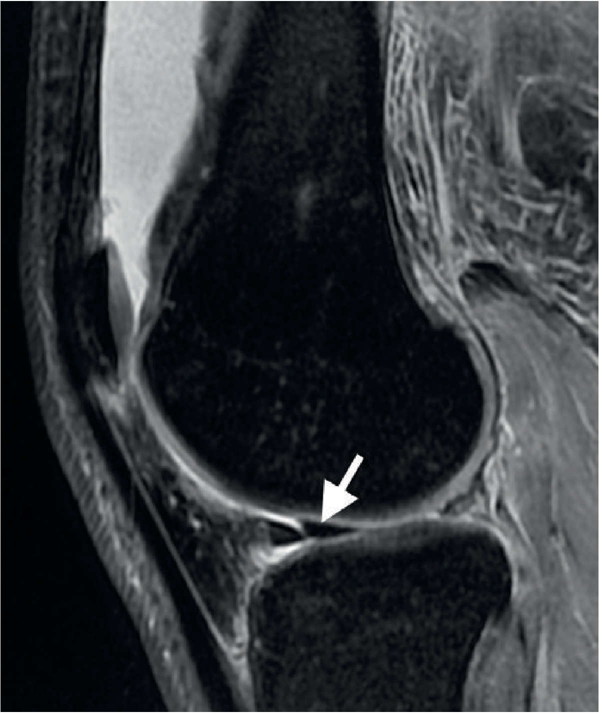
Partial tears show altered signal and/or laxity with continuity of some fibres.
Secondary (Indirect) Signs of ACL Tear:
| Sign | Description |
|---|---|
| Bone marrow "kissing" contusions | Posterolateral tibial plateau + lateral femoral condyle (pivot-shift mechanism) |
| Anterior tibial translation | ≥7 mm shift of tibia relative to femur |
| PCL buckling | Posterior cruciate ligament takes on a curved/buckled shape due to anterior tibial shift |
| Deep sulcus sign | Deep indentation of the condylopatellar sulcus on the lateral femoral condyle (>1.5 mm) |
| Segond fracture | Avulsion of the lateral tibial plateau margin at the joint capsule attachment - has high association with ACL tear; may be subtle on MRI but better seen on plain X-ray |
Commonly associated injuries: medial meniscus tear, posterolateral corner injury, MCL tear (O'Donoghue's "unhappy triad" = ACL + MCL + medial meniscus).
PCL (Posterior Cruciate Ligament)
Normal PCL appears as a thick, uniform low-signal bundle on all MRI planes - the most consistently visualised knee ligament.
MRI findings:
- Partial (more common): middle-third intrasubstance thickening + increased signal while maintaining apparent continuity
- Complete tear: overt fibre discontinuity
- Contusion pattern: anterolateral tibial plateau + lateral femoral condyle (suggesting hyperextension or dashboard mechanism)
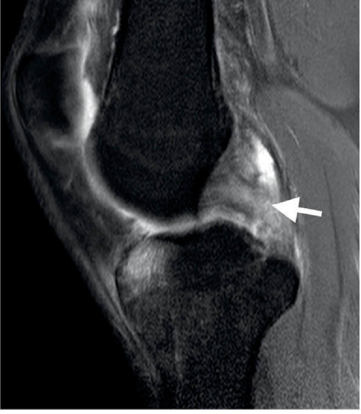
MCL (Medial Collateral Ligament)
Normal MCL is seen on coronal MRI as a thin low-signal band. The superficial component (most important for valgus stability) is longer and distinguishable from the shorter deep meniscotibial/meniscofemoral bands.
Grading system on MRI:
| Grade | MRI Finding |
|---|---|
| Grade 1 | Periligamentous oedema only; ligament intact |
| Grade 2 | Partial rupture with focal intraligamentous thickening + oedema |
| Grade 3 | Complete rupture - fibre discontinuity |
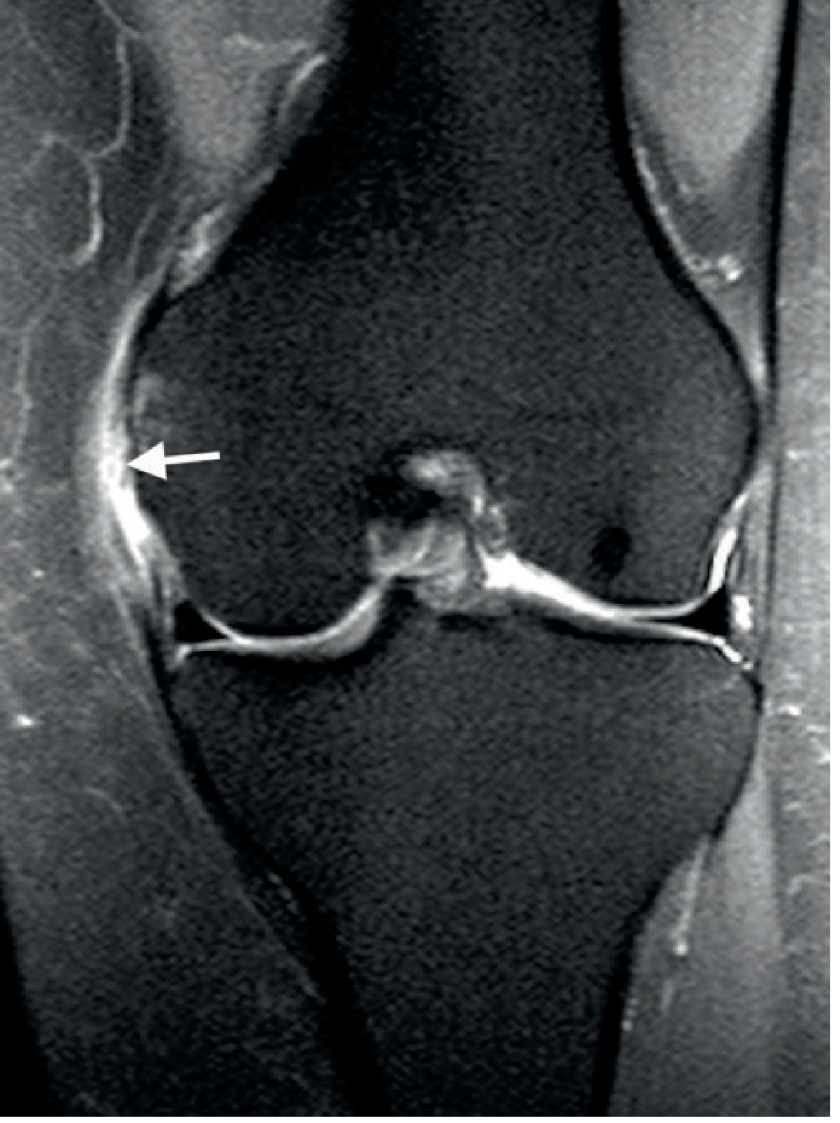
Pellegrini-Stieda lesion = chronic ossification at the proximal MCL following healed/calcified injury, visible on plain X-ray.
LCL (Lateral Collateral Ligament) & Posterolateral Corner
The LCL (fibular collateral ligament) passes from the lateral femoral condyle to the fibular head. It is part of the posterolateral corner (PLC) complex, which also includes the popliteus tendon, biceps femoris, arcuate ligament, and popliteofibular ligament.
MRI findings:
- LCL tears: intrasubstance signal change with thickening, or avulsion from the fibular head
- PLC injuries are often difficult to fully characterise on MRI due to the small size of individual structures
- Frequently associated with ACL tears - check always when ACL is torn
- Carries important surgical implications (affects ACL reconstruction outcomes if untreated)
Plain X-ray clue: Fibular head avulsion fracture = "arcuate sign" - high association with PLC injury.
Quick Reference Summary
| Ligament | Best Plane | Key Direct Signs | Key Indirect Signs |
|---|---|---|---|
| ACL | Sagittal | Fibre discontinuity, increased signal, laxity | Kissing contusions, anterior tibial translation, Segond fracture |
| PCL | Sagittal | Thickening + intrasubstance signal, discontinuity | Anterolateral tibial/lateral condyle contusions |
| MCL | Coronal | Perilig. oedema (G1), partial tear (G2), discontinuity (G3) | Pellegrini-Stieda (chronic) |
| LCL/PLC | Coronal/Axial | Intrasubstance signal, fibular head avulsion | Arcuate sign on X-ray |
Preferred sequences: Proton density fat-saturated (PD FS) and T2 fat-saturated in sagittal, coronal, and axial planes. Fat suppression markedly improves detection of bone marrow oedema and periligamentous fluid.
Source: Grainger & Allison's Diagnostic Radiology, Chapter 39 (The Knee)
This is a shared conversation. Sign in to Orris to start your own chat.