
Porphyria
Overview
- Acute (neurovisceral) porphyrias - e.g., Acute Intermittent Porphyria (AIP), Variegate Porphyria (VP), Hereditary Coproporphyria (HCP), ALA-dehydratase deficiency porphyria - present with abdominal pain, neuropathy, psychiatric symptoms, often triggered by drugs, fasting, alcohol, hormonal changes.
- Cutaneous porphyrias - e.g., Porphyria Cutanea Tarda (PCT, most common), Erythropoietic Protoporphyria (EPP), Congenital Erythropoietic Porphyria (CEP) - present with photosensitivity, blistering, skin fragility.
- Mixed - Variegate porphyria has both neurovisceral and cutaneous features.
Topic breakdown
- Heme biosynthesis pathway and enzyme defects (which enzyme = which porphyria)
- Acute Intermittent Porphyria (AIP) - presentation, triggers, diagnosis, management
- Porphyria Cutanea Tarda (PCT) - most common type, cutaneous findings, iron/HCV link
- Variegate Porphyria and Hereditary Coproporphyria
- Erythropoietic porphyrias (EPP, Congenital Erythropoietic Porphyria/Gunther disease)
- Diagnosis and lab workup (urine PBG/ALA, plasma/fecal porphyrins, genetic testing)
- Acute attack management (hemin, glucose, avoiding triggering drugs) and long-term/genetic counseling
- Drugs and precipitating factors to avoid
Which aspect of porphyria would you like to explore in depth?
Overview
- Acute (neurovisceral) porphyrias - e.g., Acute Intermittent Porphyria (AIP), Variegate Porphyria (VP), Hereditary Coproporphyria (HCP), ALA-dehydratase deficiency porphyria - present with abdominal pain, neuropathy, psychiatric symptoms, often triggered by drugs, fasting, alcohol, hormonal changes.
- Cutaneous porphyrias - e.g., Porphyria Cutanea Tarda (PCT, most common), Erythropoietic Protoporphyria (EPP), Congenital Erythropoietic Porphyria (CEP) - present with photosensitivity, blistering, skin fragility.
- Mixed - Variegate porphyria has both neurovisceral and cutaneous features.
Topic breakdown
- Heme biosynthesis pathway and enzyme defects (which enzyme = which porphyria)
- Acute Intermittent Porphyria (AIP) - presentation, triggers, diagnosis, management
- Porphyria Cutanea Tarda (PCT) - most common type, cutaneous findings, iron/HCV link
- Variegate Porphyria and Hereditary Coproporphyria
- Erythropoietic porphyrias (EPP, Congenital Erythropoietic Porphyria/Gunther disease)
- Diagnosis and lab workup (urine PBG/ALA, plasma/fecal porphyrins, genetic testing)
- Acute attack management (hemin, glucose, avoiding triggering drugs) and long-term/genetic counseling
- Drugs and precipitating factors to avoid
heme biosynthesis pathway enzyme defects porphyria diagram
porphyria cutanea tarda skin blisters dorsum of hand
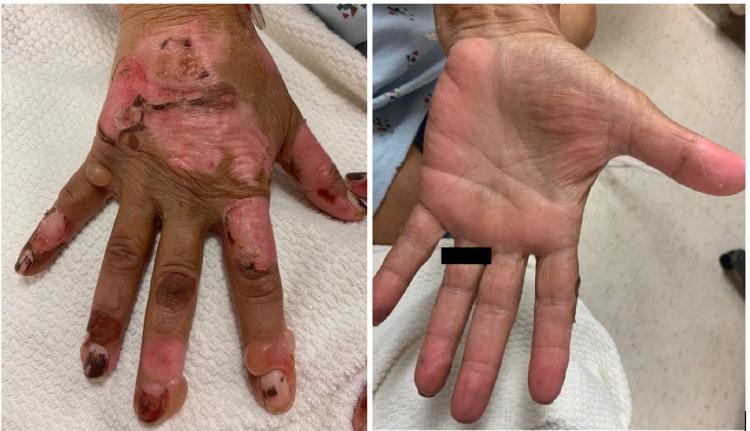
This clinical photograph displays a side-by-side comparison of the dorsal and palmar surfaces of a human hand, illustrating a bullous dermatosis. The left image (dorsal view) shows significant pathology characterized by multiple, large, tense bullae of varying sizes localized to the dorsum of the hand and fingers. Several bullae have ruptured, revealing raw, erythematous bases with overlying crusting and superficial erosions. The surrounding skin exhibits hyperpigmentation and signs of chronic inflammation or scarring. In contrast, the right image (palmar view) demonstrates a relative sparing of the palmar surface, which remains largely intact without visible blisters or erosions. This distribution—blisters on sun-exposed dorsal areas with palmar sparing—is a hallmark of porphyria cutanea tarda (PCT) or pseudoporphyria, often seen in the context of chronic kidney disease and hemodialysis. The clinical significance lies in demonstrating the photosensitive nature of these skin lesions and the typical morphological findings of fluid-filled vesicles and bullae on fragile, sun-exposed skin.
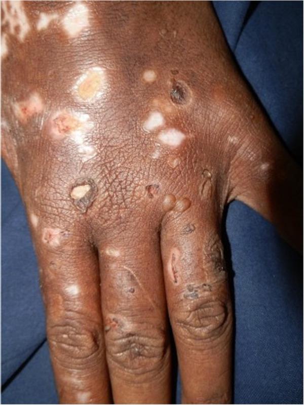
Clinical photograph showing the dorsum of a hand in a patient with darker skin (Fitzpatrick type IV-VI), demonstrating classic manifestations of Porphyria Cutanea Tarda (PCT). The image shows a polymorphic eruption characterized by multiple tense bullae (blisters), some of which have ruptured to form raw, red erosions. Chronic changes are prominent, including mottled pigmentary alterations with areas of hyperpigmentation and vitiligo-like hypopigmentation (depigmentation). Shallow, atrophic scars and evidence of skin fragility are visible across the metacarpal area and phalanges. The skin texture appears thickened with exaggerated markings (lichenification), consistent with chronic photo-induced damage. These findings illustrate the hallmark features of PCT, where porphyrin-mediated phototoxicity leads to extreme mechanical fragility and blistering in sun-exposed areas. This visual is highly relevant for medical education regarding metabolic photodermatoses and their differential diagnosis from chronic actinic dermatitis or pellagra.
porphyria treatment
Porphyria
Classification
| Category | Types | Mechanism of symptoms |
|---|---|---|
| Acute (neurovisceral) hepatic porphyrias | Acute Intermittent Porphyria (AIP), Variegate Porphyria (VP), Hereditary Coproporphyria (HCP), ALA-dehydratase deficiency porphyria | Neurotoxic accumulation of ALA/PBG |
| Cutaneous porphyrias | Porphyria Cutanea Tarda (PCT), Congenital Erythropoietic Porphyria (CEP), Erythropoietic Protoporphyria (EPP) | Phototoxic porphyrins deposited in skin, activated by UV light |
| Mixed | Variegate porphyria, Hereditary coproporphyria | Both neurovisceral attacks and photosensitive skin lesions |
- ALA dehydratase deficiency -> ALA dehydratase deficiency porphyria
- PBG deaminase (HMB synthase) deficiency -> Acute Intermittent Porphyria
- Uroporphyrinogen III synthase deficiency -> Congenital Erythropoietic Porphyria
- Uroporphyrinogen decarboxylase (UROD) deficiency -> Porphyria Cutanea Tarda
- Coproporphyrinogen oxidase deficiency -> Hereditary Coproporphyria
- Protoporphyrinogen oxidase deficiency -> Variegate Porphyria
- Ferrochelatase deficiency -> Erythropoietic Protoporphyria
Acute Intermittent Porphyria (most relevant acute type)
- Severe abdominal pain (up to 95%), nausea/vomiting, constipation (Andrews' Diseases of the Skin)
- Motor neuropathy/paresis (50-70%), seizures
- Neuropsychiatric disturbance (anxiety, hallucinations, confusion)
- Hyponatremia, hypertension, tachycardia
- No skin lesions (distinguishing it from VP and HCP)
- Urine PBG and ALA markedly elevated, urine darkens on standing (Yamada's Textbook of Gastroenterology, 7th ed; Henry's Clinical Diagnosis and Management by Laboratory Methods)
Porphyria Cutanea Tarda (most common overall)
- Photosensitivity, skin fragility, tense blisters on sun-exposed, trauma-prone areas (dorsal hands)
- Hypertrichosis, hyperpigmentation, sclerodermoid skin changes
- No acute neurovisceral attacks and normal urine PBG (helps distinguish from acute porphyrias)
Other cutaneous porphyrias
- Congenital Erythropoietic Porphyria (Gunther disease) - severe, presents in infancy with mutilating photosensitivity, hemolytic anemia, and erythrodontia
- Erythropoietic Protoporphyria - childhood onset of acute painful burning photosensitivity (not primarily blistering), due to ferrochelatase deficiency
Diagnosis
- Acute attack: spot urine PBG and ALA (markedly elevated in AIP, VP, HCP)
- Cutaneous disease: plasma and fecal porphyrin fractionation to distinguish PCT, VP, HCP, EPP
- Enzyme assays and genetic testing confirm the specific type and allow family screening
- During latency, biochemical markers can be normal, so genetic testing matters for at-risk relatives
Management
- IV hemin (heme arginate/hematin) is first-line - suppresses hepatic ALA synthase and reduces precursor overproduction
- IV glucose/carbohydrate loading (≥300 g/day) is a temporizing measure only if hemin is unavailable or the attack is mild
- Symptom control: analgesia, antiemetics, careful fluid/electrolyte monitoring (watch for hyponatremia)
- Avoid all porphyrinogenic drugs (checked against a porphyria drug safety database)
- Givosiran, an RNAi therapeutic that silences hepatic ALA synthase-1, was approved (FDA 2019, EMA 2020) for recurrent attacks in acute hepatic porphyria - a major recent addition beyond hemin