
Motor fibres of internal capsule को समझा दो। इसमें associated tract, corticospinal tract, corticonuclear, corticoponto-cerebellar tract समझा दो। Extrapyramidal tract समझा दो। इन सब की location कहाँ-कहाँ पर है और किसके damage होने से क्या-क्या, अ, problems हो सकती हैं, ये भी समझा दो।
internal capsule motor fibres corticospinal tract diagram
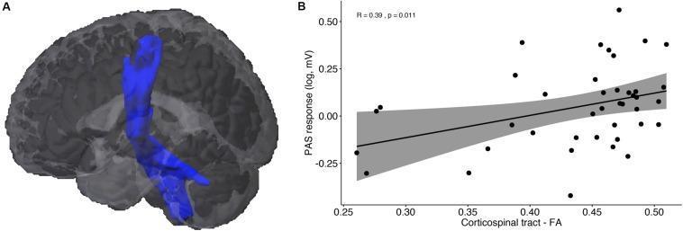
This composite educational graphic presents findings from Diffusion Tensor Imaging (DTI) and neurophysiological correlation. (A) Anatomical Diagram: A 3D volumetric rendering of the human brain (sagittal/oblique view) with a blue highlight representing probabilistic fiber tractography of the Corticospinal Tract (CST). The tract is shown descending vertically from the primary motor cortex through the internal capsule towards the brainstem. (B) Comparison Chart/Scatter Plot: A graph illustrating the correlation between white matter microstructural integrity and physiological response. The x-axis represents 'Corticospinal tract - FA' (Fractional Anisotropy), a proxy for tract integrity (range ~0.25 to 0.50). The y-axis represents 'PAS response (log, mV),' reflecting plasticity from Paired Associative Stimulation. A positive linear regression line is plotted with a gray 95% confidence interval, showing a significant positive correlation (R = 0.39, p = 0.011). This visualization demonstrates the relationship between the structural health of the motor pathways and the functional neuroplasticity of the brain.
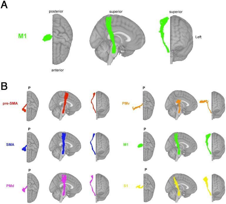
This diagnostic diagram presents theory-based biomarkers for neuroimaging research, specifically illustrating white matter fiber tracts associated with sensorimotor functions. The visual is divided into two main panels using MNI (Montreal Neurological Institute) brain templates. Panel A highlights the M1-corticospinal tract (M1-CST) in bright green across axial, sagittal, and coronal projections, showing its pathway from the primary motor cortex through the internal capsule. Panel B displays six distinct sensorimotor tract segmentations from the Sensorimotor Area Tract Template (SMATT), each color-coded for differentiation: pre-supplementary motor area (pre-SMA, red), supplementary motor area (SMA, blue), dorsal premotor cortex (PMd, magenta), ventral premotor cortex (PMv, orange), primary motor cortex (M1, green), and primary sensory cortex (S1, yellow). The sagittal views in Panel B demonstrate the anterior-to-posterior spatial arrangement, where pre-SMA is the most anterior and S1 is the most posterior. This illustration is used in clinical research to calculate lesion load on specific functional tracts, aiding in the prediction of motor recovery and chronic deficits following brain injury or stroke.
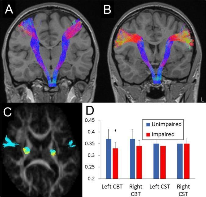
This composite educational graphic focuses on neuroimaging and diffusion tensor imaging (DTI) tractography of motor pathways. Panels A and B present coronal T1-weighted MRI scans with overlaid white matter reconstructions. Panel A depicts the corticospinal tract (CST), primarily in blue, showing its descent from the motor cortex through the internal capsule toward the brainstem. Panel B illustrates the dorsal corticobulbar tract (CBT), displaying a more varied color spectrum (red, yellow, and blue) representing different fiber orientations. Panel C provides an axial cross-section demonstrating the spatial relationship and partial overlap of the CBT (cyan) and CST (yellow) at the level of the posterior limb of the internal capsule. Panel D is a bar graph comparing fractional anisotropy (FA) values between 'Unimpaired' and 'Oromotor-Impaired' groups, highlighting a statistically significant reduction in FA within the left dorsal CBT for the impaired group. This material is designed for advanced medical education in neuroanatomy and radiology, specifically illustrating motor tract topography and the clinical significance of fiber integrity in oromotor function.
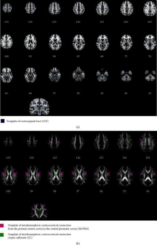
This diagnostic image displays template tracts of the human brain reconstructed via probabilistic tractography, presented across a series of axial neuroimaging slices and coronal views. Part (a) illustrates the Corticospinal Tract (CST) in blue, showing its bilateral longitudinal descent from the primary motor cortex through the posterior limb of the internal capsule and cerebral peduncles, converging toward the ventral medulla oblongata. Part (b) visualizes corticocortical connections: pink regions represent intrahemispheric connections between the primary motor cortex (M1) and the ventral premotor cortex (PMv), while green regions represent interhemispheric connections passing through the corpus callosum (CC). The visualization demonstrates the spatial distribution and anatomical pathways of white matter fibers essential for motor control. These templates, derived from healthy subjects, serve as a clinical reference for evaluating tract integrity and fractional anisotropy (FA) changes in conditions such as stroke. The image is optimized for neuroanatomical education and neuroradiological research involving diffusion-weighted imaging (DWI) and fiber tracking.
internal capsule horizontal section posterior limb anterior limb genu anatomy
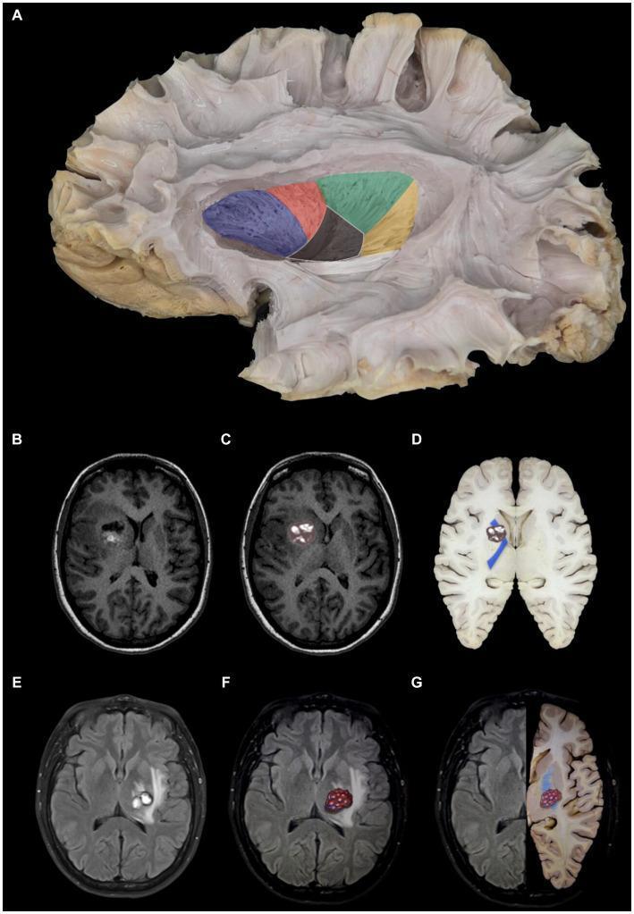
This composite educational graphic details the neuroanatomy of the internal capsule and its clinical correlation with cerebral cavernomas. Panel A features a gross anatomical white matter dissection (lateral view) with color-coded segmentation: anterior limb (blue), genu (red), posterior limb (green), retrolenticular portion (yellow), and sublenticular portion (white), alongside a partially resected lentiform nucleus. Panels B–D illustrate a cavernoma in the anterior limb of the internal capsule using axial T1-weighted MRI and anatomical overlays. The lesion demonstrates mixed signal intensity and is situated lateral to the caudate head and medial to the lentiform nucleus. Panels E–G depict a cavernoma in the posterior limb of the internal capsule via axial T2-weighted MRI and side-by-side anatomical comparisons. This lesion exhibits a heterogeneous, predominantly hyperintense signal and is localized medial to the lentiform nucleus and lateral to the thalamus. The collection serves to bridge gross anatomical segments of the internal capsule with diagnostic radiological findings and clinical pathology.
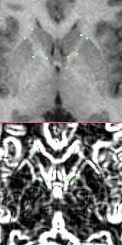
This diagnostic image demonstrates a neuroimaging segmentation protocol for the internal capsule using magnetic resonance imaging (MRI). The figure is divided into two panels: the top panel shows an axial T1-weighted enlarged view of the basal ganglia region, featuring the caudate nucleus, putamen, and the anterior limb of the internal capsule. Manual landmarks (green dots) are placed at four strategic corners to delineate the polygon of the internal capsule: the lateral anterior and posterior aspects of the caudate nucleus, and the medial anterior and posterior aspects of the putamen. The bottom panel displays the same anatomical section processed with a Sobel gradient filter. This edge-detection algorithm enhances the contrast between the gray matter of the striatum and the white matter of the internal capsule, facilitating more precise landmark placement by highlighting the boundaries of the anatomical structures. This method is utilized in clinical research, such as studies on schizophrenia, to quantify volumes and tract lengths in subcortical brain regions.
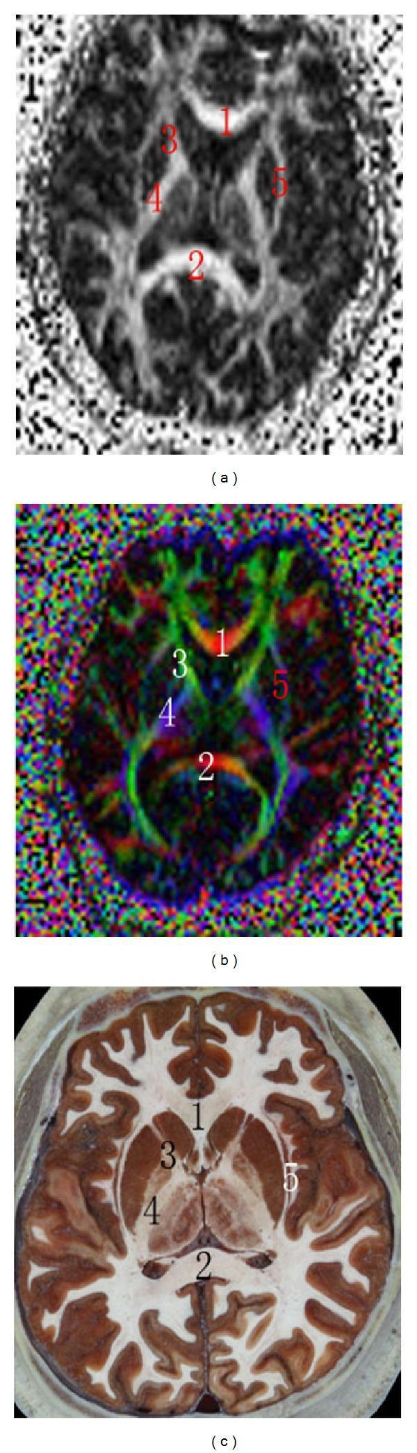
This composite figure presents a transversal comparison of human neuroanatomy at the level of the basal nuclei using three distinct modalities: (a) Fractional Anisotropy (FA) map, (b) Color Diffusion Tensor Imaging (DTI) map, and (c) a cross-sectional anatomical image. The images illustrate key white matter tracts and landmarks. Labels 1 and 2 indicate the genu and splenium of the corpus callosum, respectively; in the color DTI map, these are shown in red, signifying transverse fiber orientation. Label 3 identifies the anterior limb of the internal capsule (green in DTI, indicating anteroposterior orientation), while label 4 shows the posterior limb of the internal capsule (blue in DTI, indicating craniocaudal orientation). Label 5 highlights the region of the external and extreme capsules. The anatomical section (c) provides high-resolution morphological context, clearly delineating the boundaries between gray matter nuclei and white matter tracts that are interpreted via the FA and DTI maps. This comparison is used to teach neuroimaging correlations and the principles of tractography in clinical neurology and radiology.
Internal Capsule के Motor Fibres - Complete Guide
🧠 Internal Capsule क्या है?
- Medial side: Thalamus + Caudate nucleus
- Lateral side: Globus pallidus + Putamen (Lentiform nucleus)
📍 Internal Capsule के Parts और उनमें Fibres
| Part | Location | Fibres |
|---|---|---|
| Anterior Limb | Head of Caudate - Globus Pallidus के बीच | Frontopontine fibres, Thalamocortical (prefrontal) |
| Genu ("घुटना") | Foramen of Monro के level पर | Corticonuclear (Corticobulbar) tract |
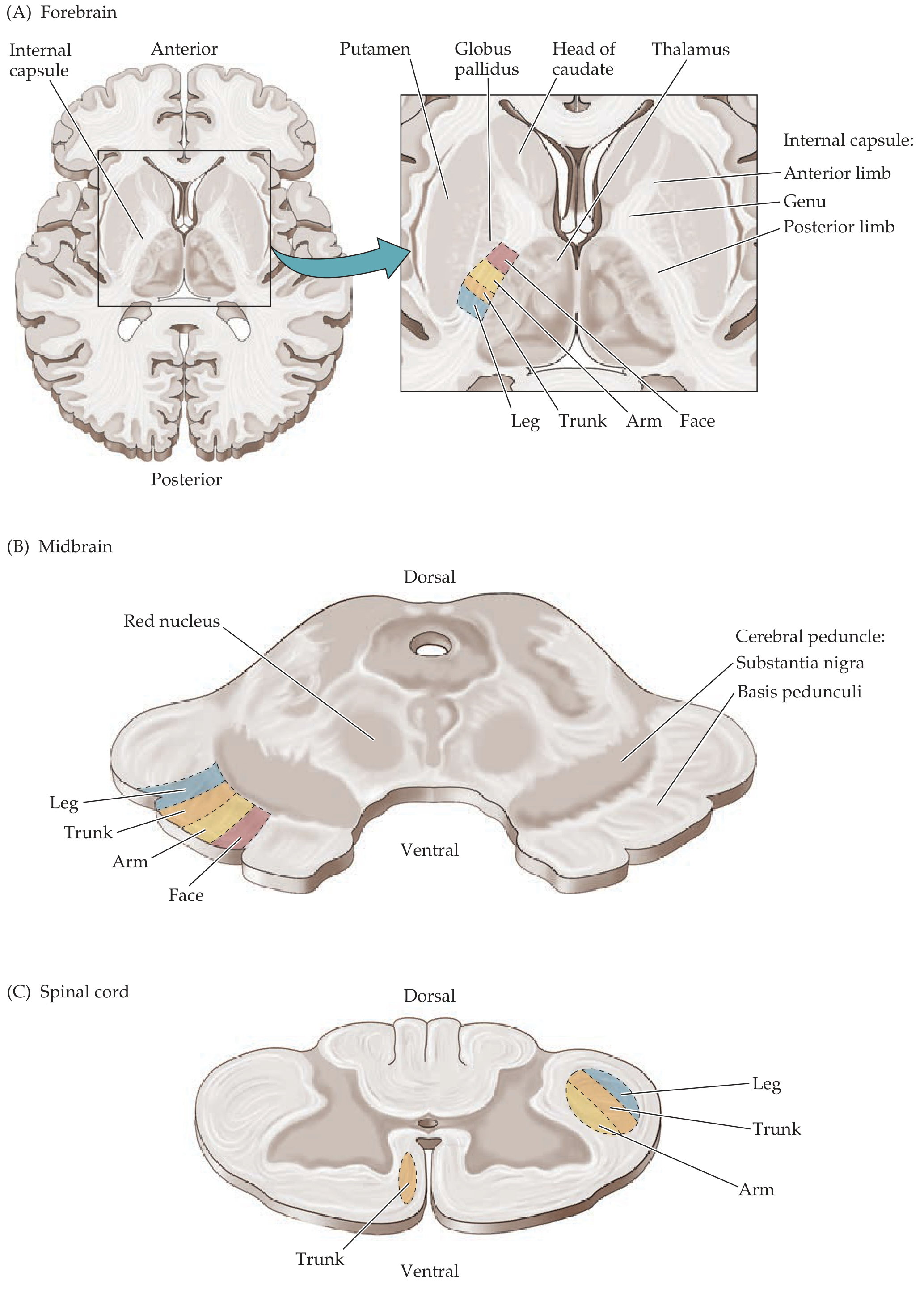
| Posterior Limb | Thalamus - Lentiform Nucleus के बीच | Corticospinal tract (Face → Arm → Trunk → Leg - Anterior से Posterior) |
| Retrolenticular part | Posterior limb के पीछे | Optic radiation, Thalamocortical fibres |
| Sublenticular part | Lentiform nucleus के नीचे | Auditory radiation, Temporopontine fibres |
1. 🔴 Corticospinal Tract (Pyramidal Tract)
Origin और Course:
- Origin: Primary Motor Cortex (Brodmann Area 4 - Precentral gyrus) से >50% fibres; बाकी Area 6 (Premotor, SMA) और Parietal lobe (Areas 3,1,2,5,7) से
- Layer 5 pyramidal cells - इनमें giant Betz cells (~3%) होते हैं जो nervous system के सबसे बड़े neurons हैं
Location at Different Levels:
Cortex → Corona Radiata → Internal Capsule (Posterior Limb)
→ Cerebral Peduncle (Basis Pedunculi - Middle 1/3)
→ Ventral Pons (Scattered fascicles)
→ Medullary Pyramids
→ Pyramidal Decussation (85% fibres cross)
→ Lateral Corticospinal Tract (Crossed - Dorsolateral column)
→ Anterior Corticospinal Tract (Uncrossed 15% - Ventral column)
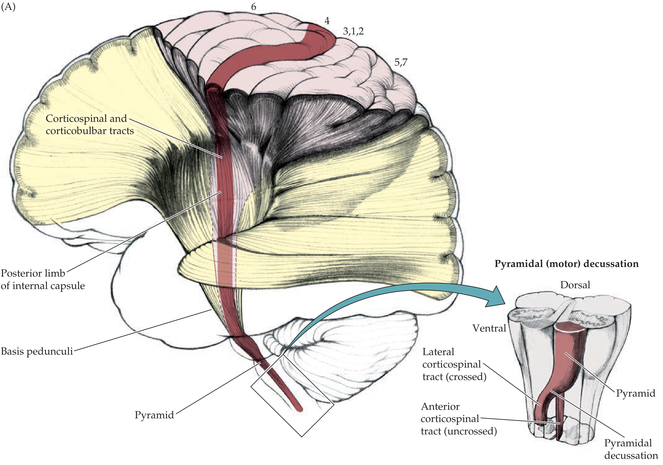
Somatotopic Organization:
- Face fibres → सबसे anterior (Genu के पास)
- Arm → उससे posterior
- Trunk → और posterior
- Leg → सबसे posterior
- Face → Medial
- Trunk → Middle
- Leg → Lateral
- Cervical fibres → Medial
- Lumbar fibres → Lateral (Lamination)
Function:
- Voluntary skilled movements, especially distal limb muscles (fine finger movements)
- Direct synapse: LMN (ventral horn) पर
- Contralateral body को control करता है
2. 🟡 Corticonuclear (Corticobulbar) Tract
Location:
Course:
Motor Cortex (Face area) → Genu of Internal Capsule
→ Basis Pedunculi का Medial 1/3
→ Brainstem → Cranial Nerve Motor Nuclei
Important Facts:
- यह Corticospinal tract के साथ चलता है लेकिन Brainstem पर end होता है (spinal cord में नहीं जाता)
- Bilateral innervation: अधिकतर cranial nerve nuclei को दोनों sides से fibres मिलते हैं
- Exception - Unilateral: Lower face (CN VII lower), CN XII (genioglossus) - ये केवल contralateral side से innervate होते हैं
Damage effects:
| Lesion | Effect |
|---|---|
| Unilateral Upper corticonuclear | Only lower face weakness (contralateral), upper face spared |
| Bilateral corticonuclear | Pseudobulbar palsy - dysarthria, dysphagia, emotional lability |
3. 🟢 Corticoponto-Cerebellar Tract (Frontopontine + Parietopontine)
Location:
- Anterior Limb: Frontopontine fibres (Frontal lobe से)
- Posterior Limb का posterior part / Sublenticular: Parietopontine, Temporopontine, Occipitopontine fibres
Course:
Frontal/Parietal/Temporal Cortex
→ Internal Capsule (Anterior Limb + Posterior Limb)
→ Basis Pedunculi (Medial 1/5 और Lateral 1/5 - Corticopontine fibres)
→ Pontine Nuclei
→ Middle Cerebellar Peduncle (Brachium Pontis)
→ Contralateral Cerebellum
Function:
- Cerebellum को voluntary motor activity की information देता है
- Coordination और smooth movement के लिए
Damage effects:
- Ipsilateral cerebellar signs (क्योंकि double crossing होती है)
- Ataxia, dysmetria, intention tremor
4. ⚪ Extrapyramidal System और Tracts
Extrapyramidal System में कौन-कौन शामिल हैं?
| Tract | Origin | Location in Spinal Cord | Function |
|---|---|---|---|
| Rubrospinal | Red nucleus (Midbrain) | Lateral column (Dorsolateral) | Distal limb flexion |
| Reticulospinal | Reticular formation (Pons + Medulla) | Anterior + Lateral column | Tone, posture, autonomic |
| Vestibulospinal | Vestibular nucleus (Lateral = Deiter's) | Anterior column | Balance, antigravity muscles |
| Tectospinal | Superior colliculus | Anterior column (Cervical) | Head turning reflexes (visual/auditory) |
Internal Capsule में Extrapyramidal Fibres:
Adams and Victor's: "Lesions in these parts can simultaneously affect both corticospinal and extrapyramidal systems, and attribution of a capsular lesion to one or the other system is not always possible."
Extrapyramidal Damage के Features:
- Rigidity (lead pipe / cogwheel)
- Resting tremor
- Bradykinesia
- Abnormal posture
- Involuntary movements (chorea, athetosis, dystonia)
- Decorticate posture (rubrospinal dominant) vs Decerebrate posture (vestibulospinal dominant)
⚡ Internal Capsule Damage - Clinical Features
Complete Capsular Lesion (e.g., MCA territory stroke):
| Feature | Description |
|---|---|
| Contralateral Hemiplegia | Face + Arm + Leg सब |
| UMN signs | Spasticity, hyperreflexia, Babinski positive |
| Contralateral hemianesthesia | Sensory fibres भी posterior limb में हैं (Thalamocortical) |
| Homonymous hemianopia | Retrolenticular part damage |
| Dysphagia/Dysarthria | Corticonuclear involvement |
Partial/Selective Damage:
| Location Damaged | Specific Deficit |
|---|---|
| Genu | Corticonuclear → Lower face weakness, dysarthria |
| Posterior Limb (anterior part) | Face + Arm weakness |
| Posterior Limb (posterior part) | Leg > Arm weakness |
| Incomplete capsular | Hemichorea-athetosis (Extrapyramidal escape) |
| Anterior Limb | Frontopontine damage → Cerebellar signs + Frontal lobe behavior |
| Retrolenticular | Optic radiation → Contralateral homonymous hemianopia |
Pure Motor Hemiplegia:
- Small lacunar infarct in posterior limb
- Only motor weakness, no sensory/visual deficit
- Classic capsular stroke
🔑 Key Points - याद रखने के लिए
GENU = corticobulbar (corticoNUclear) - "G-N याद करो"
POSTERIOR LIMB = corticospinal (Face → Arm → Trunk → Leg - Anterior to Posterior)
ANTERIOR LIMB = frontopontine + thalamocortical (prefrontal)

Internal capsule damage → CONTRALATERAL deficits (fibres अभी cross नहीं हुई)
Spinal cord damage → IPSILATERAL deficits (fibres पहले cross हो चुकी)
- Neuroanatomy through Clinical Cases, 3rd Edition - Chapter 6, Motor Systems (pp. 256-261)
- Adams and Victor's Principles of Neurology, 12th Edition - Motor System chapter
- Harrison's Principles of Internal Medicine, 22E - Pathogenesis of Motor Deficits