

answer it according to uhs standard book of gynae and obs give precise answer with pictures and convert this into pdf
female pelvic anatomy abdominal wall layers diagram
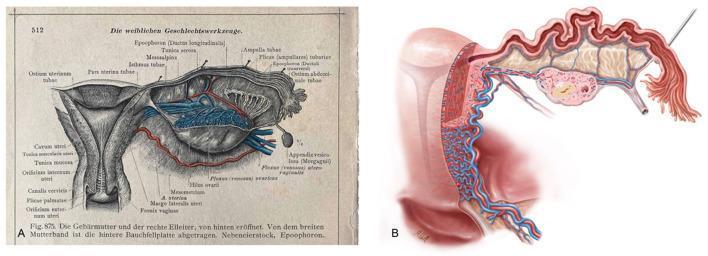
A side-by-side comparison of female pelvic anatomy illustrations. Figure A is a historical anatomical diagram from a 1903 atlas (Dr. Carl Toldt), providing a posterior view of the uterus, adnexa, and parametrium with extensive German and Latin labeling. Key structures include the uterine wall layers (Tunica muscularis and serosa), the cervical canal (Canalis cervicis), and the fallopian tube (Pars uterina, Isthmus, and Ampulla tubae). It details the epoophoron, mesosalpinx, and the fimbriated end near the ostium abdominale. Figure B is a modern medical illustration by Markus Voll, showing a coronal sectional view of the uterus and adnexa. This modern rendering emphasizes the intricate vascular supply, depicting the dense network of uterine arteries and veins within the myometrium. It also highlights the internal mucosal folds of the fallopian tube and the anatomical relationship with the ovary. Both images serve to demonstrate the consistency of anatomical knowledge and the evolution of medical visualization techniques in gynecology and pelvic surgery.
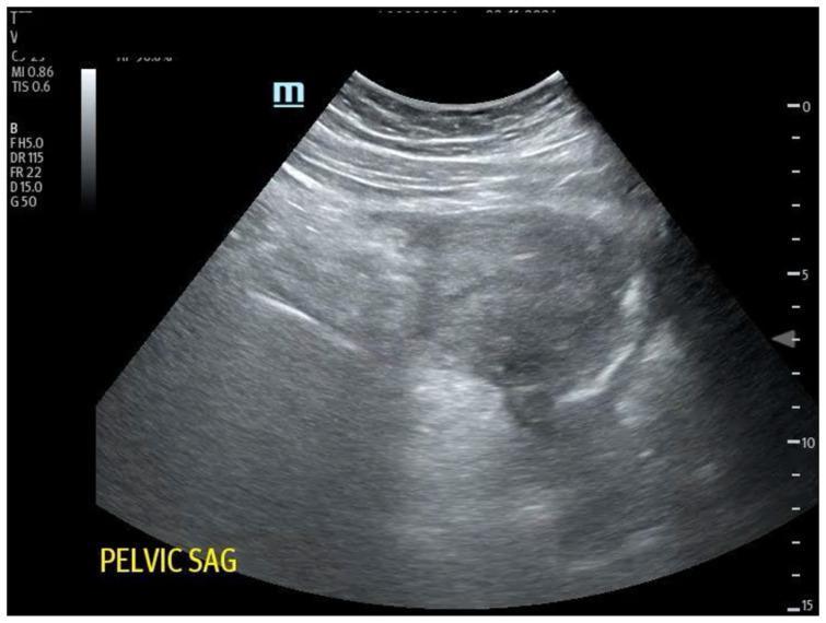
This diagnostic image is a transabdominal ultrasound of the female pelvic region, presented in a sagittal orientation as indicated by the label 'PELVIC SAG'. The ultrasound sector shows the anatomy at a depth of up to 15 cm. The central focus is a heterogeneous, largely hypoechoic to isoechoic mass or structure, potentially representing the uterus or a pelvic adnexal finding. Above this central structure, well-defined hyperechoic linear layers represent the abdominal wall and subcutaneous tissues. The internal texture of the pelvic organs appears mottled with mixed echogenicity, including darker hypoechoic areas that may suggest fluid collections or soft tissue interfaces. Clinical context suggests this image is used for educational purposes to demonstrate pelvic sonography, orientation of the endocavitary or transabdominal probe, and identification of significant pathology such as potential free fluid in cases of ectopic pregnancy or hemorrhagic shock.
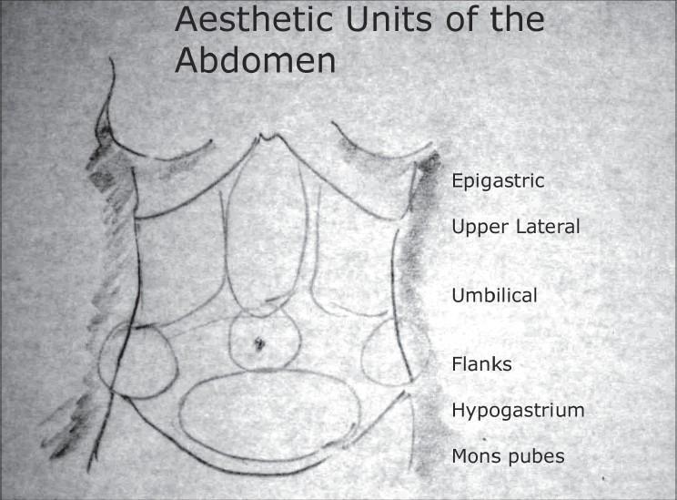
Anatomical Diagram: This is a grayscale, freehand medical sketch illustrating the aesthetic units of the human anterior abdominal wall, primarily used in plastic and reconstructive surgery planning (e.g., liposuction or abdominoplasty). The illustration outlines a female torso from the inframammary fold to the pubic region. Key anatomical landmarks depicted include the costal margins, the midline linea alba, and the umbilical center. The abdomen is segmented into specific zones of clinical interest, which are labeled on the right side. From superior to inferior, these regions include: the central Epigastric unit, bilateral Upper Lateral regions, the central Umbilical unit, bilateral Flanks (lumbar regions), the Hypogastrium (suprapubic region), and the Mons pubes. The drawing serves as an educational tool for identifying discrete topographical areas for surgical contouring and understanding the surface anatomy of the abdominal musculature and overlying adipose tissue distribution.
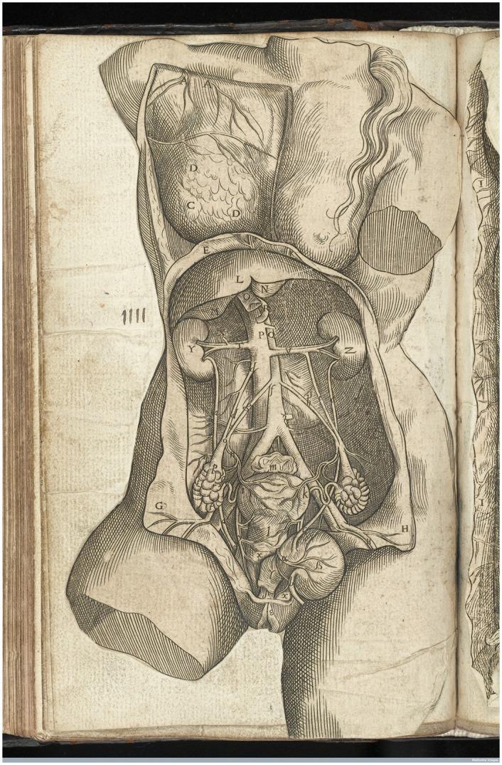
This historical anatomical engraving, based on the work of Andreas Vesalius (1543), provides a detailed frontal dissection of the female torso. The illustration focuses on the urogenital and vascular systems. In the thoracic region, a flap of the chest wall is reflected to reveal the lungs. The abdominal cavity demonstrates the kidneys (labeled Y and Z) with the renal arteries and veins connecting to the abdominal aorta and inferior vena cava. The central vascular structures bifurcate into the common iliac vessels. Significant emphasis is placed on the female reproductive anatomy, showing the uterus (central), the bladder below it, and the ovaries located laterally. The ovaries are depicted as textured, lobulated structures attached to the uterus via the fallopian tubes and ovarian ligaments. The gastrointestinal tract is partially removed to allow clear visualization of the retroperitoneal and pelvic organs. This diagram served as a foundational educational tool in early modern medical literature for understanding female internal anatomy and obstetric medicine.
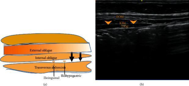
This dual-panel image features an anatomical diagram (a) and a corresponding ultrasound scan (b) demonstrating the regional anatomy for ilioinguinal and iliohypogastric nerve blocks. The diagram illustrates the layered musculature of the lateral abdominal wall, identifying the external oblique, internal oblique, and transversus abdominis muscles. It highlights the ilioinguinal and iliohypogastric nerves situated in the fascial plane between the internal oblique and transversus abdominis muscles. The ultrasound scan provides a clinical diagnostic view of the same region, showing the three muscle layers as distinct hypoechoic bands separated by bright, hyperechoic fascial planes. Specifically, the external oblique muscle (EOM) is the most superficial, the internal oblique muscle (IOM) is the intermediate layer, and the transversus abdominis muscle (TAM) is the deepest layer. Arrows on both panels indicate the target site for local anesthetic infiltration between the IOM and TAM, essential for effective regional anesthesia in the inguinal and pelvic regions.
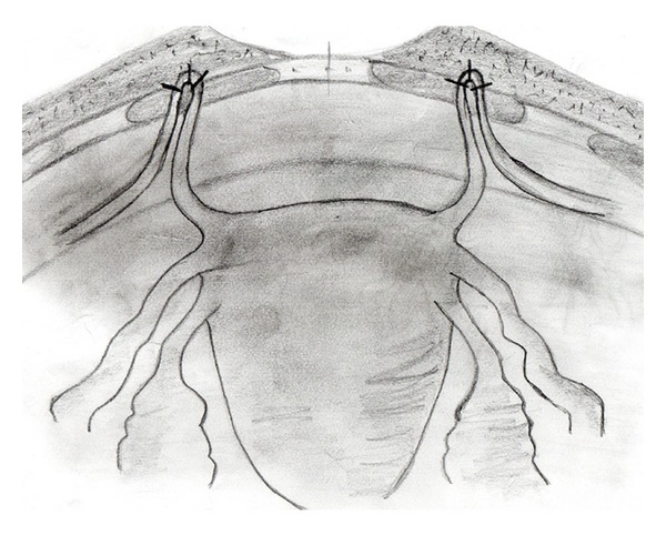
**Imaging Modality:** Medical schematic illustration (pencil/grayscale sketch). **Anatomical Region:** Female pelvic anatomy focusing on the uterus, adnexa, and anterior abdominal wall. **Observed Procedure/Landmarks:** The illustration depicts a surgical uterine suspension procedure. Key anatomical structures include the uterine fundus, fallopian tubes, and the round ligaments of the uterus. The round ligaments are shown extending bilaterally from the uterine cornua. **Characteristic Visual Features:** The schematic highlights the bilateral fixation of the round ligaments to the rectus fascia of the anterior abdominal wall. The distal portions of the round ligaments are looped through or anchored to the fascial layer, secured with visible sutures. This anatomical arrangement demonstrates the shortening or plication of the ligaments to provide superior and anterior support to the uterus. **Clinical Context:** This visual represents a surgical technique for the correction of uterine retroversion or pelvic organ prolapse. The procedure aims to reposition the uterus into an anteverted state by utilizing the native support of the round ligaments and anchoring them to the sturdy fascial layers of the abdominal wall.
female pelvis blood supply lymphatic drainage innervation perineum
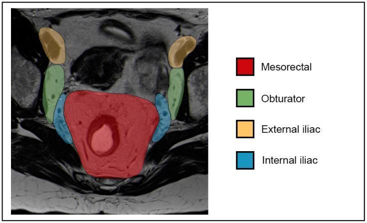
Diagnostic Image: This T2-weighted axial MR image of the female pelvis features a color-coded anatomical overlay depicting regional lymph node compartments used for oncological staging of anal and rectal cancers. The image highlights four primary regions: the centrally located Mesorectal compartment (red overlay), which displays heterogeneous signal intensity and surrounds the rectal canal; the Internal Iliac compartments (blue overlay) positioned adjacent to the lateral pelvic walls; the Obturator compartments (green overlay) located lateral and slightly anterior to the internal iliac groups; and the External Iliac compartments (yellow/orange overlay) positioned most laterally and superiorly within the pelvic field. This clinical illustration demonstrates the relevant lymphatic drainage pathways and nodal stations—specifically N1a (mesorectal, internal iliac, obturator) and N1b (external iliac) according to the 8th edition AJCC staging system. The visualization serves as a diagnostic guide for differentiating pelvic nodal anatomy in the context of pelvic malignancies like anal squamous cell carcinoma (ASCC).
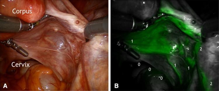
This dual-panel image displays an intraoperative laparoscopic view of the right posterior female pelvis during an indocyanine green (ICG) fluorescence lymphography procedure. Panel A is a standard white-light endoscopic photograph showing the corpus uteri, cervix, and the associated mesometrial and mesonephric compartments. Panel B shows the corresponding near-infrared fluorescence image. In Panel B, intense green ICG fluorescence is localized within the Müllerian lymphatic mesometrial network and the collecting lymph vessels of the vascular mesometrium. These pathways are labeled to indicate drainage toward the pelvic lymph nodes (1) and paraaortic lymph nodes (2). A significant negative finding is the complete absence of fluorescence (0) along the ligamentous mesometrium (uterosacral ligament region), demonstrating that the corporal uterine sub-compartment does not drain through this specific anatomical pathway. This visual comparison illustrates the compartmentalization of lymphatic drainage in gynecologic oncology, differentiating between vascular and ligamentous pathways for surgical mapping.
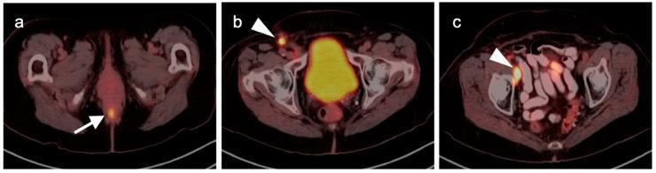
This diagnostic image consists of three axial fused 18F-FDG PET-CT slices of the pelvis demonstrating the staging of anal carcinoma. (a) An axial slice at the level of the perineum shows a focal area of intense FDG uptake (hypermetabolism) in the posterior midline, corresponding to the primary anal tumor (white arrow). (b) A more superior axial slice demonstrates a hypermetabolic right superficial inguinal lymph node (white arrowhead), indicating regional nodal spread. (c) A slice at the level of the mid-pelvis shows an intensely hypermetabolic right distal external iliac lymph node (white arrowhead), representing further lymphatic dissemination along the iliac chain. The images illustrate the typical cephalad and lateral lymphatic drainage patterns of anal malignancy. These findings are critical for oncologic staging and treatment planning, distinguishing between localized disease and regional nodal involvement. The bladder shows normal physiological tracer excretion in frames (b) and (c).
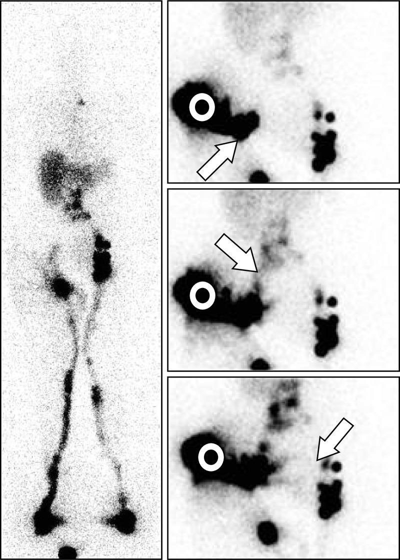
This composite diagnostic image presents 99mTc-human serum albumin nanocolloid lymphoscintigraphy findings in a 69-year-old female with secondary lower limb lymphedema (LLLE) following surgery for inguinal leiomyosarcoma. The left panel shows an anterior whole-body scan (WBS) acquired after one hour of walking. It demonstrates asymmetric lymphatic drainage: the left side displays a normal patent lymphatic vessel (LV) connecting to a complete infradiaphragmatic lymph node (LN) axis, while the right side shows an LV reaching only an inferior inguinal LN with evidence of lymphatic reflux and no visualized common iliac LNs. The right panel consists of three sequential anterior-view static images of the pelvis and abdomen following an additional intradermal injection at the external buttock. These images illustrate collateral lymphatic pathways: an oblique arrow indicates drainage toward the inguinocrural LN, a second arrow highlights drainage toward common iliac LNs, and a third identifies a prepubic pathway crossing to contralateral inguinal LNs. The visualization of these alternative drainage routes is significant for mapping collateral circulation and guiding manual lymphatic drainage therapy in patients with disrupted primary lymphatic pathways.
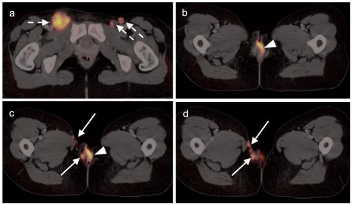
This composite diagnostic image features four axial FDG PET-CT scans (a-d) of the female pelvis demonstrating vulvar squamous cell carcinoma with metastatic spread. Image (a) shows bilateral inguinal lymphadenopathy with intense hypermetabolism; white dashed arrows highlight the metabolic activity in the right and left inguinal regions. Images (b), (c), and (d) focus on the primary vulvar carcinoma (white arrowheads), which exhibits marked FDG uptake. Progression through the axial slices (c and d) reveals local invasion of the malignancy into the adjacent right-sided subcutaneous tissue and the skin of the right medial thigh (indicated by solid white arrows). This diagnostic series illustrates the staging utility of PET-CT in identifying both the hypermetabolic primary tumor and regional nodal involvement in gynecologic oncology. The visual evidence emphasizes the typical patterns of local spread and lymphatic drainage associated with advanced vulvar malignancies.
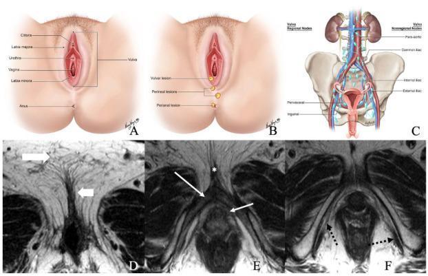
A multi-panel educational figure illustrating the normal anatomy of the female vulvar region and its lymphatic drainage. Panels A and B are anatomical illustrations showing external structures including the clitoris, labia majora and minora, urethra, vagina, and anus, with markers for typical lesion locations. Panel C is a schematic diagram of the lymphatic system, detailing regional (inguinal, perivisceral) and non-regional (external iliac, internal iliac, common iliac, and para-aortic) lymph nodes relative to the pelvis and kidneys. Panels D–F present axial non-fat saturated T2-weighted MRI scans of a 36-year-old female. Image D identifies the labia majora and labia minora through their signal intensities. Image E highlights the deep structures including the bulb of the vestibule, glans clitoris, and crus of the clitoris. Image F demonstrates the ischiocavernosus muscle. This content provides foundational anatomical context for the staging and clinical evaluation of vulvar cancer.
pelvic floor muscles anatomical landmarks perineum anatomy
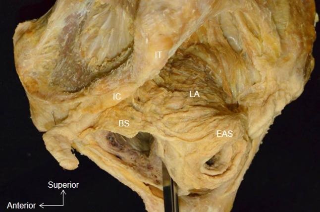
This clinical photograph displays a lateral aspect of a whole pelvis dissection, focusing on the musculature of the pelvic floor and perineum. Key anatomical landmarks are labeled: the bulbospongiosus (BS), external anal sphincter (EAS), ischiocavernosus (IC), ischial tuberosity (IT), and levator ani (LA). The image specifically demonstrates the anatomical continuity between the perineal and anal musculature, showing the muscle bundle of the bulbospongiosus (BS) attaching directly to the lateral surface of the circular external anal sphincter (EAS). Superior to these, the levator ani (LA) is visible, positioned deep to the bony ischial tuberosity (IT). The ischiocavernosus (IC) is situated anterior and superior to the bulbospongiosus. This dissection illustrates the spatial arrangement of the superficial and deep pelvic muscles, highlighting the integrated nature of the pelvic support structures and the sphincteric complex, which is critical for understanding pelvic floor functional anatomy and surgical approaches.
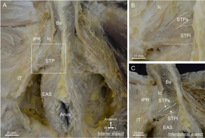
This composite of three clinical photographs displays a detailed anatomical dissection of the male perineum, focusing on the superficial perineal muscles and their spatial relationships. Panel A provides an inferior view, labeling the bulbospongiosus (Bs) centrally, the ischiocavernosus (Ic) running along the ischiopubic ramus (IPR), and the superficial transverse perineal muscle (STP) extending laterally toward the ischial tuberosity (IT). The external anal sphincter (EAS) is shown surrounding the anus. Panel B is a magnified inset of the rectangular area in A, demarcating the division of the STP into a superior bundle (STPs) and an inferior bundle (STPi). Panel C provides an inferolateral aspect with a slight pelvic tilt, illustrating the continuity between the muscle fibers of the bulbospongiosus and the external anal sphincter. The images emphasize the concentration of these muscle groups within the central region of the perineum, providing essential context for understanding pelvic floor anatomy, urogenital support, and surgical landmarks in the male pelvis.
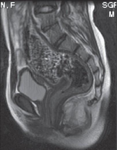
This diagnostic image is a T2-weighted sagittal MRI of the female pediatric pelvis, specifically focusing on the anorectal and urogenital anatomy. The image demonstrates a sagittal view of an 11-year-old patient with an anorectal malformation (ARM). Key anatomical landmarks include the urinary bladder, which is visible anteriorly as a hyperintense (bright), fluid-filled structure. Posterior to the bladder is the rectum, which appears distended with heterogeneous signal intensity, likely representing fecal material. The rectum descends toward the perineum, but its distal termination appears abnormal. The image serves to evaluate the path of an anovestibular fistula (AVF), looking for the communication between the terminal rectum and the vestibule, anterior to the normal anal position. Spinal vertebrae, the sacrum, and pelvic soft tissues provide spatial orientation. This imaging is clinically relevant for surgical planning in cases of delayed presentation of low-type anorectal malformations to assess the level of the rectal pouch and its relationship to the pelvic floor muscles.
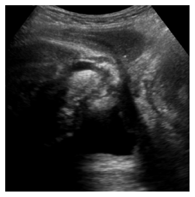
**Modality:** B-mode Ultrasound **Anatomical Region:** Male perineum, midsagittal plane. **Description:** This sonographic image visualizes the bulb of the penis (urethral bulb) and surrounding pelvic floor structures in a post-radical prostatectomy state. The ultrasound probe is positioned on the perineal skin (top of the image). Key landmarks include the echogenic urethral bulb located centrally and the anechoic (fluid-filled) urinary bladder situated in the deep field. **Characteristic Features:** - The image demonstrates the absence of the prostate gland between the bladder neck and the urethral bulb, consistent with surgical history. - The orientation shows the ventral aspect to the left and the dorsal aspect to the right. - The urethral bulb appears as a well-defined, heterogeneous, moderately echogenic structure. - Distal to the bladder, the surgical site of the vesicourethral anastomosis is visible. **Clinical Context:** Evaluation of post-prostatectomy anatomy in a patient presenting with stress urinary incontinence to assess the functional positioning and support of the bulbous urethra and pelvic floor muscles.
mkdir -p /tmp/workspace/gyn002 && ls /tmp/workspace/
cat << 'PYEOF' > /tmp/workspace/gyn002/build_pdf.py import requests import io from reportlab.lib.pagesizes import A4 from reportlab.lib.styles import getSampleStyleSheet, ParagraphStyle from reportlab.lib.units import cm from reportlab.lib import colors from reportlab.lib.enums import TA_CENTER, TA_LEFT, TA_JUSTIFY from reportlab.platypus import ( SimpleDocTemplate, Paragraph, Spacer, Table, TableStyle, HRFlowable, Image, KeepTogether ) from reportlab.platypus import PageBreak from PIL import Image as PILImage OUTPUT = "/tmp/workspace/gyn002/Gyn002_Female_AbdominoPelvic_Anatomy.pdf" doc = SimpleDocTemplate( OUTPUT, pagesize=A4, rightMargin=2*cm, leftMargin=2*cm, topMargin=2.5*cm, bottomMargin=2.5*cm, title="Gyn-002: Female Abdominal-Pelvic Anatomy", author="UHS Gynecology & Obstetrics" ) styles = getSampleStyleSheet() # Custom styles title_style = ParagraphStyle('TitleStyle', parent=styles['Title'], fontSize=20, textColor=colors.HexColor('#1a237e'), spaceAfter=6, spaceBefore=0, alignment=TA_CENTER, fontName='Helvetica-Bold') subtitle_style = ParagraphStyle('SubtitleStyle', parent=styles['Normal'], fontSize=12, textColor=colors.HexColor('#3949ab'), spaceAfter=4, alignment=TA_CENTER, fontName='Helvetica-Oblique') h1_style = ParagraphStyle('H1Style', parent=styles['Heading1'], fontSize=15, textColor=colors.HexColor('#1565c0'), spaceBefore=14, spaceAfter=6, fontName='Helvetica-Bold', borderPad=4, backColor=colors.HexColor('#e3f2fd'), leading=20) h2_style = ParagraphStyle('H2Style', parent=styles['Heading2'], fontSize=12, textColor=colors.HexColor('#1976d2'), spaceBefore=10, spaceAfter=4, fontName='Helvetica-Bold', leading=16) h3_style = ParagraphStyle('H3Style', parent=styles['Heading3'], fontSize=11, textColor=colors.HexColor('#0288d1'), spaceBefore=8, spaceAfter=3, fontName='Helvetica-BoldOblique') body_style = ParagraphStyle('BodyStyle', parent=styles['Normal'], fontSize=10, textColor=colors.HexColor('#212121'), spaceAfter=6, spaceBefore=2, leading=15, alignment=TA_JUSTIFY, fontName='Helvetica') bullet_style = ParagraphStyle('BulletStyle', parent=styles['Normal'], fontSize=10, textColor=colors.HexColor('#212121'), spaceAfter=4, spaceBefore=1, leading=14, leftIndent=20, bulletIndent=10, fontName='Helvetica', bulletFontName='Helvetica', bulletFontSize=10) caption_style = ParagraphStyle('CaptionStyle', parent=styles['Normal'], fontSize=9, textColor=colors.HexColor('#546e7a'), spaceAfter=8, alignment=TA_CENTER, fontName='Helvetica-Oblique') highlight_style = ParagraphStyle('HighlightStyle', parent=styles['Normal'], fontSize=10, textColor=colors.HexColor('#b71c1c'), spaceAfter=4, spaceBefore=4, leading=14, fontName='Helvetica-Bold', leftIndent=10, backColor=colors.HexColor('#ffebee'), borderPad=5) # ---- helper to download image ---- def get_image(url, width=14*cm): try: r = requests.get(url, timeout=15) r.raise_for_status() img_data = io.BytesIO(r.content) pil = PILImage.open(img_data) w_px, h_px = pil.size aspect = h_px / w_px img_data.seek(0) return Image(img_data, width=width, height=width*aspect) except Exception as e: print(f"Image load failed: {e}") return None story = [] # ===== COVER ===== story.append(Spacer(1, 1.5*cm)) story.append(Paragraph("GYN-002", ParagraphStyle('CodeStyle', parent=styles['Normal'], fontSize=28, textColor=colors.HexColor('#1a237e'), alignment=TA_CENTER, fontName='Helvetica-Bold', spaceAfter=4))) story.append(Paragraph("Female Abdominal-Pelvic Anatomy", title_style)) story.append(Paragraph("University of Health Sciences – Gynaecology & Obstetrics", subtitle_style)) story.append(Paragraph("Reference: Berek & Novak's Gynecology, 16th Ed.", subtitle_style)) story.append(HRFlowable(width="100%", thickness=2, color=colors.HexColor('#1565c0'), spaceAfter=10)) # Learning Objectives story.append(Paragraph("Learning Objectives", h1_style)) objectives = [ "Identify the layers and boundaries of the abdominal and pelvic walls.", "Describe the blood supply, lymphatic drainage, and innervation of the pelvis and perineum.", "Identify key anatomical landmarks in the abdomen, pelvis, and perineum.", "Identify anatomical structures that are vulnerable during common gynecological surgical procedures.", ] for obj in objectives: story.append(Paragraph(f"• {obj}", bullet_style)) story.append(Spacer(1, 0.5*cm)) story.append(HRFlowable(width="100%", thickness=1, color=colors.HexColor('#90caf9'), spaceAfter=10)) # ===== SECTION 1: ABDOMINAL WALL ===== story.append(Paragraph("1. The Abdominal Wall", h1_style)) story.append(Paragraph( "The anterior abdominal wall is bounded <b>superiorly</b> by the xiphoid process and costal cartilage of " "ribs 7–10 and <b>inferiorly</b> by the iliac crest, anterosuperior iliac spine, inguinal ligament, and " "pubic bone. It consists of skin, muscle, fascia, nerves, and vessels.", body_style)) # 1.1 Layers story.append(Paragraph("1.1 Layers of the Abdominal Wall", h2_style)) layers_data = [ ["Layer", "Details"], ["Skin", "May show striae and midline hyperpigmentation (linea nigra) in parous women."], ["Camper Fascia\n(Superficial Fascia - Fat Layer)", "Most superficial layer; contains variable fat. Continuous with superficial fatty layer of the perineum."], ["Scarpa Fascia\n(Superficial Fascia - Membranous Layer)", "Deeper membranous layer. Continuous with Colles fascia in perineum and fascia lata of the thigh."], ["Muscles + Aponeuroses", "5 muscles: External oblique, Internal oblique, Transversus abdominis, " "Rectus abdominis, Pyramidalis."], ["Transversalis Fascia", "Deep to the transversus abdominis; lines the abdominal cavity. " "Gives access to inferior epigastric vessels posteriorly."], ["Preperitoneal Fat", "Layer of extraperitoneal fat between transversalis fascia and peritoneum."], ["Peritoneum", "Innermost layer — parietal peritoneum lining the abdominal cavity."], ] t = Table(layers_data, colWidths=[5*cm, 12*cm]) t.setStyle(TableStyle([ ('BACKGROUND', (0,0), (-1,0), colors.HexColor('#1565c0')), ('TEXTCOLOR', (0,0), (-1,0), colors.white), ('FONTNAME', (0,0), (-1,0), 'Helvetica-Bold'), ('FONTSIZE', (0,0), (-1,-1), 9), ('ROWBACKGROUNDS', (0,1), (-1,-1), [colors.HexColor('#e3f2fd'), colors.white]), ('GRID', (0,0), (-1,-1), 0.5, colors.HexColor('#90caf9')), ('VALIGN', (0,0), (-1,-1), 'TOP'), ('PADDING', (0,0), (-1,-1), 5), ('WORDWRAP', (0,0), (-1,-1), True), ])) story.append(t) story.append(Spacer(1, 0.3*cm)) # 1.2 Muscles story.append(Paragraph("1.2 Muscles of the Anterolateral Abdominal Wall", h2_style)) muscles_data = [ ["Muscle", "Origin", "Insertion", "Action"], ["External Oblique", "Outer surfaces of ribs 5–12", "Anterior iliac crest, pubic tubercle, linea alba via aponeurosis", "Compresses abdomen; flexes & rotates vertebral column"], ["Internal Oblique", "Thoracolumbar fascia, anterior 2/3 iliac crest, lateral 2/3 inguinal ligament", "Inferior ribs 10–12; linea alba; pubic crest via conjoint tendon", "Compresses & supports abdominal viscera"], ["Transversus Abdominis", "Inner inferior 6 costal cartilages, thoracolumbar fascia, iliac crest", "Linea alba; pubic crest via conjoint tendon", "Compresses abdominal viscera"], ["Rectus Abdominis", "Superior pubic ramus & ligaments of symphysis pubis", "Xiphoid process & cartilage of ribs 5–7", "Tenses anterior abdominal wall; flexes trunk"], ["Pyramidalis", "Small triangular muscle anterior to rectus", "Linea alba", "Tenses linea alba; marks midline in surgery"], ] tm = Table(muscles_data, colWidths=[4*cm, 4*cm, 4*cm, 5*cm]) tm.setStyle(TableStyle([ ('BACKGROUND', (0,0), (-1,0), colors.HexColor('#0d47a1')), ('TEXTCOLOR', (0,0), (-1,0), colors.white), ('FONTNAME', (0,0), (-1,0), 'Helvetica-Bold'), ('FONTSIZE', (0,0), (-1,-1), 8), ('ROWBACKGROUNDS', (0,1), (-1,-1), [colors.HexColor('#e8eaf6'), colors.white]), ('GRID', (0,0), (-1,-1), 0.5, colors.HexColor('#9fa8da')), ('VALIGN', (0,0), (-1,-1), 'TOP'), ('PADDING', (0,0), (-1,-1), 4), ('WORDWRAP', (0,0), (-1,-1), True), ])) story.append(tm) story.append(Spacer(1, 0.3*cm)) # 1.3 Rectus Sheath story.append(Paragraph("1.3 Rectus Sheath & Arcuate Line", h2_style)) story.append(Paragraph( "The aponeuroses of external oblique, internal oblique, and transversus abdominis combine to form the " "<b>rectus sheath</b>, fusing medially at the <b>linea alba</b> and laterally at the <b>semilunar line</b>.", body_style)) story.append(Paragraph("• <b>Above arcuate line:</b> Aponeurosis of internal oblique splits into anterior and posterior lamellae, enclosing the rectus.", bullet_style)) story.append(Paragraph("• <b>Below arcuate line:</b> All three aponeuroses pass anterior to the rectus muscle; the posterior wall is only transversalis fascia.", bullet_style)) # 1.4 Blood Supply of Abdominal Wall story.append(Paragraph("1.4 Blood Supply of the Abdominal Wall", h2_style)) story.append(Paragraph("<b>Arterial supply:</b>", body_style)) story.append(Paragraph("• <b>Inferior epigastric artery</b> – branch of external iliac; runs superiorly in transversalis fascia, enters rectus sheath at arcuate line. <b>Clinical note:</b> vulnerable to damage by abdominal incisions or lateral laparoscopic ports.", bullet_style)) story.append(Paragraph("• <b>Superior epigastric artery</b> – terminal branch of internal thoracic; enters rectus sheath just below the 7th costal cartilage.", bullet_style)) story.append(Paragraph("• <b>Deep circumflex iliac artery</b> – branch of external iliac; runs between transversus abdominis and internal oblique along iliac crest.", bullet_style)) story.append(Paragraph("<b>Venous drainage:</b> Drains into the saphenous vein.", body_style)) story.append(Paragraph("<b>Lymphatic drainage:</b> Above umbilicus → axillary nodes; Below umbilicus → inguinal nodes; Subcutaneous tissues → lumbar chain.", body_style)) # 1.5 Nerves of the Abdominal Wall story.append(Paragraph("1.5 Innervation of the Abdominal Wall", h2_style)) story.append(Paragraph( "The abdominal wall is innervated by the anterior rami of thoracic and lumbar spinal nerves:", body_style)) story.append(Paragraph("• <b>T7–T11:</b> Intercostal nerves supply the upper and middle abdominal wall.", bullet_style)) story.append(Paragraph("• <b>T12 (subcostal nerve):</b> Supplies the area above the inguinal ligament.", bullet_style)) story.append(Paragraph("• <b>Iliohypogastric nerve (L1):</b> Supplies the suprapubic region.", bullet_style)) story.append(Paragraph("• <b>Ilioinguinal nerve (L1):</b> Supplies the inguinal region and inner thigh.", bullet_style)) story.append(Paragraph("• <b>Genitofemoral nerve (L1–L2):</b> Supplies the mons pubis, labia majora, and inner thigh.", bullet_style)) story.append(PageBreak()) # ===== SECTION 2: PELVIC ANATOMY ===== story.append(Paragraph("2. The Pelvis and Perineum", h1_style)) story.append(Paragraph("2.1 Boundaries of the True (Lesser) Pelvis", h2_style)) story.append(Paragraph( "The true pelvis is bounded superiorly by the pelvic inlet (linea terminalis) and inferiorly by the " "pelvic outlet. Its walls include the sacrum and coccyx posteriorly, the obturator foramen and " "ischiopubic rami laterally, and the pubic symphysis anteriorly.", body_style)) # Anatomical landmarks story.append(Paragraph("2.2 Key Anatomical Landmarks", h2_style)) landmarks = [ ("Linea terminalis (pelvic brim)", "Marks the boundary between the greater and true pelvis; runs from sacral promontory → arcuate line → iliopectineal line → pubic crest → pubic symphysis."), ("Ischial spine", "Prominent landmark on the lateral pelvic wall. Sacrospinous ligament attaches here. Important for pudendal nerve block."), ("Obturator foramen", "Formed by ischium and pubis. Covered by obturator membrane. Obturator nerve and vessels exit through the obturator canal."), ("Sacral promontory", "Anterior projecting part of the sacrum; key landmark in pelvic surgery and obstetrics."), ("Arcuate line (iliopectineal line)", "Forms part of the pelvic brim; separates the ilium from the pubis."), ("Coccyx", "Terminal portion of the vertebral column; attachment point for pelvic floor muscles."), ("Pubic symphysis", "Anterior midline joint; anterior boundary of the pelvic outlet."), ("Perineal body (central tendon)", "Dense fibromuscular mass between the anal canal and the vagina. Key anchor for multiple pelvic floor muscles."), ] for lm, desc in landmarks: story.append(Paragraph(f"<b>{lm}:</b> {desc}", bullet_style)) # 2.3 Pelvic Floor story.append(Paragraph("2.3 Pelvic Floor (Pelvic Diaphragm)", h2_style)) story.append(Paragraph( "The pelvic floor includes all structures closing the pelvic outlet from skin inferiorly to peritoneum " "superiorly. It is divided by the <b>pelvic diaphragm</b> into pelvic and perineal portions. " "The pelvic diaphragm is a hammock-like muscular sheet with a central hiatus for the urethra, vagina, and rectum.", body_style)) pf_data = [ ["Muscle", "Origin", "Insertion", "Function"], ["Levator Ani\n(Pubococcygeus,\nIliococcygeus,\nPuborectalis)", "Body of pubis, arcus tendineus levator ani, ischial spine", "Coccyx, anococcygeal raphe, perineal body", "Main pelvic floor support; resists intra-abdominal pressure; maintains urinary/fecal continence"], ["Coccygeus\n(Ischiococcygeus)", "Ischial spine", "Sacrum and coccyx", "Supports pelvic floor; flexes coccyx"], ] tpf = Table(pf_data, colWidths=[4*cm, 4*cm, 4*cm, 5*cm]) tpf.setStyle(TableStyle([ ('BACKGROUND', (0,0), (-1,0), colors.HexColor('#1b5e20')), ('TEXTCOLOR', (0,0), (-1,0), colors.white), ('FONTNAME', (0,0), (-1,0), 'Helvetica-Bold'), ('FONTSIZE', (0,0), (-1,-1), 8), ('ROWBACKGROUNDS', (0,1), (-1,-1), [colors.HexColor('#e8f5e9'), colors.white]), ('GRID', (0,0), (-1,-1), 0.5, colors.HexColor('#a5d6a7')), ('VALIGN', (0,0), (-1,-1), 'TOP'), ('PADDING', (0,0), (-1,-1), 4), ('WORDWRAP', (0,0), (-1,-1), True), ])) story.append(tpf) story.append(Spacer(1, 0.3*cm)) # Perineum story.append(Paragraph("2.4 The Perineum", h2_style)) story.append(Paragraph( "The perineum lies below the pelvic diaphragm and is bounded by the pelvic outlet. " "It is divided into two triangles by a line drawn between the ischial tuberosities:", body_style)) story.append(Paragraph("• <b>Urogenital triangle (anterior):</b> Contains external genitalia, urethra, vaginal orifice.", bullet_style)) story.append(Paragraph("• <b>Anal triangle (posterior):</b> Contains the anus, external anal sphincter, and ischiorectal fossa.", bullet_style)) story.append(Paragraph("Superficial Perineal Compartment Muscles:", h3_style)) sp_data = [ ["Muscle", "Origin", "Insertion", "Action"], ["Ischiocavernosus", "Ischial tuberosity", "Ischiopubic bone", "Compresses crura; lowers clitoris"], ["Bulbocavernosus\n(Bulbospongiosus)", "Perineal body", "Posterior aspect of clitoris (sling over dorsal vein)", "Compresses vestibular bulb and dorsal vein of clitoris"], ["Superficial Transverse Perineal", "Ischial tuberosity", "Central perineal tendon (perineal body)", "Fixes perineal body"], ] tsp = Table(sp_data, colWidths=[4.5*cm, 4*cm, 4*cm, 4.5*cm]) tsp.setStyle(TableStyle([ ('BACKGROUND', (0,0), (-1,0), colors.HexColor('#4a148c')), ('TEXTCOLOR', (0,0), (-1,0), colors.white), ('FONTNAME', (0,0), (-1,0), 'Helvetica-Bold'), ('FONTSIZE', (0,0), (-1,-1), 8), ('ROWBACKGROUNDS', (0,1), (-1,-1), [colors.HexColor('#f3e5f5'), colors.white]), ('GRID', (0,0), (-1,-1), 0.5, colors.HexColor('#ce93d8')), ('VALIGN', (0,0), (-1,-1), 'TOP'), ('PADDING', (0,0), (-1,-1), 4), ('WORDWRAP', (0,0), (-1,-1), True), ])) story.append(tsp) story.append(Spacer(1, 0.3*cm)) story.append(Paragraph("Deep Perineal Compartment:", h3_style)) story.append(Paragraph( "A fascial space between the perineal membrane (inferiorly) and a deep fascial layer (superiorly). " "Contains the <b>urogenital diaphragm</b> with the sphincter urethrae and deep transverse perineal muscle.", body_style)) story.append(Paragraph("The sphincter urethrae consists of:", body_style)) story.append(Paragraph("• <b>External urethral sphincter:</b> Surrounds the middle third of the urethra.", bullet_style)) story.append(Paragraph("• <b>Compressor urethrae:</b> Arcs across the ventral side of the urethra.", bullet_style)) story.append(Paragraph("• <b>Urethrovaginal sphincter:</b> Surrounds ventral urethra and terminates in lateral vaginal wall.", bullet_style)) story.append(PageBreak()) # ===== SECTION 3: BLOOD SUPPLY ===== story.append(Paragraph("3. Blood Supply of the Pelvis and Perineum", h1_style)) story.append(Paragraph("3.1 Arterial Supply", h2_style)) story.append(Paragraph( "The pelvic viscera are supplied mainly by branches of the <b>internal iliac artery (hypogastric artery)</b>, " "which divides into anterior and posterior divisions.", body_style)) art_data = [ ["Artery", "Branch of", "Supplies"], ["Uterine artery", "Anterior division, internal iliac", "Uterus; anastomoses with ovarian artery. Crosses OVER ureter ('water under the bridge')."], ["Ovarian artery", "Abdominal aorta (L1–L2)", "Ovary and fallopian tube; anastomoses with uterine artery."], ["Vaginal artery", "Anterior division, internal iliac", "Vagina and bladder base."], ["Internal pudendal artery", "Anterior division, internal iliac", "Perineum, vulva, clitoris, external anal sphincter."], ["Inferior epigastric artery", "External iliac artery", "Abdominal wall (rectus sheath)."], ["Superior epigastric artery", "Internal thoracic artery", "Upper rectus abdominis."], ["Obturator artery", "Anterior division, internal iliac (or external iliac)", "Medial thigh; anastomoses with inferior epigastric (corona mortis)."], ["Middle rectal artery", "Anterior division, internal iliac", "Rectum, cervix, vagina."], ["Superior vesical artery", "Anterior division, internal iliac", "Bladder and urachus."], ["Inferior vesical artery", "Anterior division, internal iliac", "Bladder base, vagina, ureter, vesical plexus."], ] ta = Table(art_data, colWidths=[4.5*cm, 5*cm, 7.5*cm]) ta.setStyle(TableStyle([ ('BACKGROUND', (0,0), (-1,0), colors.HexColor('#b71c1c')), ('TEXTCOLOR', (0,0), (-1,0), colors.white), ('FONTNAME', (0,0), (-1,0), 'Helvetica-Bold'), ('FONTSIZE', (0,0), (-1,-1), 8), ('ROWBACKGROUNDS', (0,1), (-1,-1), [colors.HexColor('#ffebee'), colors.white]), ('GRID', (0,0), (-1,-1), 0.5, colors.HexColor('#ef9a9a')), ('VALIGN', (0,0), (-1,-1), 'TOP'), ('PADDING', (0,0), (-1,-1), 4), ('WORDWRAP', (0,0), (-1,-1), True), ])) story.append(ta) story.append(Spacer(1, 0.3*cm)) # 3.2 Lymphatic Drainage story.append(Paragraph("3.2 Lymphatic Drainage of the Pelvis", h2_style)) lymph_data = [ ["Structure", "Primary Lymph Node Group", "Secondary Drainage"], ["Cervix", "External iliac, internal iliac, obturator", "Common iliac, para-aortic"], ["Uterine fundus", "Para-aortic (via ovarian vessels), superficial inguinal (via round ligament)", "—"], ["Ovary / Fallopian tube", "Para-aortic (L1–L2 level)", "Pelvic nodes"], ["Upper vagina", "Internal and external iliac", "Common iliac"], ["Lower vagina / Vulva", "Superficial inguinal, deep inguinal, femoral", "External iliac"], ["Bladder", "External and internal iliac, obturator", "Common iliac"], ["Rectum / Anal canal", "Internal iliac, inferior mesenteric", "Para-aortic"], ["Abdominal wall (above umbilicus)", "Axillary nodes", "—"], ["Abdominal wall (below umbilicus)", "Superficial inguinal nodes", "—"], ] tl = Table(lymph_data, colWidths=[5*cm, 6*cm, 6*cm]) tl.setStyle(TableStyle([ ('BACKGROUND', (0,0), (-1,0), colors.HexColor('#1a237e')), ('TEXTCOLOR', (0,0), (-1,0), colors.white), ('FONTNAME', (0,0), (-1,0), 'Helvetica-Bold'), ('FONTSIZE', (0,0), (-1,-1), 8), ('ROWBACKGROUNDS', (0,1), (-1,-1), [colors.HexColor('#e8eaf6'), colors.white]), ('GRID', (0,0), (-1,-1), 0.5, colors.HexColor('#9fa8da')), ('VALIGN', (0,0), (-1,-1), 'TOP'), ('PADDING', (0,0), (-1,-1), 4), ('WORDWRAP', (0,0), (-1,-1), True), ])) story.append(tl) story.append(Spacer(1, 0.3*cm)) # 3.3 Venous Drainage story.append(Paragraph("3.3 Venous Drainage", h2_style)) story.append(Paragraph( "Pelvic organs drain into the <b>internal iliac vein</b>, which joins the external iliac vein to form " "the <b>common iliac vein</b>. Both common iliacs join to form the <b>inferior vena cava</b>.", body_style)) story.append(Paragraph( "The ovarian veins are asymmetric: the <b>right ovarian vein</b> drains into the inferior vena cava " "and the <b>left ovarian vein</b> drains into the left renal vein.", body_style)) story.append(PageBreak()) # ===== SECTION 4: INNERVATION ===== story.append(Paragraph("4. Innervation of the Pelvis and Perineum", h1_style)) story.append(Paragraph("4.1 Autonomic Innervation", h2_style)) innervation_data = [ ["Nerve Plexus/Nerve", "Origin", "Type", "Structure Supplied"], ["Superior hypogastric plexus\n(presacral nerve)", "T10–L2 sympathetic fibers", "Sympathetic", "Descends over sacral promontory; supplies uterus, bladder, rectum"], ["Inferior hypogastric plexus\n(pelvic plexus)", "Superior hypogastric plexus + sacral parasympathetics (S2–S4)", "Mixed (sympathetic + parasympathetic)", "Uterus, cervix, vagina, bladder, rectum"], ["Pelvic splanchnic nerves\n(nervi erigentes)", "S2, S3, S4", "Parasympathetic", "Bladder, uterus, rectum, colon"], ["Uterovaginal plexus\n(Frankenhäuser plexus)", "Inferior hypogastric plexus", "Mixed", "Uterus, vagina, clitoris – responsible for uterine contractions"], ["Pudendal nerve", "S2, S3, S4 (somatic)", "Somatic", "Perineum, external genitalia, external anal/urethral sphincters"], ["Ilioinguinal nerve", "L1", "Somatic / sensory", "Mons pubis, labia majora, medial thigh"], ["Genitofemoral nerve", "L1–L2", "Mixed", "Labia majora, mons pubis, inner thigh"], ] ti = Table(innervation_data, colWidths=[4.5*cm, 3*cm, 3*cm, 6.5*cm]) ti.setStyle(TableStyle([ ('BACKGROUND', (0,0), (-1,0), colors.HexColor('#37474f')), ('TEXTCOLOR', (0,0), (-1,0), colors.white), ('FONTNAME', (0,0), (-1,0), 'Helvetica-Bold'), ('FONTSIZE', (0,0), (-1,-1), 8), ('ROWBACKGROUNDS', (0,1), (-1,-1), [colors.HexColor('#eceff1'), colors.white]), ('GRID', (0,0), (-1,-1), 0.5, colors.HexColor('#b0bec5')), ('VALIGN', (0,0), (-1,-1), 'TOP'), ('PADDING', (0,0), (-1,-1), 4), ('WORDWRAP', (0,0), (-1,-1), True), ])) story.append(ti) story.append(Spacer(1, 0.3*cm)) story.append(Paragraph("4.2 Pudendal Nerve – Key Details", h2_style)) story.append(Paragraph("The pudendal nerve (S2–S4) is the main nerve of the perineum.", body_style)) story.append(Paragraph("• Exits the greater sciatic foramen → crosses the ischial spine → re-enters through the lesser sciatic foramen → travels in the pudendal (Alcock's) canal.", bullet_style)) story.append(Paragraph("• Divides into: <b>inferior rectal nerve</b>, <b>perineal nerve</b>, and <b>dorsal nerve of the clitoris</b>.", bullet_style)) story.append(Paragraph("• <b>Clinical:</b> Blocked at the ischial spine for pudendal nerve block in obstetrics.", bullet_style)) story.append(Paragraph("4.3 Pain Transmission from Pelvic Organs", h2_style)) story.append(Paragraph("• <b>Uterus body</b> – T10–L1 (visceral afferents via superior hypogastric plexus).", bullet_style)) story.append(Paragraph("• <b>Cervix and upper vagina</b> – S2–S4 (via pelvic splanchnic nerves).", bullet_style)) story.append(Paragraph("• <b>Ovary</b> – T10 level (referred pain to umbilical region).", bullet_style)) story.append(Paragraph("• <b>Perineum</b> – S2–S4 via pudendal nerve (somatic sensation).", bullet_style)) story.append(PageBreak()) # ===== SECTION 5: VULNERABLE STRUCTURES ===== story.append(Paragraph("5. Structures Vulnerable During Gynecological Surgery", h1_style)) story.append(Paragraph( "Knowledge of vulnerable anatomical structures is essential for safe gynecological surgery. " "The ureter is the most commonly injured structure.", body_style)) vuln_data = [ ["Structure", "Site of Vulnerability", "Procedure at Risk"], ["Ureter", "1. At pelvic brim – crossed by ovarian vessels\n2. In broad ligament – lateral to uterosacral ligament\n3. At ischial spine – passes UNDER uterine artery (2–3 cm lateral to cervix)\n4. Crosses anterior upper vagina to enter bladder", "Hysterectomy (esp. laparoscopic), adnexectomy, salpingo-oophorectomy\n(~75% of ureteric injuries from gynaecological procedures)"], ["Inferior epigastric vessels", "At rectus lateral border / arcuate line", "Laparoscopic port placement, Pfannenstiel incision, retraction"], ["Uterine artery", "At level of internal cervical os, lateral to uterus", "Hysterectomy (ligation or avulsion)"], ["Pudendal nerve & vessels", "At ischial spine; in Alcock's canal", "Sacrospinous ligament fixation, vaginal vault suspension"], ["Obturator nerve & vessels", "In obturator foramen / obturator fossa", "Pelvic lymph node dissection, paravesical dissection"], ["Bladder", "Anterior to lower uterine segment, bladder dome", "Hysterectomy (esp. with previous CS), anterior colporrhaphy"], ["Rectum", "Posterior to vagina in rectovaginal space", "Posterior colporrhaphy, rectopexy, vaginal vault suspension"], ["Bowel (small intestine)", "In cul-de-sac; adhesions in endometriosis", "Laparoscopy, posterior dissection"], ["Internal iliac vein", "Posterior to internal iliac artery", "Radical hysterectomy, pelvic lymphadenectomy"], ["Femoral nerve", "Retroperitoneum at psoas major", "Excessive hip flexion with self-retaining retractors"], ] tv = Table(vuln_data, colWidths=[4*cm, 7*cm, 6*cm]) tv.setStyle(TableStyle([ ('BACKGROUND', (0,0), (-1,0), colors.HexColor('#bf360c')), ('TEXTCOLOR', (0,0), (-1,0), colors.white), ('FONTNAME', (0,0), (-1,0), 'Helvetica-Bold'), ('FONTSIZE', (0,0), (-1,-1), 8), ('ROWBACKGROUNDS', (0,1), (-1,-1), [colors.HexColor('#fbe9e7'), colors.white]), ('GRID', (0,0), (-1,-1), 0.5, colors.HexColor('#ffab91')), ('VALIGN', (0,0), (-1,-1), 'TOP'), ('PADDING', (0,0), (-1,-1), 4), ('WORDWRAP', (0,0), (-1,-1), True), ])) story.append(tv) story.append(Spacer(1, 0.3*cm)) # Ureteric injury box story.append(Paragraph( "CLINICAL PEARL – Ureteric Injury: ~75% of all iatrogenic ureteric injuries result from gynecological procedures. " "Laparoscopic hysterectomy has the highest rate; vaginal hysterectomy has the lowest. " "91% of injuries occur at the pelvic ureter. Distortions from endometriosis, fibroids, or adnexal masses " "increase risk. Always identify the ureter retroperitoneally before securing the infundibulopelvic ligament or uterine artery.", highlight_style)) story.append(PageBreak()) # ===== SECTION 6: LIGAMENTS & SPACES ===== story.append(Paragraph("6. Pelvic Ligaments and Spaces", h1_style)) story.append(Paragraph("6.1 Uterine Support Ligaments", h2_style)) lig_data = [ ["Ligament", "Composition", "Attachment", "Function / Clinical Relevance"], ["Cardinal ligament\n(Mackenrodt's)", "Dense endopelvic fascia, blood vessels, ureter", "Cervix & upper vagina → lateral pelvic wall", "Primary support for the cervix and upper vagina; contains uterine artery; ureter runs at its base."], ["Uterosacral ligament", "Smooth muscle, fibrous tissue, nerves", "Posterior cervix → sacrum (S2–S4)", "Provides posterior support; contains visceral afferent fibers from cervix; used in vault suspension."], ["Round ligament", "Smooth muscle and fibrous tissue", "Uterine cornua → inguinal canal → labia majora", "Maintains anteversion of uterus; passes through inguinal ring. No significant mechanical support."], ["Broad ligament", "Two peritoneal layers (mesentery)", "Lateral uterus → lateral pelvic wall", "Contains uterine artery, ovarian vessels, ureter, round ligament. Divided into mesosalpinx, mesovarium, mesometrium."], ["Infundibulopelvic ligament\n(suspensory lig. of ovary)", "Peritoneum + ovarian vessels + nerves", "Ovary → pelvic sidewall", "Contains ovarian artery and vein; must be ligated in oophorectomy (ureter close by)."], ["Ovarian ligament\n(proper lig. of ovary)", "Fibromuscular cord", "Ovary → uterine cornua", "Holds ovary near uterus."], ["Pubocervical fascia", "Endopelvic fascia", "Pubis → cervix / anterior vagina", "Supports anterior vaginal wall and bladder base."], ["Rectovaginal fascia\n(Denonvilliers fascia)", "Endopelvic fascia", "Posterior vaginal wall → perineal body → rectum", "Separates vagina from rectum; deficiency leads to rectocele."], ] tlig = Table(lig_data, colWidths=[4.5*cm, 3.5*cm, 3.5*cm, 5.5*cm]) tlig.setStyle(TableStyle([ ('BACKGROUND', (0,0), (-1,0), colors.HexColor('#006064')), ('TEXTCOLOR', (0,0), (-1,0), colors.white), ('FONTNAME', (0,0), (-1,0), 'Helvetica-Bold'), ('FONTSIZE', (0,0), (-1,-1), 8), ('ROWBACKGROUNDS', (0,1), (-1,-1), [colors.HexColor('#e0f7fa'), colors.white]), ('GRID', (0,0), (-1,-1), 0.5, colors.HexColor('#80deea')), ('VALIGN', (0,0), (-1,-1), 'TOP'), ('PADDING', (0,0), (-1,-1), 4), ('WORDWRAP', (0,0), (-1,-1), True), ])) story.append(tlig) story.append(Spacer(1, 0.3*cm)) story.append(Paragraph("6.2 Pelvic Spaces (Potential Spaces)", h2_style)) spaces_data = [ ["Space", "Boundaries", "Clinical Significance"], ["Space of Retzius\n(Retropubic space)", "Anterior: pubis\nPosterior: bladder\nInferior: puboprostatic ligaments", "Entered in Burch colposuspension; contains venous plexus of Santorini."], ["Paravesical space", "Medial: bladder\nLateral: obturator internus\nPosterior: cardinal ligament", "Developed to identify ureter and uterine artery in radical hysterectomy."], ["Pararectal space", "Medial: rectum\nLateral: internal iliac vessels\nFloor: sacrum", "Developed to identify ureter and define parametrium in radical hysterectomy."], ["Rectovaginal space", "Anterior: posterior vaginal wall\nPosterior: rectum", "Developed in posterior colporrhaphy and rectopexy; site of rectocele."], ["Vesicovaginal space", "Anterior: bladder\nPosterior: anterior vaginal wall", "Developed during anterior colporrhaphy, hysterectomy; avascular plane."], ["Pouch of Douglas\n(Rectouterine pouch)", "Between uterus anteriorly and rectum posteriorly", "Most dependent part of peritoneal cavity; site for culdocentesis; obliterated in endometriosis."], ["Vesicouterine pouch", "Between bladder and uterus anteriorly", "Opened during cesarean section and hysterectomy."], ] tsp2 = Table(spaces_data, colWidths=[4.5*cm, 5.5*cm, 7*cm]) tsp2.setStyle(TableStyle([ ('BACKGROUND', (0,0), (-1,0), colors.HexColor('#4a148c')), ('TEXTCOLOR', (0,0), (-1,0), colors.white), ('FONTNAME', (0,0), (-1,0), 'Helvetica-Bold'), ('FONTSIZE', (0,0), (-1,-1), 8), ('ROWBACKGROUNDS', (0,1), (-1,-1), [colors.HexColor('#f3e5f5'), colors.white]), ('GRID', (0,0), (-1,-1), 0.5, colors.HexColor('#ce93d8')), ('VALIGN', (0,0), (-1,-1), 'TOP'), ('PADDING', (0,0), (-1,-1), 4), ('WORDWRAP', (0,0), (-1,-1), True), ])) story.append(tsp2) story.append(PageBreak()) # ===== SECTION 7: IMAGES ===== story.append(Paragraph("7. Anatomical Illustrations", h1_style)) image_urls = [ ("https://cdn.orris.care/cdss_images/pmc_clinical_VQA_7e4c7a04e8fc80f952e7355e4b097e1697ad808548aa38f2aa1feb69cf946bd2.jpg", "Abdominal wall muscle layers showing external oblique (EOM), internal oblique (IOM), and transversus abdominis (TAM) with nerve block anatomy"), ("https://cdn.orris.care/cdss_images/pmc_clinical_VQA_a024a4d74a1cc3adb15063dee6f4af367676b9468a401ea6bb9d99bd6809fa35.jpg", "Female vulvar anatomy and lymphatic drainage showing inguinal, external iliac, internal iliac, common iliac, and para-aortic nodes"), ("https://cdn.orris.care/cdss_images/pmc_clinical_VQA_b187bdbe61c9ba9366c0630ec49b50d83b6f0c64f1bcd4dee0410dc70280d766.jpg", "Pelvic lymph node compartments – mesorectal (red), internal iliac (blue), obturator (green), and external iliac (yellow) nodal stations"), ("https://cdn.orris.care/cdss_images/pmc_clinical_VQA_b1c2cbe37075d1d8833c60b83f2a4652caf1e9366eb92e534f30e0c9f8f3145e.jpg", "Lateral pelvic dissection: bulbospongiosus (BS), external anal sphincter (EAS), ischiocavernosus (IC), ischial tuberosity (IT), levator ani (LA)"), ] for idx, (url, caption) in enumerate(image_urls): img = get_image(url, width=13*cm) if img: story.append(KeepTogether([ img, Paragraph(f"<b>Figure {idx+1}:</b> {caption}", caption_style), Spacer(1, 0.3*cm), ])) # ===== SECTION 8: HIGH YIELD SUMMARY ===== story.append(PageBreak()) story.append(Paragraph("8. High-Yield Summary for UHS Exams", h1_style)) summary_points = [ "The abdominal wall has 7 layers: skin → Camper fascia → Scarpa fascia → muscles/aponeuroses → transversalis fascia → extraperitoneal fat → peritoneum.", "The arcuate line marks where the posterior rectus sheath ends (all aponeuroses pass anterior below this line).", "The inferior epigastric artery is vulnerable during lateral laparoscopic port placement and rectus-transecting incisions.", "The pelvic diaphragm (levator ani + coccygeus) is the hammock of the true pelvis with a hiatus for urethra, vagina, rectum.", "Uterine artery crosses OVER the ureter ('water under the bridge') at the level of the internal os, 2–3 cm lateral to the cervix.", "~75% of iatrogenic ureteral injuries are from gynecological procedures; laparoscopic hysterectomy has the highest rate.", "The pudendal nerve (S2–S4) is the somatic nerve of the perineum; blocked at the ischial spine.", "Cardinal and uterosacral ligaments are the PRIMARY supports of the cervix and upper vagina.", "Pouch of Douglas is the most dependent part of the peritoneal cavity; obliterated in endometriosis.", "Ovarian veins are asymmetric: right drains into IVC; left drains into left renal vein.", "The round ligament maintains uterine anteversion; passes through the inguinal canal but provides minimal structural support.", "Lymphatic drainage: cervix → obturator/iliac nodes; ovary → para-aortic (L1–L2); vulva → inguinal nodes.", "Frankenhäuser plexus (uterovaginal plexus) mediates uterine contractions and cervical sensation (paracervical block target).", "The perineal body is the central anchor for pelvic floor and perineal muscles; its disruption leads to pelvic organ prolapse.", "Left lateral traction on rectus during Pfannenstiel or McBurney incisions can injure the inferior epigastric vessels.", ] for i, pt in enumerate(summary_points): story.append(Paragraph(f"<b>{i+1}.</b> {pt}", bullet_style)) # Footer story.append(Spacer(1, 1*cm)) story.append(HRFlowable(width="100%", thickness=1, color=colors.HexColor('#1565c0'), spaceAfter=6)) story.append(Paragraph( "Reference: Berek & Novak's Gynecology, 16th Edition | Prepared for UHS Gyn-002 Curriculum", caption_style)) doc.build(story) print(f"PDF created: {OUTPUT}") PYEOF python3 /tmp/workspace/gyn002/build_pdf.py
Download PDF
Gyn002 Female AbdominoPelvic Anatomy
PDF Document · PDF
Gyn-002: Female Abdominal-Pelvic Anatomy
1. Layers and Boundaries of the Abdominal Wall
- Superiorly: Xiphoid process + costal cartilage of ribs 7–10
- Inferiorly: Iliac crest, anterosuperior iliac spine, inguinal ligament, pubic bone
| Layer | Key Details |
|---|---|
| Skin | Striae / linea nigra in parous women |
| Camper fascia | Fatty layer; continuous with perineal fat |
| Scarpa fascia | Membranous layer; continuous with Colles fascia in perineum |
| Muscles + aponeuroses | External oblique, internal oblique, transversus abdominis, rectus abdominis, pyramidalis |
| Transversalis fascia | Lines the abdominal cavity; gives posterior access to inferior epigastric vessels |
| Extraperitoneal fat | Preperitoneal layer |
| Peritoneum | Innermost parietal layer |
- Above arcuate line: Internal oblique aponeurosis splits; rectus is enclosed front and back
- Below arcuate line: All three aponeuroses pass anterior to rectus; posterior wall = transversalis fascia only
2. Blood Supply of the Pelvis and Perineum
- Inferior epigastric a. (external iliac) - enters rectus sheath at arcuate line; vulnerable to lateral laparoscopic ports
- Superior epigastric a. (internal thoracic) - enters sheath below 7th costal cartilage
- Deep circumflex iliac a. (external iliac) - runs between transversus and internal oblique
- Uterine artery → supplies uterus; crosses OVER ureter ("water under the bridge")
- Ovarian artery → from aorta at L1-L2; anastomoses with uterine artery
- Internal pudendal artery → perineum, vulva, clitoris
- Vaginal, vesical, middle rectal arteries → respective pelvic organs
- Cervix → obturator / external + internal iliac nodes
- Uterine fundus → para-aortic + superficial inguinal (round ligament)
- Ovary → para-aortic nodes (L1–L2 level)
- Vulva / lower vagina → superficial inguinal nodes
3. Innervation of the Pelvis and Perineum
| Nerve | Origin | Supplies |
|---|---|---|
| Superior hypogastric plexus | T10–L2 sympathetic | Uterus, bladder, rectum (over sacral promontory) |
| Pelvic splanchnic nerves (S2–S4) | Parasympathetic | Bladder, uterus, colon |
| Frankenhäuser plexus | Inferior hypogastric | Uterus, cervix, vagina - mediates contractions |
| Pudendal nerve (S2–S4) | Somatic | Perineum, external sphincters, clitoris |
| Ilioinguinal (L1) | Somatic/sensory | Mons pubis, labia majora |
4. Key Anatomical Landmarks
- Ischial spine - attachment of sacrospinous ligament; landmark for pudendal nerve block
- Arcuate line - where posterior rectus sheath ends
- Perineal body - central anchor for pelvic floor muscles
- Pouch of Douglas - most dependent part of peritoneal cavity (obliterated in endometriosis)
- Linea terminalis - divides false from true pelvis
5. Structures Vulnerable in Gynecological Surgery
| Structure | Site of Risk | Procedure |
|---|---|---|
| Ureter | Under uterine artery; 2–3 cm lateral to cervix | Hysterectomy (75% of ureteral injuries are gynecological) |
| Inferior epigastric a. | At arcuate line / lateral rectus border | Laparoscopic port placement |
| Pudendal nerve | At ischial spine / Alcock's canal | Sacrospinous vault suspension |
| Bladder | Anterior to lower uterine segment | Hysterectomy, anterior repair |
| Obturator nerve | Obturator fossa | Pelvic lymph node dissection |
| Rectum | Rectovaginal space | Posterior colporrhaphy |
High-Yield Mnemonics & Pearls
- "Water under the bridge" - Uterine artery (bridge) crosses OVER the ureter (water) at the level of the internal cervical os
- Arcuate line - below this, the posterior rectus sheath is absent (only transversalis fascia)
- Pudendal nerve = S2, S3, S4 = "keeps the pelvic floor off the floor"
- Cardinal + uterosacral ligaments = PRIMARY cervical support
- Laparoscopic hysterectomy has the HIGHEST rate of ureteral injury; vaginal hysterectomy the LOWEST