
Give me the answer for mucocele for ent subject of mbbs
frontal sinus mucocele CT scan ENT
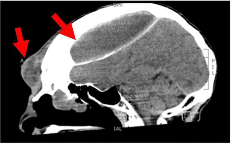
This diagnostic image is a non-contrast sagittal CT scan of the head and facial bones illustrating a large frontal mucocele with significant intra- and extracranial extension. Red arrows highlight two key components of the pathology: a localized bulging mass in the subcutaneous forehead tissue and a massive, biconvex-shaped intracranial collection. The frontal sinus appears opacified and expanded, with clear evidence of bony erosion and destruction of both the anterior and posterior cortical tables of the frontal bone. The intracranial portion of the mucocele occupies a substantial volume of the anterior cranial vault, exerting a significant mass effect on the underlying frontal lobes, characterized by downward displacement and compression of the brain parenchyma. The density of the collection is heterogeneous, suggesting inspissated mucus or secondary infection (Pott's puffy tumor clinical presentation). This scan demonstrates the potential for paranasal sinus pathology to result in advanced cranial complications and structural remodeling.
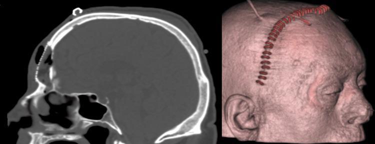
This composite clinical image displays a postoperative evaluation of a patient who underwent surgical management for a frontal sinus mucocele. Left: A sagittal non-contrast CT scan of the head demonstrates the reconstruction of the frontal sinus anterior wall. A high-density, linear, beaded structure representing a titanium mesh is visible, spanning the osseous defect in the frontal bone to restore the cranial contour. The underlying sinus appears obliterated with soft-tissue density material, consistent with the reported fat graft and fascia lata packing. Right: A 3D surface rendering of the patient’s head in profile shows the external cosmetic results and surgical site. A distinct, reddish-brown corrugated line follows a coronal trajectory across the scalp, representing a surgical incision site closed with metallic staples or sutures. The reconstruction provides a smooth forehead contour without significant depression, demonstrating the efficacy of the cranioplasty. This image is an educational resource for neurosurgery and otolaryngology, illustrating surgical reconstruction of the frontal sinus following bony erosion.
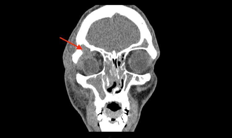
This diagnostic image is a coronal Computed Tomography (CT) scan of the head, presented in a soft tissue window. It illustrates a frontal sinus mucocele with orbital extension. Key features include a homogenous soft tissue density mass originating from the opacified right frontal sinus and herniating through a bony defect in the superolateral orbital roof, indicated by a red arrow. This mass causes inferior and medial displacement of the right globe and extraocular muscles. Additionally, there is evidence of diffuse periorbital soft tissue swelling and complete opacification of the bilateral ethmoid and maxillary sinuses. The image serves as a clinical example of sinus pathology complications, specifically highlighting the erosion of the orbital wall and the resulting mechanical effect on orbital contents. It is relevant for medical education regarding otorhinolaryngology, ophthalmology, and radiology, demonstrating the diagnostic utility of CT in assessing chronic sinusitis and its extrasinus manifestations.
Mucocele - ENT (MBBS)
Definition
- Scott-Brown's Otorhinolaryngology Head & Neck Surgery, p. 1155
- Cummings Otolaryngology Head and Neck Surgery, p. 919
Sites of Involvement (in Order of Frequency)
| Rank | Sinus | Approximate Frequency |
|---|---|---|
| 1st | Frontal | ~43-45% |
| 2nd | Ethmoid (including fronto-ethmoid) | ~14-28% |
| 3rd | Sphenoid | ~10-18% |
| 4th | Maxillary | ~6-25% |
Frontal sinus is most common because of its complex, narrow drainage pathway (frontonasal duct) which is easily obstructed.
Aetiology / Causes
- Previous sinus surgery - Most common cause (66-86%): FESS, Caldwell-Luc, Lynch-Howarth fronto-ethmoidectomy
- Trauma - 10-14% of cases; mean interval of 9 years after injury
- Spontaneous - 15-17% (due to chronic inflammation/polyposis causing ostial obstruction)
- Benign tumours - e.g., osteoma causing frontal recess obstruction
- Malignancy - rare (1-5%)
- Fibrous dysplasia, Paget disease - rare
The Lynch-Howarth operation is a particularly high-risk procedure: 78% of post-surgical mucoceles were associated with this and/or Caldwell-Luc operations vs only 1.5% after endonasal surgery.
Pathogenesis
- Obstruction of sinus outflow tract - prevents mucus drainage
- Inflammatory process within the sinus - drives mucus secretion
- IL-1 (Interleukin-1)
- Tumour Necrosis Factor (TNF)
- Prostaglandins
Clinical Features
General Features
- Slowly expanding, painless swelling (months to years)
- Visible mass over forehead, medial canthus, or gingivobuccal sulcus/cheek
Ophthalmic Features (Most Common)
- Periorbital swelling and pain
- Proptosis (exophthalmos) - due to orbital wall erosion
- Diplopia - displacement of orbital contents
- Limited ocular motility
- Visual disturbance
- Optic neuropathy - in up to 18% (especially from posterior ethmoid/sphenoid mucoceles)
- Epiphora - if dacryocele is present
Rhinological Features
- Nasal obstruction
- Headache
- Nasal discharge (if infected - pyocele)
Neurological Features (from intracranial extension)
- Headache, meningism (if eroded through posterior table of frontal sinus)
- Rare: CSF leak, meningitis
By Sinus
| Sinus | Key Symptom |
|---|---|
| Frontal | Forehead swelling, proptosis, headache |
| Ethmoid | Medial canthal swelling, proptosis |
| Maxillary | Cheek swelling, inferior orbital displacement |
| Sphenoid | Headache, visual loss, optic neuropathy, diplopia |
Radiology / Investigations
CT Scan (First Choice)
- Homogeneous, isodense soft-tissue lesion within an expanded sinus
- Bony remodelling (expansion, thinning, or erosion) of sinus walls
- No contrast enhancement in simple mucocele
- Contrast enhancement = pyocele (infected mucocele)
- Bony defect of lamina papyracea in fronto-ethmoid mucoceles
- Globe displacement (lateral and/or inferior) on axial images
- Erosion of posterior table of frontal sinus in advanced cases
MRI Scan
- Excellent for assessing intracranial or intraorbital extension
- Signal varies based on mucocele contents (protein concentration):
- T1: variable (low to high)
- T2: typically hyperintense
- Distinguishes dural involvement/intracranial extension
Differential Diagnosis
- Benign or malignant tumours of paranasal sinuses
- Allergic fungal sinusitis / other fungal disease
- Cholesterol granuloma (rare)
- Encephalocele (if posterior table is eroded)
- Dermoid/epidermoid cyst
Treatment
Endoscopic (Preferred/First-Line)
- Endoscopic marsupialization (nasalization) - wide opening of the sac into the nasal cavity
- Achieves permanent drainage without complete removal of the sac
- Recurrence rates: 0-2.5%
- For simple frontal/ethmoid mucoceles: Draf IIa procedure (3.6% restenosis)
- Draf IIb: 23% restenosis (higher risk when extensive drilling performed)
- Modified Endoscopic Lothrop Procedure (MELP) - for complex frontal sinus mucoceles, especially where there is loss of lateral support in frontal recess
Open Approaches (When Endoscopic Not Sufficient)
- Combined endoscopic + external approach for laterally located mucoceles
- Frontal sinus obliteration - used in specific cases (e.g., with significant posterior table erosion)
- External fronto-ethmoidectomy (now rarely used as primary approach)
Key Points on Management
- Pre-op CT + MRI mandatory to assess extent
- Cosmetic deformities (bony expansion) often resolve with time as bony remodelling occurs after drainage
- Infected mucocele (pyocele) requires antibiotics plus surgical drainage
Pyocele (Mucopyocele)
- An infected mucocele
- Contains pus + mucus
- CT shows contrast enhancement (distinguishes from simple mucocele)
- More urgent management - risk of orbital cellulitis/abscess, meningitis, intracranial abscess
- Treatment: IV antibiotics + immediate surgical drainage/marsupialization
Summary for Exam (Quick Recall)
| Feature | Key Point |
|---|---|
| Definition | Epithelium-lined, mucus-filled, expanding sinus cyst |
| Most common site | Frontal sinus |
| Most common cause | Previous sinus surgery (FESS/Lynch-Howarth) |
| Pathogenesis | Obstruction + inflammation + IL-1/TNF bone erosion |
| Most common symptoms | Proptosis, periorbital swelling, pain, diplopia |
| CT appearance | Homogeneous isodense, expanded sinus, bony remodelling; NO contrast enhancement |
| Infected = pyocele | CT shows contrast enhancement |
| Treatment | Endoscopic marsupialization (wide drainage into nose) |
| Surgery to avoid restenosis | Preserve mucosa, Draf IIa > Draf IIb |
Give answers such that to write in exam to fetch good marks
MUCOCELE OF PARANASAL SINUSES
Definition
- When infected, it is called a Pyocele (Mucopyocele)
Sites of Occurrence
- Frontal sinus - Most common (43-45%)
- Ethmoid sinus - Second most common
- Sphenoid sinus
- Maxillary sinus - Least common
Reason frontal is most common: Its drainage pathway (frontonasal duct) is narrow and tortuous, making it easily obstructed.
Aetiology / Predisposing Factors
| Cause | Notes |
|---|---|
| Previous sinus surgery | Most common (66-86%); FESS, Caldwell-Luc, Lynch-Howarth operation |
| Trauma | 10-14%; mucocele may develop 9 years after injury |
| Spontaneous | 15-17%; due to chronic rhinosinusitis/polyposis |
| Benign tumour | e.g., Osteoma obstructing frontal recess |
| Malignancy | Rare (1-5%) |
| Fibrous dysplasia / Paget disease | Rare |
Pathogenesis (Important for viva/theory)
- Obstruction of sinus ostium - mucus cannot drain
- Chronic inflammation within the sinus
- IL-1 (Interleukin-1)
- Tumour Necrosis Factor (TNF)
- Prostaglandins
Clinical Features
A. General
- Slow, progressive, painless swelling over months to years
- Swelling visible over forehead (frontal), medial canthus (ethmoid), cheek/gingivobuccal sulcus (maxillary)
- Becomes painful if infected (pyocele)
B. Ophthalmic Features (Most Common - Examiner Favourite)
- Proptosis (exophthalmos) - hallmark feature due to orbital wall erosion
- Diplopia - from orbital contents displacement
- Periorbital swelling and pain
- Limited ocular movements
- Visual loss / Optic neuropathy - seen in up to 18% (sphenoid/posterior ethmoid mucoceles)
- Epiphora - if nasolacrimal duct involved (dacryocele)
C. Rhinological Features
- Nasal obstruction
- Chronic headache
- Purulent nasal discharge (if pyocele)
D. Neurological Features (if intracranial extension)
- Severe headache
- Meningism
- Rarely: meningitis, intracranial abscess
By Sinus - Specific Symptoms
| Sinus | Characteristic Presentation |
|---|---|
| Frontal | Forehead swelling, proptosis, headache |
| Ethmoid | Medial canthal swelling, proptosis, diplopia |
| Sphenoid | Headache, visual loss, diplopia, optic neuropathy |
| Maxillary | Cheek swelling, downward globe displacement |
Investigations
1. CT Scan (Investigation of Choice)
- Homogeneous isodense soft-tissue lesion within an expanded sinus
- Bony remodelling (expansion/thinning/erosion) of sinus walls
- No contrast enhancement in simple mucocele
- Contrast enhancement = PYOCELE (infected) ← exam favourite fact
- Bony defect of lamina papyracea in fronto-ethmoid mucoceles
- Erosion of posterior table of frontal sinus in advanced cases
2. MRI Scan
- Best for assessing intracranial and intraorbital extension
- Signal varies with mucus protein content:
- T2: typically hyperintense
- T1: variable
- Distinguishes dural involvement and brain compression
Differential Diagnosis
- Benign/malignant tumours of paranasal sinuses
- Allergic fungal sinusitis
- Cholesterol granuloma
- Encephalocele (if posterior table eroded)
- Dermoid/epidermoid cyst
Treatment (Most Important for Exam)
Principle
1. Endoscopic Surgery (Treatment of Choice / First Line)
- Endoscopic Marsupialization - wide opening of sac into nasal cavity
- Recurrence rate: 0-2.5% (excellent results)
- Draf IIa procedure for frontal mucoceles (3.6% restenosis)
- Modified Endoscopic Lothrop Procedure (MELP) - for complex/recurrent frontal sinus mucoceles
2. Combined (Endoscopic + External) Approach
- For laterally placed or large mucoceles where endoscope alone is insufficient
3. Open Surgery (Rarely needed now)
- External fronto-ethmoidectomy (Lynch-Howarth) - largely abandoned as primary treatment
- Frontal sinus obliteration in selected cases
Pyocele Management
- IV antibiotics + immediate surgical drainage and marsupialization
- Urgent due to risk of: orbital cellulitis, intracranial abscess, meningitis
Pyocele vs Mucocele - Quick Comparison (Viva Point)
| Feature | Mucocele | Pyocele |
|---|---|---|
| Contents | Sterile mucus | Infected mucus (pus) |
| Pain | Absent/mild | Present, severe |
| CT enhancement | Absent | Present (ring enhancement) |
| Urgency | Elective surgery | Urgent drainage + antibiotics |
Key Exam Points to Remember
"FEMS" - Frequency of sinus involvement: Frontal > Ethmoid > Maxillary > Sphenoid (Note: some texts put sphenoid before maxillary - just remember frontal is #1)
Most common cause = Previous sinus surgery (especially Lynch-Howarth operation)
Most common symptom = Proptosis/ophthalmologic complaints
Investigation of choice = CT scan of PNS with contrast
Treatment of choice = Endoscopic marsupialization
CT with NO enhancement = Mucocele; CT WITH enhancement = Pyocele
Bone erosion is caused by IL-1, TNF, Prostaglandins
Lined by pseudostratified/low-columnar respiratory epithelium (NOT a separate membrane like a cyst)
Model Exam Answer Template
Explain each line to understand the topic
paranasal sinus anatomy diagram frontonasal duct
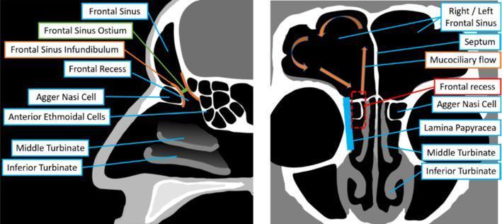
This medical anatomical diagram illustrates the paranasal sinus anatomy, specifically focusing on the frontal sinus, via two comparative cross-sectional views: a sagittal plane (left) and a coronal plane (right). The sagittal view highlights the vertical relationship between the frontal sinus and the nasal cavity, identifying key drainage structures including the frontal sinus ostium, frontal sinus infundibulum, and the frontal recess. It further labels the agger nasi cell, anterior ethmoidal cells, and the middle and inferior turbinates. The coronal view demonstrates bilateral symmetry, showing the right and left frontal sinuses divided by a midline septum. This view identifies the frontal recess within a dashed red box and illustrates mucociliary flow patterns with orange arrows showing drainage from the superior sinus toward the infundibulum. Key lateral boundaries like the lamina papyracea are labeled alongside the turbinates and agger nasi cells. This educational graphic is designed to aid in understanding the complex drainage pathways of the frontal sinus, essential for functional endoscopic sinus surgery (FESS) and rhinological diagnosis.
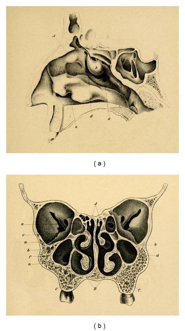
This historical anatomical diagram by Emil Zuckerkandl (1882) illustrates the paranasal sinuses and nasal anatomy in two planes. Image (a) is a sagittal section of the nasal cavity, detailing the complex arrangement of the superior, middle, and inferior turbinates (conchae) along the lateral wall. It visualizes the frontal sinus superiorly, the sphenoid sinus posteriorly, and the relationship between the nasal cavity and the hard palate. Image (b) presents a frontal (coronal) section, showcasing the bilateral symmetry of the mid-facial anatomy. Key features include the central nasal septum, the convoluted turbinates projecting into the nasal passages, and the expansive maxillary sinuses situated laterally within the maxilla bones. Small ethmoid air cells are visible medially to the orbits and superior to the maxillary sinuses. This anatomical illustration serves as a foundational reference for otorhinolaryngology and neurosurgical approaches, particularly regarding the development of transsphenoidal surgery for pituitary lesions.
MUCOCELE - LINE BY LINE EXPLANATION
🔷 DEFINITION (Explained)
Line: "A mucocele is an epithelium-lined, mucus-filled sac"
- Epithelium-lined = The inner wall of the balloon is made of the same kind of cells that normally line your nose and sinuses (called respiratory epithelium - pseudostratified columnar cells). These cells normally produce mucus.
- Mucus-filled = The balloon fills up with the mucus that these cells keep producing.
- Sac = It is a closed pouch - like a bag. The mucus goes in but cannot come out.
Simple picture: Imagine a plastic bag that keeps getting filled with water but has no outlet. It slowly fills up and expands. That is a mucocele.
Line: "...that completely fills and slowly expands a paranasal sinus"
Line: "...formed due to obstruction of the sinus outflow tract"
Line: "It is lined by pseudostratified or low-columnar epithelium, NOT a separate membrane like a cyst"
| Feature | Mucocele | Cyst |
|---|---|---|
| Lining | Respiratory epithelium (same as sinus wall - continuous) | Has a distinct separate membrane of its own |
| Origin | Sinus wall stretches and expands | Separate structure forms |
Line: "When infected, it is called a Pyocele (Mucopyocele)"
- The mucus inside the mucocele is normally sterile (no bacteria).
- If bacteria enter, the mucus turns to pus (infected mucus).
- Pyo = pus in Latin. So pyocele = pus-filled sac.
- This is more dangerous - it can burst and spread infection to the orbit or brain.
🔷 SITES (Explained)
Line: "Frontal sinus - Most common site"
- Very narrow
- Surrounded by multiple small cells (agger nasi, ethmoid cells)
- Easily blocked by swelling, scar tissue, or surgery
Line: "Frontal > Ethmoid > Sphenoid > Maxillary (in descending frequency)"
- Ethmoid sinus - second most common. Many small cells with multiple tiny openings, easily blocked.
- Sphenoid sinus - deep behind eyes; when blocked, causes severe symptoms like vision loss.
- Maxillary sinus - has a wide opening (antrum); harder to completely block, so mucocele is least common here.
🔷 AETIOLOGY (Explained)
Line: "Previous sinus surgery is the most common cause (66-86%)"
- Scar tissue forms around the narrow frontal drainage pathway
- The scarring slowly closes the drain over months to years (average 1-19 years after surgery)
- The sinus then becomes blocked and a mucocele forms
Line: "Trauma causes 10-14% of mucoceles; mean interval of 9 years"
Line: "Spontaneous - 15-17%; due to chronic rhinosinusitis/polyposis"
- Chronic sinusitis = long-standing sinus infection/inflammation
- Nasal polyps (small grape-like growths inside the nose) can block sinus openings
- Even without surgery or injury, these can slowly obstruct the sinus outflow and cause a mucocele
Line: "Benign tumour e.g. osteoma obstructing frontal recess"
🔷 PATHOGENESIS (Explained)
Line: "Two factors are essential: obstruction of outflow tract + chronic inflammation"
- Obstruction alone - just causes stagnation but may not progress to full mucocele
- Inflammation alone - causes mucus production but it can still drain
- BOTH together - mucus production continues but cannot drain, leading to progressive accumulation and expansion
Line: "Osteolytic cytokines - IL-1, TNF, Prostaglandins cause bone erosion"
- The epithelial cells lining the mucocele are not just passive bystanders. They are angry and inflamed.
- They release chemical signals (cytokines): IL-1, TNF (Tumour Necrosis Factor), and Prostaglandins
- These chemicals act like acid on the surrounding bone - they tell osteoclasts (bone-destroying cells) to break down bone
- This is called osteolysis (lysis = breaking down)
🔷 CLINICAL FEATURES (Explained)
Line: "Slow, progressive, painless swelling over months to years"
- Slow - bone erosion by cytokines takes months to years. This is NOT an overnight swelling.
- Painless - because there is no acute infection, no pus, no fever initially. The pressure builds slowly so the body adapts.
- When you see a patient with a slowly growing, painless swelling near the forehead or eye - think mucocele.
Line: "Visible swelling over forehead (frontal), medial canthus (ethmoid), cheek (maxillary)"
- Frontal sinus is in the forehead → swelling appears there
- Ethmoid sinus is between the eyes → swelling appears at the medial canthus (inner corner of the eye)
- Maxillary sinus is in the cheek → swelling appears over the cheek or inside the mouth (gingivobuccal sulcus)
Line: "Ophthalmic symptoms are the most common presenting complaints"
Line: "Proptosis (exophthalmos) - the eye is pushed forward/outward"
- The mucocele occupies space inside the orbit
- This pushes the eyeball forward (proptosis) and/or downward/sideways
- From outside, the patient's eye looks like it is bulging out
Analogy: Imagine squeezing a grape into a small box from the side - the box pushes the contents forward. That is what happens to the eyeball.
Line: "Diplopia - seeing double images"
Line: "Optic neuropathy in up to 18% - due to compression of the optic nerve"
- The optic nerve (Cranial Nerve II) carries vision signals from the eye to the brain
- It runs right next to the sphenoid and posterior ethmoid sinuses
- A mucocele in these sinuses can directly press on the optic nerve
- This causes blurred vision, reduced vision, or even blindness if not treated urgently
Line: "Neurological symptoms - headache, meningism - if intracranial extension occurs"
- The frontal sinus is separated from the brain by the posterior table (back wall) of the frontal sinus
- If the mucocele erodes through this wall, it enters the anterior cranial fossa (the part of the skull that holds the frontal lobe of the brain)
- The expanding sac then presses on the brain → severe headache
- If it ruptures, infection spreads to the meninges (brain coverings) → meningitis
🔷 RADIOLOGY (Explained)
Line: "CT scan - investigation of choice"
- The bones very clearly (whether they are thinned, eroded, or expanded)
- The sinus contents (whether they are filled with soft tissue/mucus)
- The extent of the mucocele into orbit or skull
Line: "Homogeneous isodense soft-tissue lesion within an expanded sinus"
- Homogeneous = the contents look uniform, same density throughout (because it is just mucus - no chunks, no blood, no calcification)
- Isodense = it appears the same density as soft tissue (grey) on CT - not bright white like bone, not black like air
- Expanded sinus = the sinus walls are pushed outward - the sinus is bigger than normal
Line: "Bony remodelling - expansion, thinning, or erosion of sinus walls"
- Remodelling = the bone slowly changes shape because of the pressure and cytokines
- Early stage: bone thins and bows outward (like a balloon stretching the box walls)
- Late stage: bone completely erodes away, leaving holes (defects)
Line: "No contrast enhancement in simple mucocele; contrast enhancement = PYOCELE"
- Contrast enhancement on CT means increased blood supply / active infection / inflammation at the wall
- A simple mucocele is NOT infected - its wall has minimal blood supply = NO enhancement
- A pyocele (infected) has inflamed, vascular walls = ring enhancement on contrast CT
Memory trick: No infection = No enhancement. Pus = Picks up contrast.
Line: "MRI - best for intracranial and intraorbital extension"
- MRI does not use X-rays - it uses magnetic fields
- It shows soft tissues (brain, dura, orbital muscles) much better than CT
- It can tell whether the mucocele has pushed into the brain or is compressing the optic nerve
- Signal on MRI varies with the protein content of the mucus:
- Thin watery mucus = low T1, high T2
- Thick inspissated mucus = high T1 (bright on T1)
🔷 TREATMENT (Explained)
Line: "Treatment of choice - Endoscopic Marsupialization"
Analogy: Instead of removing the blocked drain and replacing all the plumbing, you just create a wide new opening so that water can flow freely. The old blocked pipe is left in place but it no longer matters.
Line: "Endoscopic surgery - through the nose, with a camera (endoscope)"
- The surgeon inserts a thin tube with a camera and light (endoscope) through the nostril
- No cuts on the face
- Instruments are passed alongside the scope to open the mucocele
- The mucocele is widely marsupialized into the nose so it drains permanently
- No external scar
- Lower risk of damaging dura or orbital contents (because you can see exactly where you are going)
- Low recurrence: 0-2.5%
Line: "Open surgery (Lynch-Howarth) - now rarely used as primary treatment"
- Left a scar
- Removed bone that supported the frontal drainage area
- Ironically, this bone removal caused MORE mucoceles later (that is why post-surgical mucocele is now the #1 cause)
Line: "Pyocele management - IV antibiotics + urgent surgical drainage"
- IV antibiotics target the bacterial infection
- BUT antibiotics alone cannot reach inside the thick pus-filled closed sac
- So the sac MUST be surgically opened and drained urgently
- If not treated, pus can spread to the orbit (orbital abscess → blindness) or brain (intracranial abscess → death)
🔷 PYOCELE vs MUCOCELE - UNDERSTANDING THE DIFFERENCE
| Mucocele | Pyocele | |
|---|---|---|
| Contents | Clean mucus | Pus (infected mucus) |
| Smell | None | Foul smelling pus if drained |
| Pain | Absent or mild | Severe, throbbing |
| Fever | No | Yes |
| CT | No enhancement | Ring enhancement |
| Risk | Slow orbital/cranial compression | Rapid life-threatening spread |
| Treatment | Elective surgery | Urgent surgery + IV antibiotics |
🔷 FINAL MEMORY MAP
BLOCKED SINUS DRAIN
        ↓
Mucus accumulates (can't escape)
        ↓
Epithelial cells get inflamed
        ↓
Release IL-1, TNF, Prostaglandins
        ↓
Bone slowly erodes (osteolysis)
        ↓
Sac expands into...
    ├── ORBIT → Proptosis, Diplopia, Visual loss
    ├── SKULL → Headache, Meningitis
    └── FACE → Visible swelling
        ↓
Treatment:
Endoscopic Marsupialization (WIDE OPEN DRAINAGE)
Clinical features and management of vestibulitis for ent subject of mbbs with explanation in brief like you are explaining to very dull student with basics
nasal vestibulitis furuncle nose infection
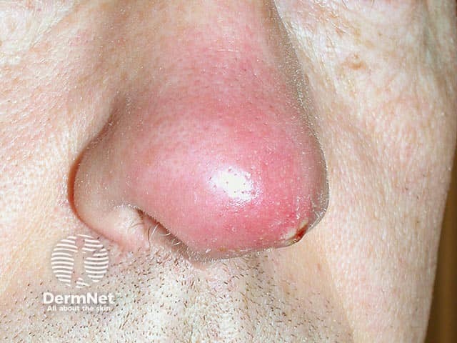
Clinical photography of a single nasal skin lesion showing an acute furuncle (boil) on the nasal vestibule/dorsum. The image depicts a solitary, erythematous, dome-shaped nodule with a shiny surface and surrounding mild edema. A palpable tenderness is suggested by the lesion’s conspicuous inflamed halo. A central pustule or crust may be present at the core, consistent with progression toward abscess formation within a pilosebaceous follicle. The lesion is located on the external nasal skin near the nostril, with frontal/anterior perspective offering clear visualization of the nasal bridge region. The clinical diagnosis is furunculosis of the nose, most often caused by Staphylococcus aureus; in this case PVL (Panton-Valentine leukocidin)–producing strains are implicated, which are associated with more aggressive skin and soft tissue infections and a higher risk of complications. Imaging is not radiologic; this is a dermatologic/clinical photograph used for documentation, teaching, and correlating with microbiology culture results. Management implications include incision and drainage if fluctuant, culture to guide antibiotics, and consideration of toxin-producing strains. The image serves educational purposes for dermatology, infectious disease, microbiology, and medical training in recognizing PVL-associated nasal furunculosis and differentiating it from impetigo, cellulitis, and Sty.
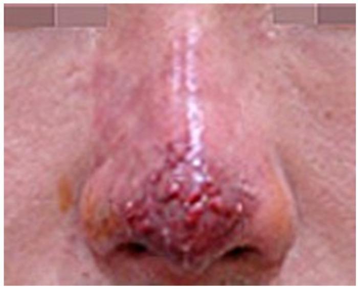
This clinical photograph shows a close-up view of the human nose exhibiting signs of vascular compromise and secondary infection following a dermatological procedure. The primary pathology is localized to the nasal tip and dorsum, where a cluster of small, raised, erythematous pustules is prominently visible. These lesions are characterized by a rounded morphology and a deep red to violaceous hue, suggesting intense underlying inflammation and possible infection. The skin of the nasal bridge appears edematous and displays a shiny texture with diffuse erythema extending towards the glabella. In contrast, the skin on the adjacent cheeks maintains a more normal tone and texture, highlighting the localized nature of the inflammatory process. The visual findings are consistent with an infection state and impending tissue necrosis, likely resulting from vascular occlusion post-filler injection. This image serves as a clinical reference for identifying early progression from ischemic stages to pustule formation and potential eschar development in aesthetic medicine complications.
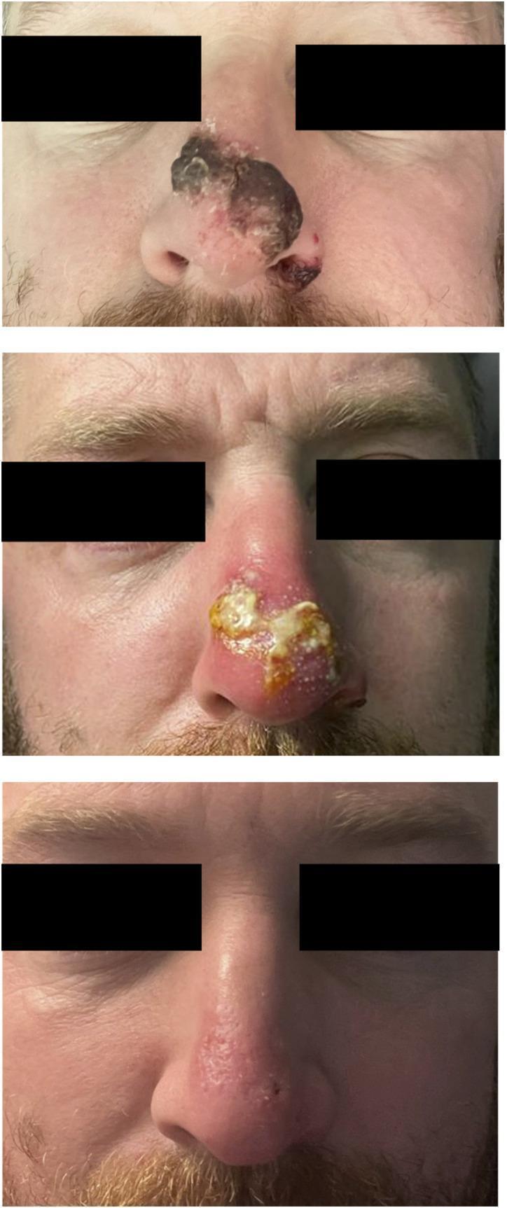
A series of three clinical photographs arranged vertically, documenting the progression and resolution of a secondary bacterial infection complicating a primary mpox lesion on the nose. The top image (Day -1) shows a large, dark, necrotic-appearing plaque with irregular borders and central ulceration covering the nasal bridge and tip. The middle image (Day 5) illustrates the inflammatory phase, characterized by intense peri-lesional erythema, edema, and a thick, yellow purulent exudate/crust indicative of secondary infection. The bottom image (Day 13) demonstrates significant clinical resolution following treatment, with the nose showing only residual mild erythema and subtle textural irregularities where the primary lesion was located. This timeline emphasizes the evolution from acute necrotic infection and secondary bacterial complication to successful healing and tissue remodeling in the context of viral dermatopathology.
NASAL VESTIBULITIS - ENT (MBBS)
Explained Simply - Like Teaching a Beginner
FIRST - UNDERSTAND THE BASICS: What is the Nasal Vestibule?
Think of your nose like a house. The vestibule is the doorway/entrance before you actually enter the house (nasal cavity).
- It is lined by skin (NOT mucous membrane like the rest of the nose)
- Because it is skin, it has hair follicles (the vibrissae - the coarse hairs inside your nose)
- Hair follicles can get infected - just like a pimple on your face
DEFINITION
Super simple version: Vestibulitis = A pimple/boil inside the nostril entrance.
CAUSES / RISK FACTORS
1. Digital Trauma (Nose Picking - Most Common!)
- When you pick your nose, your fingernail scratches the delicate skin inside the vestibule
- This creates tiny cuts/breaks in the skin
- Bacteria (Staph. aureus) enter through these breaks
- Infection starts in the hair follicle
Analogy: Just like how scratching a pimple introduces bacteria and makes it worse - nose picking does the same thing inside the nostril.
2. Excessive Nose Blowing
- Repeated, forceful nose blowing rubs and irritates the vestibular skin
- This causes micro-abrasions (tiny scratches)
- Bacteria get in and cause infection
- Common in patients with colds/allergic rhinitis who blow their nose constantly
3. Chronic Rhinorrhoea (Runny Nose)
- Constant dripping of mucus out of the nose keeps the vestibular skin wet
- Moist, macerated skin = easy entry for bacteria
4. Chemotherapy (Important additional cause)
- Chemotherapy drugs damage the epithelial cells in the nose → dryness, cracking → secondary bacterial infection
- Seen in >70-80% of patients receiving taxanes (paclitaxel) or bevacizumab
5. Non-infective causes
- Eczema / contact dermatitis
- Trauma (e.g., after nasal cautery for nosebleed)
- Irritants
CLINICAL FEATURES (Symptoms & Signs)
Symptoms:
| Symptom | Simple Explanation |
|---|---|
| Anterior nasal pain | Pain at the very entrance of the nostril - hurts when you touch the nose tip |
| Swelling | The skin inside the nostril looks puffy and red |
| Tenderness | Moving the nose (like pressing the tip) hurts a lot |
| Crusting | Dried mucus/pus forms a crust at the nostril opening |
| Pustules | Small pus-filled spots (like a whitehead pimple) inside the nostril |
| Nasal obstruction | If swelling is severe, it partially blocks the nose |
Signs on Examination:
- Redness and swelling of the vestibular skin
- Possible pustules (pus spots) over hair follicles
- Nasal septum is usually SPARED - because the infection stays limited to the vestibular skin/hair follicles, not the deeper cartilage or mucosa
Key exam point: "The nasal septum is often spared" - because disease is limited to the vestibular skin hair follicles, NOT the deeper mucosa.
TYPES OF VESTIBULITIS
1. Simple Vestibulitis / Folliculitis
- Infection is superficial - just in the hair follicle
- Mild pain, redness, crusting, maybe a tiny pus spot
- No serious risk
2. Furunculosis (Furuncle = Boil)
- Infection goes deeper into the hair follicle and surrounding tissue
- Forms a boil (furuncle) - a tender, red, pus-filled lump
- Signs: perifollicular erythema (redness around the follicle) and oedema (swelling)
- May form an abscess (collection of pus)
Analogy: Simple vestibulitis is like a blackhead. Furunculosis is like a full boil on the skin.
3. Carbuncle
- When multiple furuncles join together
- More extensive, more dangerous
WHY IS FURUNCULOSIS DANGEROUS? - THE FEARED COMPLICATION
⚠️ CAVERNOUS SINUS THROMBOSIS
Imagine this: A drain pipe in your bathroom connects directly to your house's main water tank. If something dirty gets into the drain, it can flow backward straight into the clean water tank. That is exactly what happens here - infection from the nose can travel backward through veins straight into the brain.
- A furuncle forms in the nasal vestibule
- Patient squeezes/presses it (trying to pop it like a pimple)
- Bacteria enter the bloodstream through the veins
- These veins have no valves → bacteria travel backward into the skull
- They reach the cavernous sinus (a large venous space behind the eye socket)
- A blood clot + infection (septic thrombosis) forms there
- High fever, severe headache
- Proptosis (eye bulging out)
- Painful, swollen eye
- Ophthalmoplegia (eye cannot move)
- Chemosis (swollen conjunctiva)
- Can be fatal if not treated
TREATMENT / MANAGEMENT
A. Simple/Non-Infective Vestibulitis
- Vaseline (petroleum jelly) - applied to the vestibule to moisturize and protect the skin
- Mild topical corticosteroid - to reduce inflammation
- Saline nasal sprays - especially for chemotherapy-related vestibulitis
B. Infective Vestibulitis (Bacterial - Folliculitis Stage)
| Treatment | Details |
|---|---|
| Warm compresses | A warm cloth on the nose 3-4 times a day - brings blood flow, helps the body fight infection naturally and can drain small pustules |
| Topical antibiotics | Applied directly inside the nostril as ointment |
- Mupirocin (Bactroban nasal) - First choice, very effective against Staph. aureus and MRSA
- Naseptin (Chlorhexidine + Neomycin) - Also effective; applied 3 times a day for 5 days
- Polymyxin / Bacitracin / Neomycin / Retapamulin - Other options
Note on Naseptin: Contains arachis (peanut) oil - DO NOT use in patients with peanut allergy. Also contraindicated in pregnancy.
Note on Mupirocin: Used as second-line for MRSA eradication. After 5 days of treatment, a nasal swab is taken 2 days later to check if MRSA is cleared.
C. Furunculosis (Deep Boil) Management
| Treatment | Why |
|---|---|
| Warm compresses | Brings pus to a head, helps localize and drain it |
| Oral anti-staphylococcal antibiotics | Because infection is deep - topical alone won't reach; need oral route |
| Topical antimicrobials | In addition to oral |
| Incision and Drainage (I&D) | If abscess forms - surgeon makes a small cut to drain the pus |
- Flucloxacillin / Dicloxacillin (anti-staphylococcal penicillins)
- Amoxicillin-clavulanate
- Co-trimoxazole (for MRSA)
- Clindamycin
NEVER squeeze a nasal furuncle - risk of cavernous sinus thrombosis (explained above)
QUICK SUMMARY TABLE
| Feature | Simple Vestibulitis | Furunculosis |
|---|---|---|
| Depth | Superficial (skin/follicle) | Deep (perifollicular + tissue) |
| Appearance | Redness, crusting, pustules | Boil, abscess, swelling |
| Pain | Mild | Severe |
| Risk | Low | Cavernous sinus thrombosis |
| Treatment | Warm compress + topical antibiotics | Warm compress + ORAL antibiotics + I&D if abscess |
MEMORY TRICKS FOR EXAM
Organism: Almost always Staphylococcus aureus
Cause: Nose picking (#1) + Nose blowing
Septum is SPARED (disease stays in skin/hair follicles)
Dreaded complication: Cavernous Sinus Thrombosis (from danger triangle of face)
Never squeeze a nasal boil - tell this to every viva examiner with confidence
Naseptin = Chlorhexidine + Neomycin (avoid in peanut allergy + pregnancy)
Mupirocin = MRSA agent (second line for MRSA eradication)
FLOW CHART TO REMEMBER
Nose picking / Nose blowing
        ↓
Skin scratch in nasal vestibule
        ↓
Staph. aureus enters
        ↓
Infects hair follicle
        ↓ (superficial)              ↓ (deep)
FOLLICULITIS                      FURUNCULOSIS
(simple vestibulitis)              (Boil/Abscess)
        ↓                                  ↓
Topical antibiotics           Oral antibiotics + I&D
+ warm compress               + warm compress
                                           ↓ (if squeezed!)
                             CAVERNOUS SINUS THROMBOSIS
                                  ← DANGEROUS! →