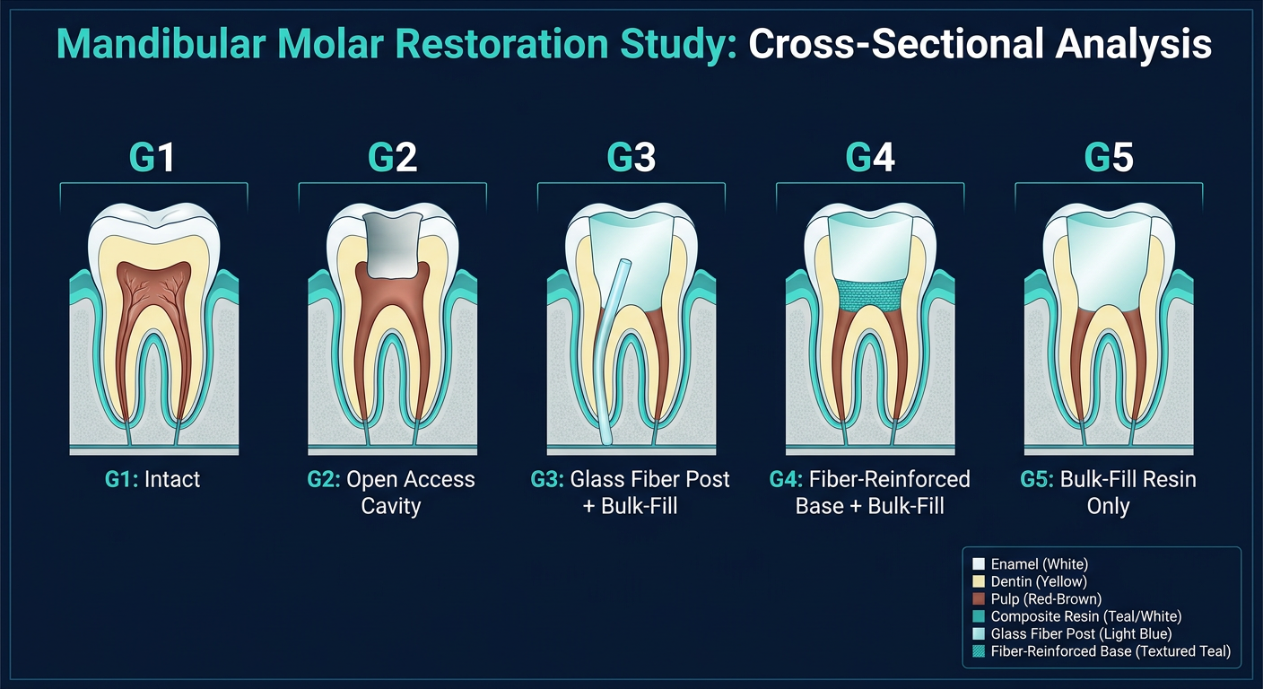
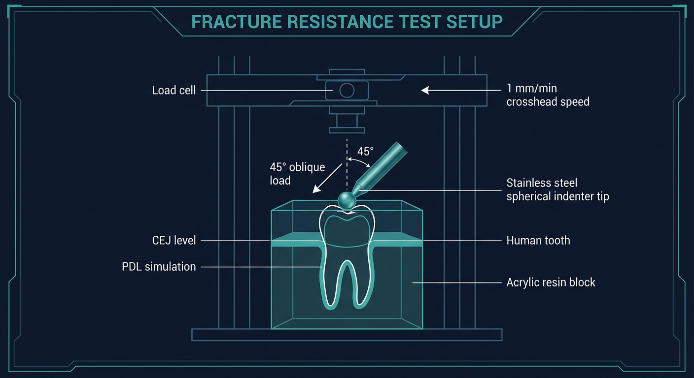
convert this into2-3 image slides for my presentation;A total of 100 intact human mandibular first molars extracted for periodontal reasons were used for this study. All teeth were free from caries, defects, restoration and cracks and were used in compliance with ethical guidelines of Gazi University Faculty of Dentistry Clinical Research Ethics Committee (ethical protocol no. 2021.19/6). Plaque, attached periodontal tissues and calculus deposits were removed by hand-scaling and then stored in 0.1% thymol solution until use. The bucco-lingual and mesio-distal dimensions of the teeth were measured using a digital caliper (Insize 1112 − 150, Insize, Jiangsu, China). The teeth were divided into five groups so that there was no difference in the oneway analysis of variance (ANOVA) test between buccolingual and mesio-distal widths (p > 0.05) [32, 33]. The materials used in the study and their contents are listed in Table 1. Before preparing the cavities in the teeth, the distance between the cusps was measured using the digital caliper. To prepare a conservative access cavity, digital radiography was used to determine the borders of the access cavity according to the dimensions of the teeth. Then, the canal entrances were confirmed by considering the length of the periodontal probe according to the notches of the periodontal probe on the radiograph. The gingival step of the cavity Table 1 The composition of the materials used in the study Material (CEJ). The conservative mesio-occlusal (MO) endodontic access cavity was applied to all teeth except the control group (intact teeth) using a straight fissure carbide bur (Hicare Medical Co. Ltd., Guangzhou, China) using a highspeed handpiece under water cooling and all Cavo surface margins prepared at 90° with internal line angles rounded. The bur was changed after every five cavity preparations, and cavity dimensions were measured during the preparation using a digital caliper to ensure standardization. Following the endodontic access cavity, the working length was determined using #10 K-files (Shenzhen Perfect Medical Instruments Co. Ltd., Shenzhen, China), and the root canals were instrumented using ProTaper rotary files (Endoart, İstanbul, Turkey) up to sizes F3 with the crowndown method. During instrumentation, the root canals were irrigated with 1 mL of 2.5% NaOCl before each file was introduced into the canal and finally with distilled water. The canals were dried with paper points (Diadent Group International, Almere, the Netherlands) and obturated with gutta-percha cones and an AH Plus sealer (Dentsply De Trey, Konstanz, Germany) using the single cone technique. Excess gutta-percha cones were cut 1 mm apically from the canal orifices with a gutta cutter (C-Blade, Coxo, Guangdong Province, China) and covered with resin-modified glass ionomer cement (Ketac Cem Plus, 3 M, St. Paul, MN, USA). All teeth were examined after root canal treatment using periapical radiography to ensure accurate root canal treatment.To simulate the clinical conditions, each specimen was embedded in a block of self-curing acrylic cylinders at a level of 2.0 mm below the CEJ. The periodontal ligaments were simulated using a base plate wax at a 0.3 mm (Efes Dental, Bursa, Turkey). The distal cavity was determined and standardized according to the distal marginal ridge ness and periapical radiography. On the mesial side, the distance between the gingival margin and the CEJ was prepared at 1 mm. Care was taken to ensure a thickness of approximately 2 mm between the buccal and lingual walls and the interproximal cavity walls using the digital caliper. The buccolingual size of the cavity was 4 mm, and this width was measured above the cavities [34]. Clearfil S3 Bond Universal (Kuraray, Osaka, Japan) was applied using total etch mode with 35% orthophosphoric acid gel (Scotchbond Universal Etchant, 3 M ESPE, St. Paul, MN, USA). Acid gel was applied and left on enamel for 30 s and on dentin for 15 s, before being rinsed with water and gently dried with air to create a moist dentin surface. Clearfil S3 Bond Universal (Kuraray, Osaka, Japan) was applied using a micro-brush for 20 s and light-cured for 10 s with an LED light curing unit (D-Light Pro, GC, Leuven, Belgium). Upon completion of root canal treatment, the restorative steps for each group were as follows (Figs. 1 and 2): ● Group 1 (positive control group): Intact teeth without any cavity preparation were used as the positive control. ● Group 2 (negative control group): The mesial endodontic access was prepared, and coronal restoration was not applied. ● Group 3: A 4 mm glass fiber post (Cytec Blanco, Hannerkratt, Germany) was cut with a diamond fissure bur. The post was then fixed to the buccal and lingual walls of the cavity with a flowable resin composite (Filtek Ultimate Flowable, 3 M Espe, St. Paul, MN, USA). For this purpose, a flowable resin composite was applied to both ends of the post and adapted to the buccal and lingual cavity walls in the middle one-third of the cavity. The overflowing flowable resin composite was removed using a sond. Then, the flowable resin composite was polymerized at the connection points of the post with the cavity using an LED light device for 20 s. Then, bulk-fill resin composite (Filtek One Bulk Fill Restorative, FOB; 3 M ESPE, St. Paul, MN, USA) was applied in 4-mm layers for the restoration of the entire cavity and polymerized on the occlusal, buccal and lingual sides for 10 s using an LED Light Device. ● Group 4: A 4-mm thick fiber-reinforced composite (EverX Posterior, EXP; GC, Tokyo, Japan) was applied to the first half of the cavity and polymerized with an LED light device for 20 s. Then, a 4-mm thick bulk-fill resin composite (Filtek One Bulk Fill Restorative, FOB; 3 M ESPE, St. Paul, MN, USA) was applied to the upper part of EverX Posterior, and the restoration was completed by polymerizing the occlusal, buccal and lingual sides for 10 s using an LED Light Device from. ● Group 5: Coronal restoration completed with FOB using the bulk technique. The preparation and restoration of all specimens were performed by the same operator. Teeth were stored in distilled water for 24 h at 37 °C to prevent dehydration of the teeth and complete the post polymerization. They were then thermocycled at 5 °C and 55 °C for 10,000 cycles with a 30-s dwell time (MTE-101, MOD Dental, Ankara, Turkey).To determine fracture resistance, a 5 mm stainless steel spherical tip mounted on a universal testing machine (Shimadzu IG-IS, Tokyo, Japan) and a 45° oblique compressive load were applied to the central fossa of the teeth at a crosshead speed of 1 mm/min until a fracture occurred. The maximum load before fracture was recorded in Newtons (N). The fractured specimens were examined by two different operators, indicating the fracture mode as restorable (fracture above the CEJ or within 1 mm apical to the CEJ
ls -la /home/daytona/workspace/ 2>/dev/null || echo "workspace empty or missing"
~/molar-study-pptx/create_slides.js
const pptxgen = require("pptxgenjs");

const pres = new pptxgen();
pres.layout = "LAYOUT_16x9";
pres.title = "Mandibular First Molar Fracture Resistance Study";

// ─── PALETTE ───────────────────────────────────────────────
const DARK_BG   = "0D1B2A";   // deep navy
const MID_BG    = "1B2A3B";   // slightly lighter navy
const ACCENT1   = "00B4D8";   // bright teal
const ACCENT2   = "48CAE4";   // lighter teal
const WHITE     = "FFFFFF";
const LIGHT_GRY = "B8D4E8";
const WARM      = "F4A261";   // warm orange for highlights
const CARD_BG   = "152535";   // card surface

// ─── HELPERS ───────────────────────────────────────────────
function addHeader(slide, label, title, subtitle) {
  // top bar
  slide.addShape(pres.ShapeType.rect, {
    x: 0, y: 0, w: 10, h: 1.05,
    fill: { color: MID_BG }, line: { color: MID_BG }
  });
  // teal left accent
  slide.addShape(pres.ShapeType.rect, {
    x: 0, y: 0, w: 0.08, h: 1.05,
    fill: { color: ACCENT1 }, line: { color: ACCENT1 }
  });
  // label chip
  slide.addShape(pres.ShapeType.rect, {
    x: 0.22, y: 0.17, w: 1.05, h: 0.35,
    fill: { color: ACCENT1 }, line: { color: ACCENT1 },
    rounding: true
  });
  slide.addText(label, {
    x: 0.22, y: 0.17, w: 1.05, h: 0.35,
    fontSize: 9, bold: true, color: DARK_BG, align: "center", valign: "middle", margin: 0
  });
  // title
  slide.addText(title, {
    x: 1.45, y: 0.1, w: 7.8, h: 0.52,
    fontSize: 22, bold: true, color: WHITE, valign: "middle", margin: 0
  });
  // subtitle
  if (subtitle) {
    slide.addText(subtitle, {
      x: 1.45, y: 0.62, w: 7.8, h: 0.35,
      fontSize: 11, color: ACCENT2, valign: "middle", margin: 0, italic: true
    });
  }
}

function addCard(slide, x, y, w, h) {
  slide.addShape(pres.ShapeType.rect, {
    x, y, w, h,
    fill: { color: CARD_BG },
    line: { color: ACCENT1, width: 0.5 },
    shadow: { type: "outer", color: "000000", opacity: 0.35, blur: 6, offset: 3, angle: 45 }
  });
}

function addSectionLabel(slide, x, y, w, text) {
  slide.addShape(pres.ShapeType.rect, {
    x, y, w, h: 0.3,
    fill: { color: ACCENT1 }, line: { color: ACCENT1 }
  });
  slide.addText(text, {
    x, y, w, h: 0.3,
    fontSize: 9, bold: true, color: DARK_BG, align: "center", valign: "middle", margin: 0, charSpacing: 1.5
  });
}

// ═══════════════════════════════════════════════════════════
// SLIDE 1 — Study Design & Specimen Preparation
// ═══════════════════════════════════════════════════════════
{
  const s = pres.addSlide();
  s.background = { color: DARK_BG };

  addHeader(s, "SLIDE 01", "Study Design & Specimen Preparation",
    "100 intact human mandibular first molars — Gazi University Ethics Protocol 2021.19/6");

  // ── big stat circle ──
  s.addShape(pres.ShapeType.ellipse, {
    x: 0.22, y: 1.25, w: 2.1, h: 2.1,
    fill: { color: MID_BG }, line: { color: ACCENT1, width: 2 }
  });
  s.addText([
    { text: "100", options: { fontSize: 36, bold: true, color: ACCENT1, breakLine: true } },
    { text: "MOLARS", options: { fontSize: 10, color: LIGHT_GRY, bold: true, charSpacing: 2 } }
  ], { x: 0.22, y: 1.6, w: 2.1, h: 1.3, align: "center", valign: "middle", margin: 0 });

  // ── inclusion criteria card ──
  addCard(s, 2.6, 1.2, 3.5, 2.2);
  addSectionLabel(s, 2.6, 1.2, 3.5, "INCLUSION CRITERIA");
  s.addText([
    { text: "✓  ", options: { color: ACCENT1, bold: true } },
    { text: "Caries-free, no restorations", options: { color: WHITE } },
    { text: "\n✓  ", options: { color: ACCENT1, bold: true } },
    { text: "No cracks or defects", options: { color: WHITE } },
    { text: "\n✓  ", options: { color: ACCENT1, bold: true } },
    { text: "Extracted for periodontal reasons", options: { color: WHITE } },
    { text: "\n✓  ", options: { color: ACCENT1, bold: true } },
    { text: "Compliant with ethical guidelines", options: { color: WHITE } },
  ], { x: 2.7, y: 1.55, w: 3.3, h: 1.75, fontSize: 11, valign: "top", margin: 4 });

  // ── preparation steps card ──
  addCard(s, 6.3, 1.2, 3.5, 2.2);
  addSectionLabel(s, 6.3, 1.2, 3.5, "PREPARATION STEPS");
  s.addText([
    { text: "①  ", options: { color: WARM, bold: true } },
    { text: "Plaque & calculus removed by hand-scaling", options: { color: WHITE } },
    { text: "\n②  ", options: { color: WARM, bold: true } },
    { text: "Stored in 0.1% thymol solution", options: { color: WHITE } },
    { text: "\n③  ", options: { color: WARM, bold: true } },
    { text: "Bucco-lingual & mesio-distal dims measured (digital caliper)", options: { color: WHITE } },
    { text: "\n④  ", options: { color: WARM, bold: true } },
    { text: "Divided into 5 groups (ANOVA — no significant diff, p > 0.05)", options: { color: WHITE } },
  ], { x: 6.4, y: 1.55, w: 3.3, h: 1.75, fontSize: 10.5, valign: "top", margin: 4 });

  // ── bottom row: 5 groups ──
  const groups = [
    { num: "G1", label: "Positive\nControl", sub: "Intact teeth" },
    { num: "G2", label: "Negative\nControl", sub: "No restoration" },
    { num: "G3", label: "Fiber Post\n+ Bulk Fill", sub: "FOB + glass fiber" },
    { num: "G4", label: "FRC\n+ Bulk Fill", sub: "EverX + FOB" },
    { num: "G5", label: "Bulk Fill\nOnly", sub: "FOB alone" },
  ];
  groups.forEach((g, i) => {
    const bx = 0.22 + i * 1.92;
    addCard(s, bx, 3.6, 1.82, 1.75);
    s.addShape(pres.ShapeType.ellipse, {
      x: bx + 0.55, y: 3.7, w: 0.72, h: 0.72,
      fill: { color: ACCENT1 }, line: { color: ACCENT1 }
    });
    s.addText(g.num, {
      x: bx + 0.55, y: 3.7, w: 0.72, h: 0.72,
      fontSize: 11, bold: true, color: DARK_BG, align: "center", valign: "middle", margin: 0
    });
    s.addText(g.label, {
      x: bx + 0.05, y: 4.45, w: 1.72, h: 0.55,
      fontSize: 9.5, bold: true, color: WHITE, align: "center", valign: "top", margin: 0
    });
    s.addText(g.sub, {
      x: bx + 0.05, y: 5.0, w: 1.72, h: 0.3,
      fontSize: 8, color: ACCENT2, align: "center", valign: "top", margin: 0, italic: true
    });
  });

  // footer
  s.addShape(pres.ShapeType.rect, { x: 0, y: 5.42, w: 10, h: 0.21, fill: { color: MID_BG }, line: { color: MID_BG } });
  s.addText("Gazi University Faculty of Dentistry  |  Ethics Protocol No. 2021.19/6", {
    x: 0, y: 5.42, w: 10, h: 0.21, fontSize: 7.5, color: LIGHT_GRY, align: "center", valign: "middle", margin: 0
  });
}

// ═══════════════════════════════════════════════════════════
// SLIDE 2 — Cavity Preparation & Root Canal Treatment
// ═══════════════════════════════════════════════════════════
{
  const s = pres.addSlide();
  s.background = { color: DARK_BG };

  addHeader(s, "SLIDE 02", "Cavity Preparation & Root Canal Treatment",
    "Conservative mesio-occlusal (MO) endodontic access cavity — standardised protocol");

  // ── left: cavity prep timeline ──
  addCard(s, 0.18, 1.18, 4.4, 4.15);
  addSectionLabel(s, 0.18, 1.18, 4.4, "CAVITY PREPARATION");

  const cavitySteps = [
    { n: "1", t: "Digital radiography used to define access cavity borders" },
    { n: "2", t: "Canal entrances confirmed with periodontal probe" },
    { n: "3", t: "Gingival step at CEJ; MO cavity with straight fissure carbide bur" },
    { n: "4", t: "High-speed handpiece under water cooling; 90° cavosurface margins" },
    { n: "5", t: "Bur changed every 5 preparations; dimensions verified with caliper" },
    { n: "6", t: "Cavity: 4 mm buccolingual, ~2 mm buccal/lingual wall thickness; 1 mm mesial to CEJ" },
  ];
  cavitySteps.forEach((st, i) => {
    const ty = 1.55 + i * 0.6;
    s.addShape(pres.ShapeType.ellipse, {
      x: 0.32, y: ty + 0.04, w: 0.38, h: 0.38,
      fill: { color: ACCENT1 }, line: { color: ACCENT1 }
    });
    s.addText(st.n, {
      x: 0.32, y: ty + 0.04, w: 0.38, h: 0.38,
      fontSize: 10, bold: true, color: DARK_BG, align: "center", valign: "middle", margin: 0
    });
    s.addText(st.t, {
      x: 0.8, y: ty, w: 3.65, h: 0.52,
      fontSize: 10, color: WHITE, valign: "middle", margin: 2
    });
  });

  // ── right: RCT protocol ──
  addCard(s, 4.78, 1.18, 5.05, 4.15);
  addSectionLabel(s, 4.78, 1.18, 5.05, "ROOT CANAL TREATMENT PROTOCOL");

  const rctRows = [
    { icon: "⚙", label: "Instrumentation", val: "ProTaper rotary files to F3 (crown-down)" },
    { icon: "💧", label: "Irrigation", val: "1 mL 2.5% NaOCl before each file + final distilled water" },
    { icon: "📄", label: "Drying", val: "Paper points" },
    { icon: "🪤", label: "Obturation", val: "Gutta-percha + AH Plus sealer (single cone)" },
    { icon: "✂", label: "Excess GP", val: "Cut 1 mm apical to orifice; covered with RMGIC (Ketac Cem Plus)" },
    { icon: "🔬", label: "Verification", val: "Periapical radiograph confirming accurate RCT" },
  ];
  rctRows.forEach((r, i) => {
    const ty = 1.55 + i * 0.6;
    s.addShape(pres.ShapeType.rect, {
      x: 4.88, y: ty + 0.05, w: 0.42, h: 0.38,
      fill: { color: MID_BG }, line: { color: ACCENT2, width: 0.5 }
    });
    s.addText(r.icon, {
      x: 4.88, y: ty + 0.05, w: 0.42, h: 0.38,
      fontSize: 13, align: "center", valign: "middle", margin: 0
    });
    s.addText(r.label + ": ", {
      x: 5.38, y: ty + 0.04, w: 1.15, h: 0.4,
      fontSize: 9.5, bold: true, color: ACCENT2, valign: "middle", margin: 0
    });
    s.addText(r.val, {
      x: 6.5, y: ty + 0.04, w: 3.2, h: 0.4,
      fontSize: 9.5, color: WHITE, valign: "middle", margin: 0
    });
  });

  // bottom specimen note
  s.addShape(pres.ShapeType.rect, {
    x: 0.18, y: 5.1, w: 9.65, h: 0.33,
    fill: { color: MID_BG }, line: { color: WARM, width: 1 }
  });
  s.addText(
    "Specimens embedded in self-curing acrylic 2 mm below CEJ  |  PDL simulated with base-plate wax (0.3 mm)  |  Stored 24 h at 37 °C → Thermocycled 10,000 cycles (5 °C / 55 °C)",
    { x: 0.22, y: 5.1, w: 9.55, h: 0.33, fontSize: 8.5, color: WARM, align: "center", valign: "middle", margin: 0 }
  );

  // footer
  s.addShape(pres.ShapeType.rect, { x: 0, y: 5.43, w: 10, h: 0.2, fill: { color: MID_BG }, line: { color: MID_BG } });
  s.addText("Conservative MO access | Clearfil S3 Bond Universal — total-etch mode | LED light-curing unit", {
    x: 0, y: 5.43, w: 10, h: 0.2, fontSize: 7.5, color: LIGHT_GRY, align: "center", valign: "middle", margin: 0
  });
}

// ═══════════════════════════════════════════════════════════
// SLIDE 3 — Restorative Groups & Fracture Resistance Testing
// ═══════════════════════════════════════════════════════════
{
  const s = pres.addSlide();
  s.background = { color: DARK_BG };

  addHeader(s, "SLIDE 03", "Restorative Groups & Fracture Resistance Testing",
    "5 groups — universal testing machine — 45° oblique load to central fossa");

  // ── 5 group cards ──
  const grpData = [
    {
      id: "G1", color: LIGHT_GRY,
      title: "Positive Control",
      lines: ["Intact teeth", "No cavity / RCT", "Baseline fracture\nresistance"]
    },
    {
      id: "G2", color: "#E63946",
      title: "Negative Control",
      lines: ["MO endodontic access", "No coronal\nrestoration applied"]
    },
    {
      id: "G3", color: WARM,
      title: "Fiber Post + Bulk Fill",
      lines: ["4 mm glass fiber post", "Fixed to buccal/lingual\nwalls (flowable RC)", "Bulk-fill RC over (FOB, 4 mm)"]
    },
    {
      id: "G4", color: ACCENT1,
      title: "FRC + Bulk Fill",
      lines: ["EverX Posterior 4 mm\n(1st half of cavity)", "Filtek One Bulk Fill\n4 mm (upper half)"]
    },
    {
      id: "G5", color: ACCENT2,
      title: "Bulk Fill Only",
      lines: ["Filtek One Bulk Fill\n(FOB) entire cavity", "Bulk technique\nno substructure"]
    },
  ];

  grpData.forEach((g, i) => {
    const bx = 0.18 + i * 1.93;
    // card
    s.addShape(pres.ShapeType.rect, {
      x: bx, y: 1.15, w: 1.8, h: 3.05,
      fill: { color: CARD_BG },
      line: { color: g.color, width: 1.5 }
    });
    // colored header band
    s.addShape(pres.ShapeType.rect, {
      x: bx, y: 1.15, w: 1.8, h: 0.42,
      fill: { color: g.color }, line: { color: g.color }
    });
    s.addText(g.id, {
      x: bx, y: 1.15, w: 0.48, h: 0.42,
      fontSize: 11, bold: true, color: DARK_BG, align: "center", valign: "middle", margin: 0
    });
    s.addText(g.title, {
      x: bx + 0.5, y: 1.15, w: 1.3, h: 0.42,
      fontSize: 9, bold: true, color: DARK_BG, align: "left", valign: "middle", margin: 0
    });
    // body text
    g.lines.forEach((ln, li) => {
      s.addText(ln, {
        x: bx + 0.08, y: 1.65 + li * 0.72, w: 1.64, h: 0.65,
        fontSize: 9, color: WHITE, valign: "top", margin: 2, align: "left"
      });
    });
  });

  // ── bottom: testing method ──
  addCard(s, 0.18, 4.3, 9.65, 1.02);
  addSectionLabel(s, 0.18, 4.3, 9.65, "FRACTURE RESISTANCE TESTING");

  const testCols = [
    { h: "Apparatus", v: "Universal Testing Machine\n(Shimadzu IG-IS, Tokyo)" },
    { h: "Load Type", v: "45° oblique compressive load\nvia 5 mm stainless steel spherical tip" },
    { h: "Speed", v: "1 mm/min crosshead\nspeed until fracture" },
    { h: "Outcome", v: "Maximum load recorded in\nNewtons (N) at fracture" },
    { h: "Fracture Mode", v: "Restorable (≥ CEJ / ≤ 1 mm apical)\nvs. Non-restorable" },
  ];
  testCols.forEach((tc, i) => {
    const tx = 0.28 + i * 1.93;
    s.addText(tc.h, {
      x: tx, y: 4.62, w: 1.8, h: 0.25,
      fontSize: 8.5, bold: true, color: ACCENT1, align: "center", valign: "middle", margin: 0
    });
    s.addText(tc.v, {
      x: tx, y: 4.87, w: 1.8, h: 0.42,
      fontSize: 8.5, color: WHITE, align: "center", valign: "top", margin: 0
    });
    if (i < 4) {
      s.addShape(pres.ShapeType.line, {
        x: tx + 1.82, y: 4.35, w: 0, h: 0.92,
        line: { color: MID_BG, width: 1 }
      });
    }
  });

  // footer
  s.addShape(pres.ShapeType.rect, { x: 0, y: 5.43, w: 10, h: 0.2, fill: { color: MID_BG }, line: { color: MID_BG } });
  s.addText("FOB = Filtek One Bulk Fill (3M ESPE)  |  EXP = EverX Posterior (GC)  |  FRC = Fiber-Reinforced Composite", {
    x: 0, y: 5.43, w: 10, h: 0.2, fontSize: 7.5, color: LIGHT_GRY, align: "center", valign: "middle", margin: 0
  });
}

// ── WRITE FILE ───────────────────────────────────────────
pres.writeFile({ fileName: "/home/daytona/workspace/molar-study-pptx/Molar_Study_Slides.pptx" })
  .then(() => console.log("DONE: /home/daytona/workspace/molar-study-pptx/Molar_Study_Slides.pptx"))
  .catch(e => { console.error(e); process.exit(1); });
cd /home/daytona/workspace/molar-study-pptx && node create_slides.js
Molar Study Slides
Presentation · PPTX
- Large "100 Molars" stat circle as a visual anchor
- Inclusion criteria card (caries-free, no restorations, periodontal extractions, ethics compliance)
- Preparation steps card (scaling, thymol storage, dimension measurement, 5-group ANOVA balancing)
- Bottom row showing all 5 groups with short labels (G1-G5)
- Step-by-step cavity prep protocol (6 numbered steps with radiography, probe confirmation, MO access, bur specs, dimensions)
- RCT protocol table with icons (ProTaper to F3, NaOCl irrigation, AH Plus sealer, RMGIC covering, radiographic verification)
- Highlighted note bar on specimen mounting (2 mm below CEJ, PDL simulation, thermocycling 10,000 cycles)
- 5 color-coded group cards (each group clearly summarized: positive control, negative control, fiber post + bulk-fill, FRC + bulk-fill, bulk-fill only)
- Testing method summary bar (Shimadzu UTM, 45° oblique load, 1 mm/min, Newtons, fracture mode classification)
Create 2-3 images for presentation
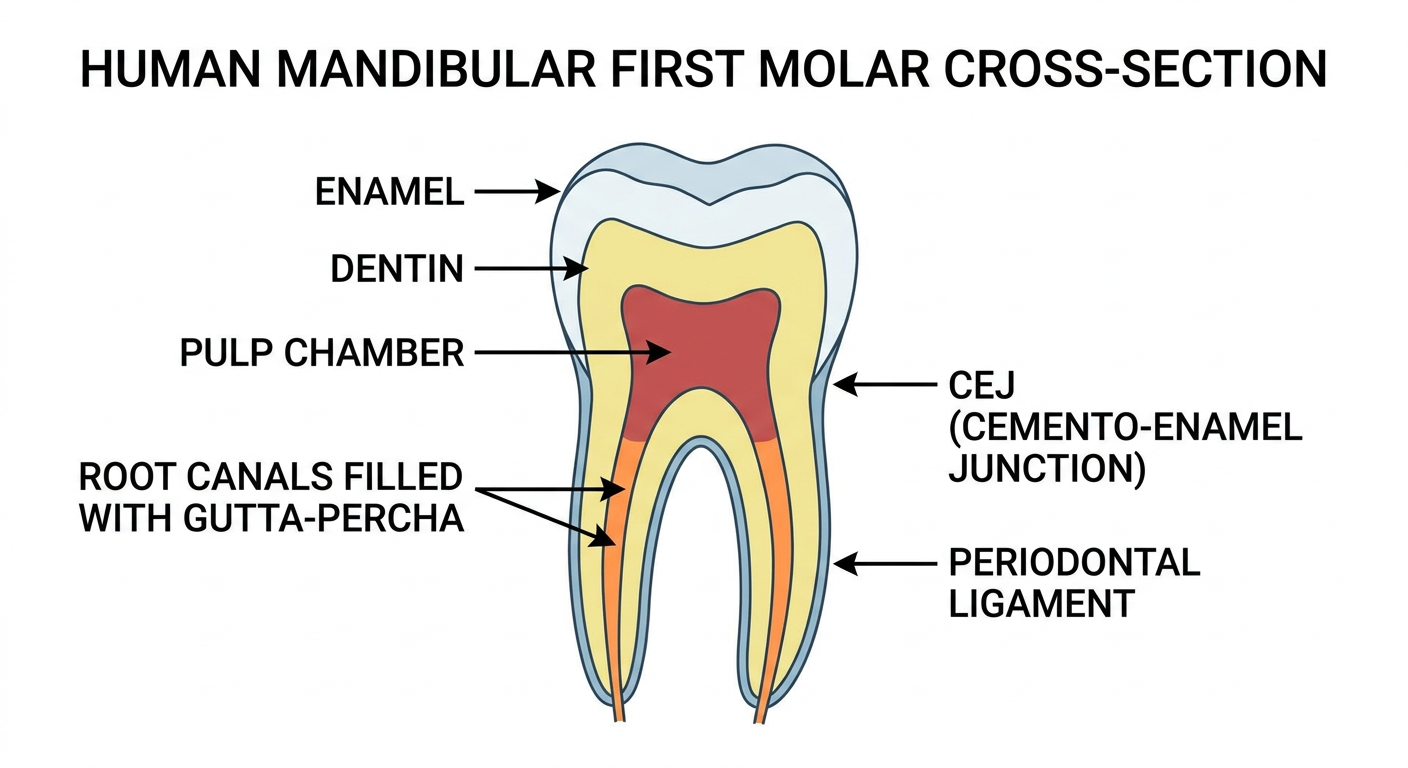
Scientific illustration of a human mandibular first molar tooth cross-section showing root canal anatomy, with labeled parts: enamel, dentin, pulp chamber, root canals filled with gutta-percha, CEJ (cemento-enamel junction), and periodontal ligament. Clean white background, medical textbook style, detailed anatomical diagram with clear labels and color coding.
Dental research laboratory illustration showing five groups of mandibular molar teeth side by side in cross-section, each group labeled G1 through G5. G1 is intact, G2 has an open endodontic access cavity, G3 shows a glass fiber post with bulk-fill composite resin, G4 shows fiber-reinforced composite base with bulk-fill on top, G5 shows bulk-fill resin only. Clean scientific diagram style, dark navy background, teal and white labels, professional dental research presentation.
Scientific illustration of a universal testing machine applying a 45-degree oblique compressive load to a human molar tooth embedded in acrylic resin block. The tooth shows a fracture resistance test setup with a stainless steel spherical tip pressing at 45 degrees onto the central fossa. Labels show: 45° oblique load, 1 mm/min crosshead speed, CEJ level, acrylic embedding, PDL simulation. Dark navy background with teal accents, clean engineering diagram style, professional research presentation.