
Explain near muscular junction andtransmission
Reading File
Finding Sources
Searching Images
neuromuscular junction structure diagram synaptic cleft acetylcholine receptors
Reading File
Reading File
The Neuromuscular Junction (NMJ) and Neuromuscular Transmission
1. Structure of the Neuromuscular Junction
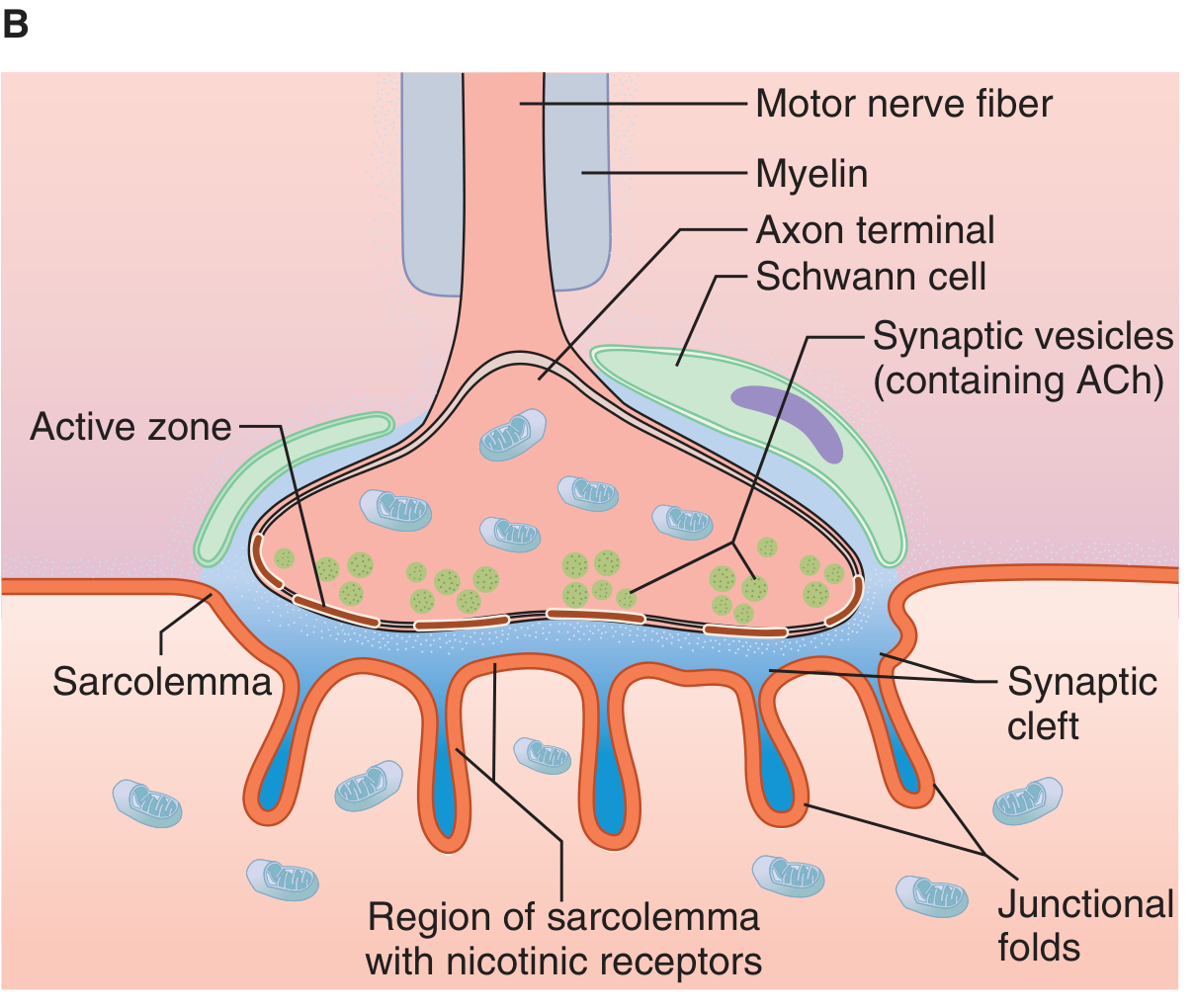
The neuromuscular junction is the specialized synapse where a motor nerve terminates on a skeletal muscle fiber. As the motor axon approaches the muscle, it loses its myelin sheath and branches into multiple terminal boutons. Key structural components:
- Presynaptic terminal: contains many small, clear synaptic vesicles filled with acetylcholine (ACh), the neurotransmitter at this junction.
- Motor endplate: the thickened region of muscle membrane where the nerve terminal sits, containing junctional folds (depressions) that increase surface area.
- Synaptic cleft: the space between the nerve terminal and the muscle membrane, analogous to the cleft at a neuron-to-neuron synapse.
- Each endplate receives input from only a single nerve fiber.
2. Sequence of Events During Transmission
- A nerve impulse (action potential) arrives at the motor nerve terminal and increases its permeability to Ca2+.
- Ca2+ enters the nerve ending and triggers exocytosis of ACh-containing synaptic vesicles into the synaptic cleft.
- ACh diffuses across the cleft and binds to nicotinic cholinergic (N_M) receptors concentrated at the tops of the junctional folds on the endplate membrane.
- Receptor binding increases Na+ and K+ conductance; the net Na+ influx produces a local depolarization called the endplate potential (EPP).
- The current sink from this local potential depolarizes the adjacent muscle membrane to its firing threshold.
- Action potentials are generated on both sides of the endplate and propagate in both directions along the muscle fiber, triggering muscle contraction (via excitation-contraction coupling).
- ACh is rapidly cleared from the synaptic cleft by acetylcholinesterase, an enzyme present in high concentration at the junction, terminating the signal.
(Ganong's Review of Medical Physiology, 26th Ed., p. 141)
3. Quantal Release of Transmitter
-
Each nerve impulse releases ACh from roughly 60 synaptic vesicles, each containing about 10,000 molecules of ACh.
-
At rest, small random "packets" (quanta) of ACh are released spontaneously, producing tiny depolarizations called miniature endplate potentials (~0.5 mV).
-
Quantal size varies directly with Ca2+ and inversely with Mg2+ concentration at the endplate.
-
When a nerve impulse arrives, quantal release increases by orders of magnitude, producing a large EPP that exceeds the firing threshold.
-
This same quantal release mechanism also occurs at noradrenergic, glutamatergic, and other synapses.
-
Ganong's Review of Medical Physiology, 26th Ed., p. 141-142
4. Clinical Relevance
Disorders and drugs that disrupt this process include:
- Myasthenia gravis - autoantibodies against nicotinic ACh receptors reduce endplate sensitivity, causing fatigable weakness.
- Lambert-Eaton myasthenic syndrome - antibodies against presynaptic voltage-gated Ca2+ channels reduce ACh release.
- Neuromuscular blocking drugs (e.g., succinylcholine, rocuronium, vecuronium) act at nicotinic receptors on the endplate and are widely used in anesthesia to produce muscle relaxation.
- Botulinum toxin blocks Ca2+-mediated vesicle exocytosis, preventing ACh release entirely.
- Organophosphates/nerve agents and some pesticides inhibit acetylcholinesterase, causing prolonged ACh action and overstimulation.
If you're studying this for a specific purpose (e.g., pharmacology of neuromuscular blockers, or the pathophysiology of myasthenia gravis/Lambert-Eaton syndrome), let me know and I can go deeper into that specific area.
This is a shared conversation. Sign in to Orris to start your own chat.