
Histology of Elastic& Muscular arteries
elastic artery histology wall layers microscopy
Low-power brightfield light microscopy of a formalin-fixed, paraffin-embedded arterial cross-section stained with Hematoxylin and Eosin. The specimen shows a circular vessel with a well-defined, multilayered wall. The lumen is patent and lined by a narrow endothelial layer. The tunica media exhibits concentric layers of eosinophilic smooth muscle cells and occasional nuclei aligned along the wall, giving a dense, pink stippled appearance. The internal elastic lamina is not clearly resolved at this magnification. The tunica adventitia is relatively inconspicuous with sparse connective tissue. There is no overt atheromatous plaque, calcification, hemorrhage, or inflammatory infiltrate noted. Overall architecture is preserved, suggesting a normal or nonpathologic arterial segment. This image is suitable for illustrating normal vascular histology and can serve as a reference for comparison with arteriosclerotic lesions in teaching, research, and diagnostic practice. Potential clinical utility includes training in recognition of arterial layering, identifying pathologies such as atherosclerosis, vasculitis, and aneurysm formation, and correlating histology with imaging findings from ultrasound or CT angiography. The dataset supports queries for vascular histology, arterial wall ultrastructure, smooth muscle organization, and H&E staining patterns in a cross-sectional artery. The image exemplifies typical histologic cues used in vascular pathology curricula and can anchor comparative discussions of disease progression and treatment implications.
This is a light microscopy image of a hematoxylin and eosin (H&E) stained vascular cross-section. The specimen appears to be a muscular artery or arteriole embedded in loose connective tissue. The lumen is relatively round to oval and patent, with a pale, eosinophilic lumen border and surrounding concentric layers. The tunica media displays densely packed, spindle-shaped smooth muscle cells with eosinophilic cytoplasm arranged in parallel bundles. The internal elastic lamina is not clearly delineated at this low magnification, while the adventitia contains sparse connective tissue and scattered inflammatory cells. The vascular wall demonstrates intact architecture without obvious dissection, necrosis, or thrombosis. Cytologic atypia is absent in the wall, and there is no evident marrow-like or sinusoidal pattern. Extracellular matrix components—collagen fibers—are visible in the adventitia, contributing to wall rigidity. The image highlights normal histologic vascular morphology suitable for educational comparison with pathologic vessels showing vasculitis, atherosclerotic changes, or aneurysmal dilation. Clinically, this morphology supports differential diagnoses ranging from normal anatomic variation to early arteriosclerosis, depending on patient age and comorbidity. In research contexts, this slide serves as a reference standard for vessel wall histology and for developing image-recognition models in vascular pathology.
muscular artery histology cross section microscopy
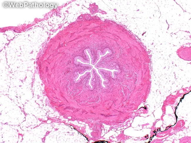
Imaging modality and technique: Bright-field light microscopy on a formalin-fixed, paraffin-embedded tissue section stained with Hematoxylin and Eosin (H&E). Specimen shows a transverse cross-section of a vascular structure within subcutaneous adipose and connective tissue. Anatomical context: vascular tissue from soft tissue; a small to medium-sized muscular artery or arteriolar segment; wall demonstrates layered tunica media with concentric lamination, an intact adventitia, and a single patent lumen visible as a bright white cavity. The lumen appears narrowed by inward folding of the intima and by mural thickening, while surrounding adipose tissue provides a pale pink backdrop. Notable features include radiating inward extensions within the lumen that may reflect physiologic folds or artifact from sectioning, not true septa. Normal variants such as mural elasticity and preserved architectural organization are evident; no dense inflammatory infiltrate, necrosis, or calcification is clearly seen. Diagnostic significance is limited by single high-magnification view; if clinically indicated, this finding could correspond to normal vascular morphology, hypertensive arteriopathy, or early atherosclerotic change depending on additional context. Clinical correlation with age, blood pressure, lipid status, and history of cardiovascular disease is recommended. This image is relevant for education on vascular histology and for differential diagnosis in soft-tissue pathology. Educational use.
This is a light microscopy image of a hematoxylin and eosin (H&E) stained vascular cross-section. The specimen appears to be a muscular artery or arteriole embedded in loose connective tissue. The lumen is relatively round to oval and patent, with a pale, eosinophilic lumen border and surrounding concentric layers. The tunica media displays densely packed, spindle-shaped smooth muscle cells with eosinophilic cytoplasm arranged in parallel bundles. The internal elastic lamina is not clearly delineated at this low magnification, while the adventitia contains sparse connective tissue and scattered inflammatory cells. The vascular wall demonstrates intact architecture without obvious dissection, necrosis, or thrombosis. Cytologic atypia is absent in the wall, and there is no evident marrow-like or sinusoidal pattern. Extracellular matrix components—collagen fibers—are visible in the adventitia, contributing to wall rigidity. The image highlights normal histologic vascular morphology suitable for educational comparison with pathologic vessels showing vasculitis, atherosclerotic changes, or aneurysmal dilation. Clinically, this morphology supports differential diagnoses ranging from normal anatomic variation to early arteriosclerosis, depending on patient age and comorbidity. In research contexts, this slide serves as a reference standard for vessel wall histology and for developing image-recognition models in vascular pathology.
elastic artery aorta tunica media elastic lamellae histology
Histology of Elastic & Muscular Arteries
Quick Comparison Table
| Feature | Elastic Artery (Large) | Muscular Artery (Medium) |
|---|---|---|
| Diameter | >10 mm | 2-10 mm |
| Examples | Aorta, pulmonary trunk, brachiocephalic | Femoral, radial, coronary, renal |
| Tunica Intima | Endothelium + thick subendothelial CT + smooth muscle; IEM indistinct | Endothelium + thin subendothelial CT + prominent IEM |
| Tunica Media | Smooth muscle + 40-70 fenestrated elastic lamellae | Almost entirely smooth muscle (10-40 layers); little elastic tissue |
| Tunica Adventitia | Thinner than media; CT + elastic fibers; vasa vasorum | ~Same thickness as media; CT + EEM (external elastic membrane) |
| Dominant stain finding | Wavy, concentric elastic lamellae (Weigert/resorcin-fuchsin) | Prominent IEM + EEM; circular smooth muscle |
| Function | Windkessel / pressure dampening | Active vasoconstriction, blood flow regulation |
ELASTIC ARTERIES (Large Arteries)
Diagram + Photomicrograph
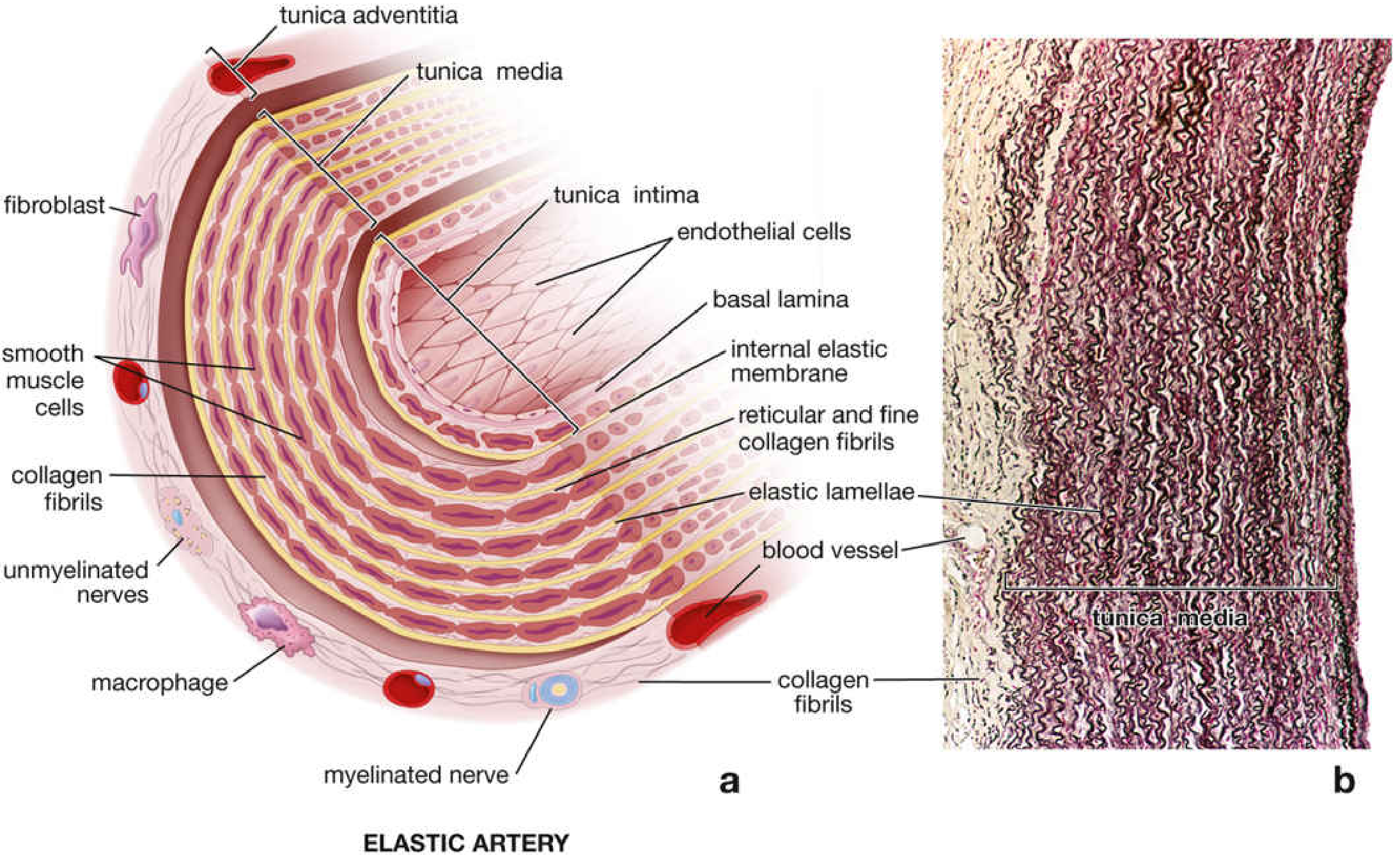
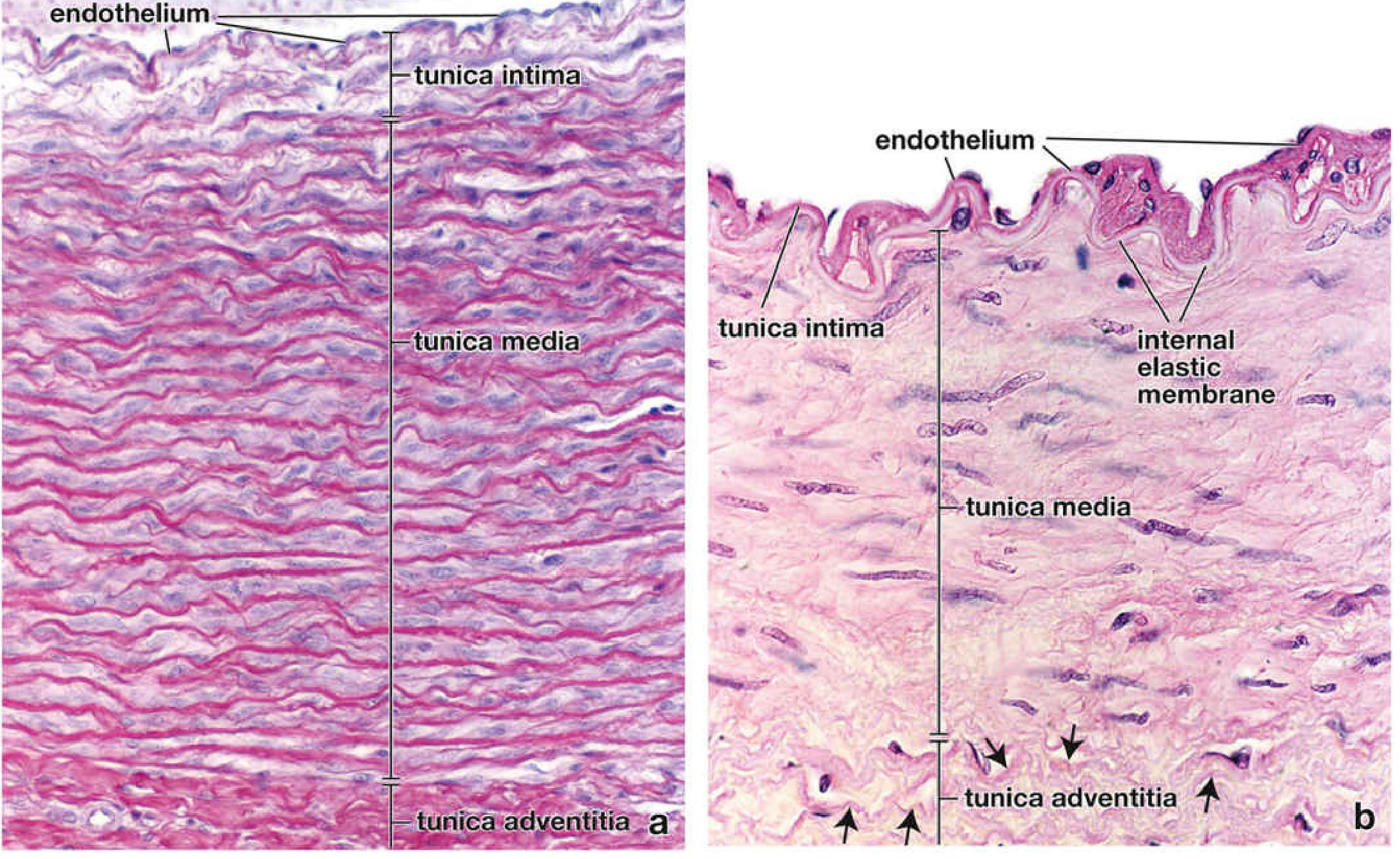
Tunica Intima
- Endothelium: flat, elongated cells oriented parallel to blood flow; joined by tight junctions and gap junctions
- Cells contain Weibel-Palade bodies (rod-shaped organelles storing von Willebrand factor and P-selectin)
- Subendothelial layer: connective tissue with smooth muscle cells, macrophages, collagen, and elastic fibers
- Internal elastic membrane (IEM): not well defined in elastic arteries - it is represented by the innermost elastic lamella, blending into the tunica media
Tunica Media (Thickest Layer - the hallmark)
- 40-70 concentric, fenestrated elastic lamellae arranged as wavy sheets (best seen with Weigert resorcin-fuchsin or orcein stain)
- Smooth muscle cells lie between the lamellae, arranged in a low-pitch spiral
- Fenestrations in the lamellae allow diffusion of nutrients through the thick wall
- Number and thickness of lamellae correlate with blood pressure and age: the aorta is nearly devoid of lamellae at birth but gains 40-70 in adults; hypertension increases both number and thickness
- Collagen and ground substance (proteoglycans) produced by smooth muscle cells fill the spaces
- No fibroblasts in the tunica media
Tunica Adventitia
- Thinner than the tunica media
- Loose connective tissue: collagen fibers, elastic fibers, fibroblasts
- Contains vasa vasorum (small vessels supplying the arterial wall) and unmyelinated + myelinated nerves
- No distinct external elastic membrane
Function
Side-by-side Photomicrographs (Elastic vs. Muscular)
MUSCULAR ARTERIES (Medium Arteries)
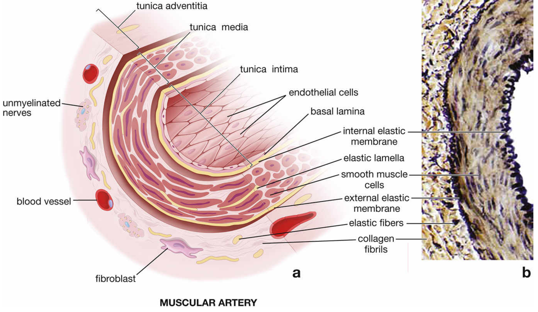
Diagram + Photomicrograph
Tunica Intima
- Endothelium with basal lamina
- Sparse subendothelial layer - in some vessels so thin that the endothelial basal lamina almost contacts the IEM directly
- Prominent Internal Elastic Membrane (IEM) - the defining feature; appears as a bright, wavy/undulating, refractile band in H&E sections (because the smooth muscle in the media is contracted, pushing folds into it)
- Intima thickness varies with age: very thin in children, ~1/6 of wall thickness in young adults, may widen with fatty streaks in older adults
Tunica Media (Dominant Layer)
- Composed almost entirely of circularly arranged smooth muscle cells (up to 40 layers in the largest muscular arteries)
- Only relatively little elastic material - scattered elastic fibers and a few elastic lamellae (not the sheets seen in elastic arteries)
- Collagen fibers and ground substance fill the intercellular spaces
- Smooth muscle cells have elongated nuclei; when contracted, they show a corkscrew appearance histologically
- Smooth muscle cells are arranged in a spiral; in cross-section they appear circular
- Gap junctions connect adjacent smooth muscle cells
- No fibroblasts
Tunica Adventitia
- Approximately same thickness as the tunica media (relatively thicker than in elastic arteries)
- Contains fibroblasts, collagen (principal component), elastic fibers, and scattered adipose cells
- A concentration of elastic material at the border with the media forms the External Elastic Membrane (EEM) - a second distinct elastic boundary
- Nerves and vasa vasorum travel here and penetrate into the media in larger muscular arteries
Function
Key Histological Distinguishing Points
| Distinguishing Feature | Elastic | Muscular |
|---|---|---|
| IEM | Indistinct (= innermost elastic lamella) | Thick, wavy, prominent, highly refractile |
| EEM | Absent | Present and distinct |
| Tunica media character | Elastic lamellae > smooth muscle | Smooth muscle > elastic material |
| Best stain for media | Weigert resorcin-fuchsin (elastic) | H&E (smooth muscle nuclei) |
| Smooth muscle nuclei in cross-section | Oval, less contracted | Corkscrew appearance when contracted |
| Vasa vasorum | Present in media of largest vessels | In adventitia only |