
Radiology of cns (sonography,contrast radiological methods,radionuclide sctinigraphy,ct,mri) advantages disadvantages indications Include a Anatomy and physiology of cns in short
"neuroimaging"[MeSH Terms] AND "central nervous system"[MeSH Terms]
CT and MRI of the brain comparison axial T1 T2 FLAIR
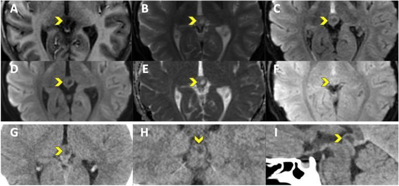
This multi-modal neuroimaging series displays a pineal cyst (indicated by yellow arrowheads) using various MRI sequences and CT reconstructions for clinical comparison. The MRI sequences (A-F) include T1-FLAIR, T2, T2-FLAIR, DWI, ADC, and T2* EPIMIX. The cyst appears as a well-circumscribed, oval lesion in the pineal region, demonstrating characteristic CSF-like signal intensity: markedly hyperintense on T2 (B) and ADC (E), while appearing hypointense to isointense on T1-FLAIR (A) and suppressed on T2-FLAIR (C). There is no evidence of restricted diffusion on DWI (D). The CT component (G-I) shows the lesion in axial, coronal, and sagittal planes using a soft tissue window, where the cyst presents as a focal area of low attenuation (hypodensity) relative to the adjacent brain parenchyma. The series illustrates the utility of multi-planar imaging in localizing midline intracranial structures and distinguishing benign pineal cysts from solid neoplasms by evaluating signal consistency across sequences.
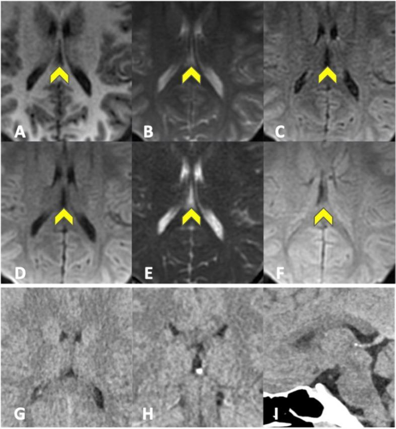
This composite diagnostic image presents a multi-modal neuroradiological evaluation of the brain, comparing different MRI sequences with CT scans to highlight midline cerebrospinal fluid (CSF)-filled cavities, specifically a cavum septum pellucidum or cavum vergae. Panels A-F display images from an EPIMix sequence, including T1-FLAIR (A), T2-weighted (B), T2-FLAIR (C), Diffusion-Weighted Imaging (DWI - D), Apparent Diffusion Coefficient (ADC - E), and T2* (F). In these MRI sequences, yellow arrowheads point to the CSF-filled space between the lateral ventricles. The fluid demonstrates characteristic signal intensities: hypointense on T1-FLAIR and T2-FLAIR, and hyperintense on T2 and ADC, consistent with simple fluid properties without restricted diffusion. Panels G-I provide CT correlation in axial (G), coronal (H), and sagittal (I) planes using soft tissue windows, where the cavities appear as hypodense (dark) areas along the midline. This comparison illustrates how small anatomical variants, often considered incidental findings, are visualized across different imaging physics and plane orientations for diagnostic classification in clinical neurology.
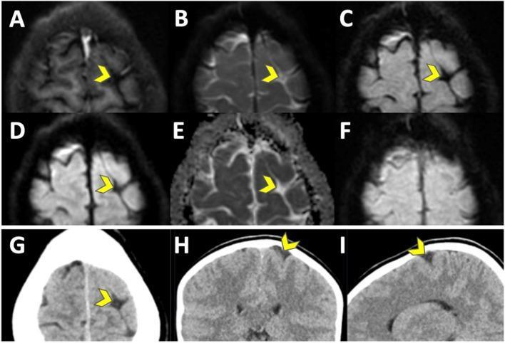
This composite diagnostic image displays a comparison between EPIMIX MRI sequences and CT scans of the human brain in a clinical neuroimaging context. Panels A-F represent various MR imaging modalities: T1-FLAIR (A), T2-weighted (B), T2-FLAIR (C), Diffusion-Weighted Imaging (DWI) (D), Apparent Diffusion Coefficient (ADC) map (E), and T2* gradient-echo (F). Panels G-I show non-contrast CT scans in axial (G), coronal (H), and sagittal (I) planes. The primary finding, indicated by a yellow arrowhead across all modalities, is a prominently widened sulcus in the superior frontal region of the left hemisphere. The visual contrast of the cerebrospinal fluid (CSF) within the sulcus varies by modality: it appears dark (hypointense) on T1-FLAIR, DWI, and T2*, and bright (hyperintense) on T2, T2-FLAIR, and ADC. On the CT images, the widened sulcus is consistently hypodense relative to the adjacent gray and white matter. This case illustrates the radiological appearance of a wide sulcus, an incidental finding sometimes associated with differential diagnoses like focal cortical dysplasia or focal atrophy.
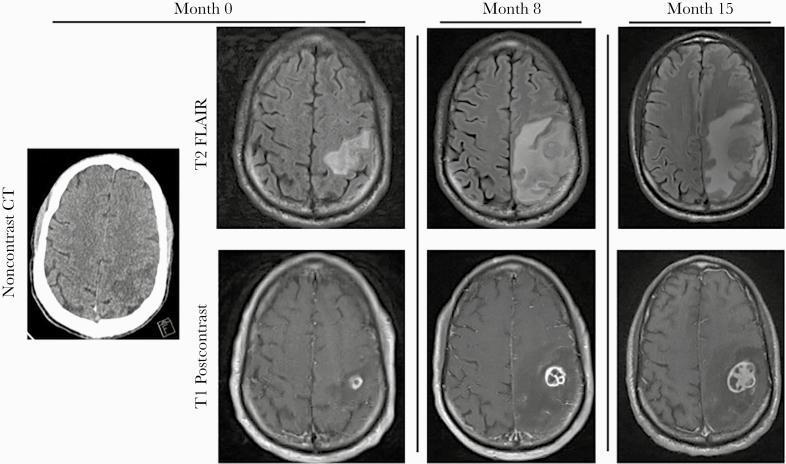
A longitudinal comparison of brain imaging across three time points (Month 0, 8, and 15) using noncontrast head CT, T2-weighted fluid-attenuated inversion recovery (FLAIR) MRI, and T1-weighted post-contrast MRI. The axial images focus on a progressive intracranial lesion in the left parietal lobe. At Month 0, the CT and MRI show a small rim-enhancing lesion with mild surrounding vasogenic edema. By Month 8, the T2 FLAIR sequence demonstrates a significant increase in hyperintense signal indicating worsening vasogenic edema, while the T1 post-contrast scan reveals a larger, multiloculated rim-enhancing mass. By Month 15, the lesion exhibits further expansion with a complex, internal honeycomb-like multilocular enhancement pattern. The associated T2 FLAIR hyperintensity shows extensive progression across the left hemisphere, causing mass effect with sulcal effacement and a developing midline shift. This radiographic series illustrates the evolution of a parietal abscess and secondary inflammatory changes, such as Immune Reconstitution Inflammatory Syndrome (IRIS), in the context of advanced HIV/AIDS.
Radiology of the CNS: modalities, indications, advantages and disadvantages
1. Anatomy and physiology of the CNS (brief)
- Brain:
- Cerebrum: frontal, parietal, temporal and occipital lobes.
- Basal ganglia and thalamus.
- Diencephalon: hypothalamus, pituitary, pineal.
- Brainstem: midbrain, pons, medulla.
- Cerebellum: in the posterior fossa.
- Spinal cord:
- Runs from the foramen magnum to the conus medullaris (about L1-L2 in adults).
- Below this is the cauda equina.
- Gray matter is central and white matter is peripheral.
- Coverings: Dura, arachnoid and pia mater (the meninges). The subarachnoid space contains CSF.
- Ventricular system:
- Two lateral ventricles, the third ventricle (via the foramina of Monro), the aqueduct, and the fourth ventricle.
- CSF leaves the fourth ventricle through the foramina of Luschka and Magendie.
- CSF is made by the choroid plexus and absorbed by arachnoid granulations into the venous sinuses.
- Blood supply:
- Anterior circulation: internal carotid arteries (ACA, MCA).
- Posterior circulation: vertebral and basilar arteries (PCA, cerebellar arteries).
- The circle of Willis joins them.
- Venous drainage is through the dural sinuses to the internal jugular veins.
- Protective bony cover: Skull and vertebral column.
- Blood-brain barrier (BBB):
- Tight endothelial junctions keep iodinated and gadolinium contrast inside the vessels.
- Contrast enhancement therefore means the BBB is broken (tumor, abscess, demyelination, subacute infarct).
- It also means the lesion is highly vascular, or the structure lies outside the BBB (pituitary, choroid plexus, dura).
- Energy use:
- The brain is about 2% of body weight but uses about 20% of cardiac output and oxygen.
- It depends on glucose, which is why FDG-PET and perfusion SPECT work.
- Cerebral blood flow is autoregulated, and neuronal activity is coupled to flow (the basis of fMRI).
- Water content:
- Gray matter has more water and white matter has more myelin lipid. This produces the different densities on CT and signals on MRI.
- Edema (vasogenic or cytotoxic) is the commonest tissue change imaged.
- CSF: About 500 mL is produced per day, with 100-150 mL present at any time. Disturbed flow gives hydrocephalus. Leaks are shown by cisternography.
2. Imaging modalities
A. Sonography (ultrasound, including Doppler)
- Neonates and infants through the anterior fontanelle: Intraventricular and germinal matrix hemorrhage, periventricular leukomalacia, hydrocephalus, congenital malformations, and some infections.
- Neonatal spine: Tethered cord, low conus, dysraphism, and sacral dimple screening. This works only in the first few months, before the posterior elements ossify.
- Carotid and vertebral duplex: Atheromatous stenosis, dissection, and occlusion.
- Transcranial Doppler:
- Vasospasm after subarachnoid hemorrhage.
- Sickle-cell stroke risk screening.
- Detection of emboli.
- Supportive testing for brain death.
- Intraoperative ultrasound: Localizing tumors and spinal lesions.
- Prenatal ultrasound: Screening for neural tube and brain anomalies.
- Portable and bedside.
- Real-time imaging.
- No ionizing radiation.
- No sedation is usually needed.
- Cheap and widely available.
- Doppler gives hemodynamic information.
- Skull blocks the beam once the fontanelle closes.
- Operator dependent.
- Limited field of view and tissue contrast, especially for the posterior fossa and brain convexity.
- Poor detection of subtle parenchymal lesions.
- Needs follow-up with CT or MRI if abnormal.
B. Contrast radiological methods
| Method | Contrast | Main indications |
|---|---|---|
| Plain radiographs of skull and spine | None | Trauma and fractures, bone destruction, congenital craniovertebral anomalies, scoliosis, and instability (flexion/extension views). Rarely used for the brain itself. |
| Myelography / CT myelography | Intrathecal non-ionic iodinated contrast (e.g. iohexol) | Cord and root compression when MRI is contraindicated or unavailable, root avulsion in brachial plexus injury, CSF leak, post-operative spine with metal artifact |
| Cisternography (CT) | Intrathecal iodinated contrast | CSF rhinorrhea or otorrhea, arachnoid cysts |
| Conventional catheter angiography (DSA) | Intra-arterial iodinated contrast | Aneurysm, AVM and dural AV fistula, vasculitis, stenosis and dissection, and planning or performing endovascular therapy (coiling, thrombectomy, embolization) |
| CT angiography / CT venography | IV iodinated | Acute stroke, aneurysm, vessel occlusion, venous sinus thrombosis |
| Contrast-enhanced CT | IV iodinated | Tumor, abscess, infection, subacute vascular lesions, and suspected BBB disruption when MRI is unavailable |
| Contrast-enhanced MRI / MRA | IV gadolinium | Tumor, metastases, demyelination (MS), infection (meningitis, abscess, discitis, epidural abscess), post-operative spine |
| Ventriculography and pneumoencephalography | Air or contrast | Obsolete; replaced by CT and MRI |
- Contrast shows lesions that are isodense or isointense on plain studies.
- It shows whether the BBB is intact.
- It gives vessel detail.
- DSA has the highest spatial resolution for small vessels and allows treatment in the same session.
- Iodinated contrast:
- Allergic reactions.
- Contrast-induced nephropathy.
- Contraindicated in severe prior reaction, renal failure, and uncontrolled thyrotoxicosis.
- Thyroid and metformin precautions.
- Gadolinium:
- Nephrogenic systemic fibrosis with older agents in severe renal impairment (eGFR below 30).
- Possible tissue deposition.
- Used cautiously in pregnancy.
- Myelography: It is invasive. Complications are post-lumbar-puncture headache, infection, nerve root injury, arachnoiditis, and seizures if ionic contrast reaches the intracranial space.
- DSA: It is invasive. It carries a small risk of stroke (roughly 0.5-1%), groin hematoma, vessel dissection, and radiation to patient and operator. It is reserved for when CTA or MRA cannot answer the question or when treatment is planned.
C. Radionuclide scintigraphy (nuclear medicine: planar, SPECT, PET)
| Technique | Tracer | Indications |
|---|---|---|
| Brain perfusion SPECT | Tc-99m HMPAO or ECD | Epilepsy focus localization (ictal hyperperfusion), stroke and vasospasm, dementia pattern (Alzheimer, frontotemporal, vascular), brain death |
| Radionuclide cisternography | In-111 DTPA (intrathecal) | CSF leak, normal-pressure hydrocephalus, shunt patency |
| Dopamine transporter scan | I-123 ioflupane (DaTscan) | Parkinsonian syndromes versus essential tremor, dementia with Lewy bodies |
| FDG-PET | F-18 fluorodeoxyglucose | Interictal hypometabolism in epilepsy, dementia, tumor grading, recurrence versus radiation necrosis, and lymphoma |
| Amyloid and tau PET | Amyloid or tau ligands | Alzheimer disease diagnosis |
| Tumor-seeking agents | Tl-201, Ga-67 | Differentiating CNS lymphoma from toxoplasmosis in AIDS (older use) |
| Bone scan | Tc-99m MDP | Spinal metastases, occult fracture, osteoid osteoma, infection |
| Tc-99m DTPA / pertechnetate flow study | Tc-99m | Brain death: absent intracranial perfusion ("hollow skull" sign) |
- Shows physiology and metabolism, often before structural change appears.
- Whole-body survey is possible (bone scan, PET).
- Highly sensitive.
- Quantifiable.
- Non-invasive, apart from intrathecal injection.
- Poor spatial resolution and little anatomical detail, so it is usually fused with CT or MRI.
- Radiation exposure.
- Findings are often non-specific.
- Costly and not widely available.
- Short-lived isotopes need a cyclotron (PET).
- Long acquisition times.
- Pregnancy is a relative contraindication.
D. Computed tomography (CT)
- Head trauma: Fractures, extradural, subdural and contusional hemorrhage. This is the first-line study.
- Acute stroke: To exclude hemorrhage before thrombolysis. CT perfusion and CTA are added for large vessel occlusion and penumbra.
- Subarachnoid hemorrhage: Acute blood is dense and CT is highly sensitive in the first 6 hours.
- Raised intracranial pressure, mass effect, herniation, and acute hydrocephalus.
- Suspected CNS infection or mass lesion when MRI is not immediately available. Harrison's states CT should be done emergently in that situation (Harrison's, Brain Imaging).
- Bone and calcification detail: Skull base and spinal fractures, calcified tumors, and spinal canal stenosis.
- Cerebral angiography and venography (CTA/CTV).
- Guidance for stereotactic biopsy and drainage.
- Spine: Trauma and bony involvement by tumor or degenerative disease. Rosen's describes CT as essential for these because it defines bone better (Rosen's Emergency Medicine).
- Fast (seconds to minutes), widely available, and relatively cheap.
- Excellent for acute hemorrhage, bone, and calcification.
- Suitable for unstable or uncooperative patients.
- Compatible with pacemakers, metal implants, and life-support equipment.
- Multiplanar reformatting and 3D reconstruction.
- Ionizing radiation (a head CT is about 2 mSv). The concern is greater in children and pregnancy.
- Soft-tissue contrast is lower than MRI.
- Early and small infarcts, brainstem and posterior fossa lesions (beam-hardening artifact), and small demyelinating plaques are missed.
- Metal and dental artifact.
- Iodinated contrast risks (see above).
- Axial acquisition is native; other planes are reconstructed.
E. Magnetic resonance imaging (MRI)
- T1: Anatomy; fat, subacute blood and gadolinium are bright; CSF is dark.
- T2: Pathology; CSF and edema are bright.
- FLAIR: T2 with CSF suppressed. Best for periventricular and cortical lesions (MS, small infarcts).
- DWI/ADC: Restricted diffusion. Detects acute infarct within minutes, plus abscess and some tumors.
- SWI/GRE: Blood products, microbleeds, calcification.
- MRA/MRV: Non-contrast or gadolinium vascular imaging.
- Advanced: Perfusion, spectroscopy, tractography, fMRI.
- Preferred modality for most CNS disease:
- Brain tumors, including posterior fossa and pituitary.
- Demyelination (MS).
- Encephalitis and meningitis.
- Abscess.
- Epilepsy. MRI is superior to CT for epileptogenic lesions such as hippocampal sclerosis and cortical malformations (Harrison's).
- Dementia and degenerative disease.
- Acute ischemic stroke: DWI detects infarct earlier and with greater sensitivity than CT.
- Spinal cord disease: Compression, myelopathy, syrinx, tumor, infection, disc disease, and trauma with cord injury. Rosen's names MRI as the preferred modality because it clearly defines the cord and soft tissues and shows edema and hemorrhage in the cord (Rosen's).
- Congenital malformations and pediatric neuroimaging (no radiation).
- Cranial nerves: Internal auditory canal, orbits.
- Vascular lesions: Cavernomas, venous thrombosis, MRA for aneurysm and stenosis.
- Pre-surgical mapping: fMRI and tractography.
- Best soft-tissue contrast, so gray and white matter and small lesions are clearly seen.
- No ionizing radiation.
- Direct multiplanar imaging in any plane (Neuroscience: Exploring the Brain, p. 593).
- No bone artifact, so the posterior fossa, brainstem and cord are well seen.
- Functional, vascular and metabolic information (fMRI, MRA, spectroscopy) in one examination.
- Long scan time (10 minutes to an hour). The patient must lie still. Sedation or anesthesia is often needed in children.
- Noisy, and claustrophobia is common.
- Absolute or relative contraindications:
- Older pacemakers and defibrillators (many newer devices are MR-conditional).
- Cochlear implants.
- Ferromagnetic intracranial aneurysm clips.
- Metallic foreign bodies in the eye.
- Some neurostimulators.
- Grainger notes that magnetic field strength may exceed 60,000 times that of the Earth and is always on, so screening is mandatory (Grainger & Allison's).
- Poor for acute hemorrhage in some sequences, cortical bone, and calcification (versus CT).
- Poor for unstable, ventilated or monitored patients.
- More expensive and less available than CT.
- Gadolinium precautions (renal function, pregnancy).
- Metal artifact from implants.
3. Quick comparison
| Ultrasound | CT | MRI | Contrast angiography/myelography | Nuclear medicine | |
|---|---|---|---|---|---|
| Radiation | None | Yes | None | Yes | Yes |
| Soft-tissue detail | Low | Moderate | Excellent | Vessels/canal only | Low (functional) |
| Bone / calcification / acute blood | Poor | Excellent | Poor | Not applicable | Not applicable |
| Speed | Real-time | Seconds | Slow | Slow | Slow |
| Best for | Neonatal brain, carotids | Trauma, acute stroke, SAH | Almost everything else, spine, posterior fossa | Aneurysm/AVM, intervention, root avulsion | Epilepsy, dementia, parkinsonism, CSF leaks, brain death |
| Key limit | Acoustic window | Poor posterior fossa, radiation | Implants, time, claustrophobia | Invasive, contrast risks | Poor resolution |
4. Practical first choices
- Acute head trauma, suspected hemorrhage, or acute stroke: Non-contrast CT first (plus CTA/CT perfusion if thrombectomy is considered).
- Neonate with a suspected intracranial bleed or hydrocephalus: Cranial ultrasound.
- Suspected cord compression, MS, tumor, encephalitis, or epilepsy work-up: MRI (with gadolinium when needed).
- Aneurysm or AVM treatment planning: CTA or MRA, then DSA.
- MRI contraindicated, spinal canal question: CT myelography.
- Functional question (focus localization, dementia type, parkinsonism, CSF leak, brain death): SPECT, PET, or cisternography.