
Classify and describe the etiology, pathogenesis, manifestations, radiologic and morphologic features and complications of Paget's disease of the bone
Reading File
This is a broad visual/morphologic question. I'll run parallel searches across headings and paragraphs for Paget's disease of bone.
Searching sources...
Finding Sources
Finding Sources
Excellent. I have strong hits from Robbins, Harrison's, Rheumatology (Elsevier), Goldman-Cecil, and Firestein & Kelley. Now I'll read the most comprehensive sections in parallel.
Reading File
Reading File
Reading File
Excellent data. Now I'll read the Robbins morphology section and the Goldman-Cecil section for clinical/radiologic features, and fetch key images in parallel.
Reading File
Reading File
Reading File
Now let me read the Robbins morphology section and get the Goldman-Cecil main content, plus fetch images.
Reading File
Reading File
Loading Image
Now I have comprehensive data from Robbins, Harrison's, Goldman-Cecil, and Rheumatology textbooks. Let me fetch the key Robbins morphology/phase diagram image and read the Goldman-Cecil clinical section for complications.
Loading Image
Reading File
I now have all the key information needed. Let me also fetch the histologic mosaic pattern image and the pelvis X-ray image from Harrison's.
Finding Sources
Loading Image
I now have comprehensive information from multiple authoritative sources. Let me fetch two more key images - the histologic mosaic pattern and the tibia X-ray.
Loading Image
Reading File
I now have all the data needed for a comprehensive answer. Let me fetch the final key image showing the histologic mosaic pattern alongside the tibia X-ray.
Loading Image
I now have everything needed for a comprehensive, well-illustrated answer. Let me compile it.
Paget's Disease of Bone (Osteitis Deformans)
Classification
Paget's disease is classified as a focal, metabolic bone disorder characterized by markedly increased but structurally disorganized bone remodeling. It is subdivided by extent:
- Monostotic (~15% of cases): one bone affected
- Polyostotic (~85% of cases): multiple, non-contiguous skeletal sites involved
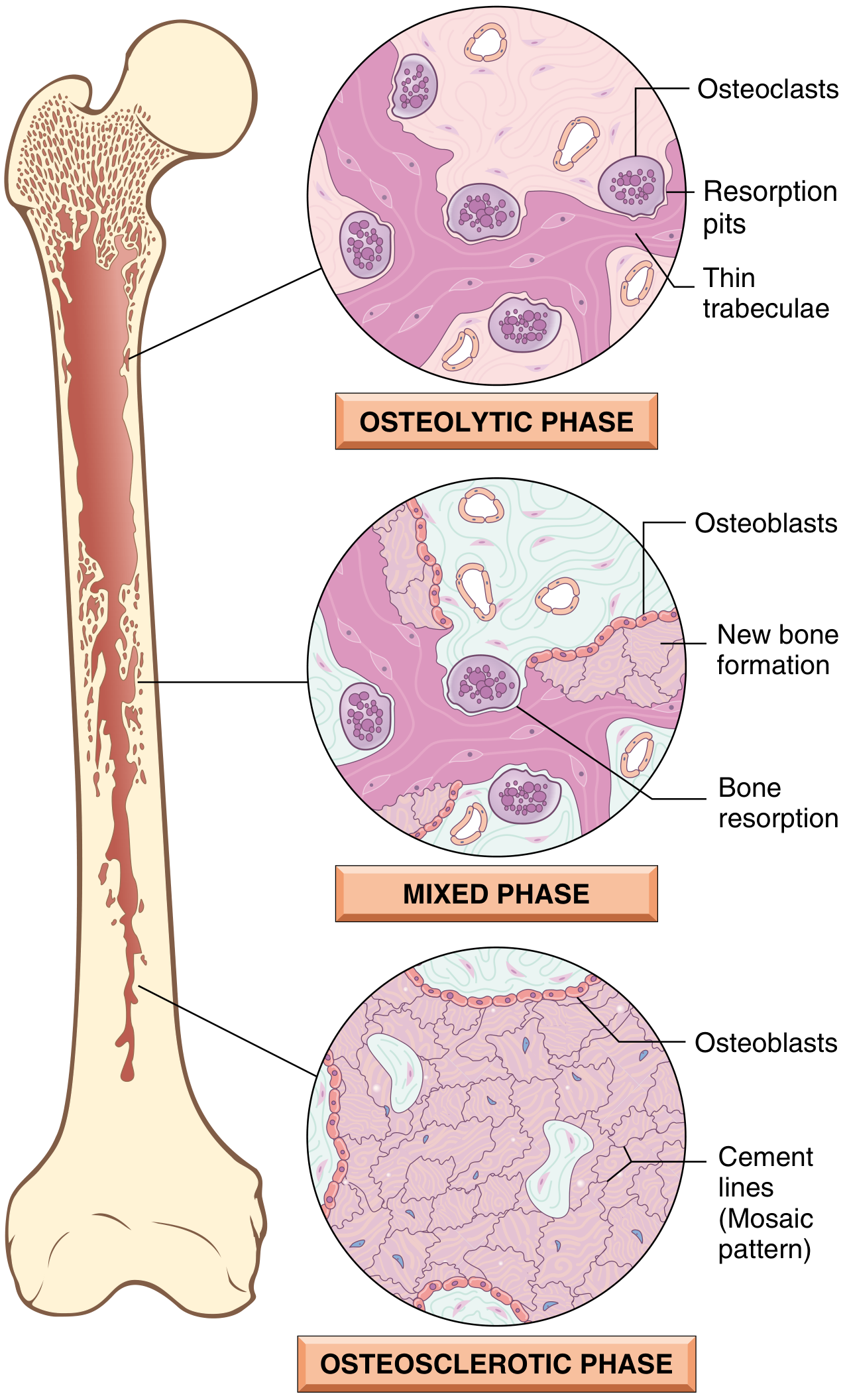
The disease follows three sequential phases:
- Initial osteolytic (resorptive) phase
- Mixed osteoclast-osteoblast phase
- Final osteosclerotic (burned-out) phase
Epidemiology
- Prevalence ~0.5-1% in the UK and US; autopsy studies reveal ~3% in those over age 40
- More common in males (ratio ~1.4-1.7:1), rising with age
- High prevalence in Western Europe (UK, France, Germany), Australia, New Zealand, and among European immigrants
- Rare in Scandinavia, East Asia, sub-Saharan Africa, and native populations of the Americas
- Prevalence and severity have decreased in recent decades for unclear reasons
Etiology
The cause remains incompletely understood; both genetic and environmental factors contribute.
Genetic Factors
- SQSTM1 (sequestosome-1/p62) mutation is the most important: found in ~60% of familial cases and ~10-16% of sporadic cases; more than 20 mutations described in or near the ubiquitin-associated (UBA) domain. p62 is a scaffold protein regulating NF-κB signaling, the master pathway for osteoclast differentiation. Mutations increase NF-κB activity and thus osteoclast activity.
- RANK (TNFRSF11A) activating mutations: cause familial expansile osteolysis and early-onset Paget disease
- OPG (TNFRSF11B) inactivating mutations: loss of osteoprotegerin leads to uncontrolled osteoclastogenesis; homozygous deletions cause juvenile Paget disease (familial idiopathic hyperphosphatasia)
- VCP mutations: cause multisystem proteinopathy - inclusion body myopathy + Paget disease + frontotemporal dementia (± ALS)
- ZNF687 mutations: rare syndrome of Paget disease with giant cell tumors
- PFN1 (profilin-1) mutations: early-onset disease with predisposition to osteosarcoma
- Genome-wide association studies identified 7 additional loci near genes including CSF1 (M-CSF), TM7SF4 (DC-STAMP), OPTN (optineurin), and RIN3 - all converging on osteoclast function and autophagy
- Autosomal dominant transmission with variable penetrance; positive family history in 15-25% of patients (raises prevalence 7-10-fold in first-degree relatives)
Environmental / Viral Factors
- In vitro studies raised the possibility of chronic paramyxoviral infection (measles virus, canine distemper virus, respiratory syncytial virus) of osteoclast precursors - nuclear inclusions in pagetic osteoclasts resemble nucleocapsids of paramyxoviruses
- Geographic variation and declining incidence over time support an environmental trigger
- Mechanical loading, skeletal injuries, rural exposures, and toxins have also been proposed
Pathogenesis
The central defect is abnormal osteoclast behavior driven by the interaction of genetic susceptibility (especially SQSTM1/NF-κB dysregulation) with environmental triggers.
Key mechanisms:
- Osteoclast hyperactivation: pagetic osteoclasts are increased in number, markedly enlarged, and hypernucleated (up to 100 nuclei vs. normal 3-5). They show hypersensitivity to RANKL and 1,25(OH)₂D₃
- RANKL/OPG imbalance: marrow stromal cells from pagetic lesions overexpress RANKL; osteoclast precursor recruitment is amplified by IL-6 (elevated in blood of active disease)
- Osteoblastic response: compensatory osteoblastic new bone formation is markedly increased, exceeding the amount resorbed - producing enlarged, deformed bones
- Woven bone: the new bone is deposited in a disorganized (woven) pattern with impaired mechanical strength
- Autophagy defects: nuclear inclusions in pagetic osteoclasts may represent aggregates of undegraded proteins due to defects in autophagy; proteins encoded by VCP, OPTN, and SQSTM1 all regulate autophagy
- Marrow fibrosis and hypervascularity: increased vascularity causes warmth overlying lesions; marrow is replaced by loose fibrovascular tissue in active phases
- Focal nature: possibly explained by somatic mutations in affected bones causing local osteoclast hyperactivity, or by early-life mechanical injury creating a focus for remodeling
Clinical Manifestations
An estimated 7-16% of patients come to medical attention; ~20% of those are completely asymptomatic, with Paget disease detected incidentally on lab work (elevated ALP) or imaging.
Symptoms
| System | Manifestations |
|---|---|
| Bone pain | Most common symptom; from microfractures, increased bone turnover, or secondary joint disease |
| Skull | Headache, skull enlargement (hat size increase), leontiasis ossea (lion-face), platybasia (invagination of skull base), compression of posterior fossa |
| Cranial nerves | Sensorineural or conductive deafness (most common CN complication; from temporal bone involvement or cochlear nerve compression), vertigo, facial nerve palsy, vision changes |
| Spine | Back pain, spinal stenosis, nerve root compression, kyphosis, radiculopathy, myelopathy |
| Pelvis | Pelvic bowing, hip pain from secondary osteoarthritis, protrusio acetabuli |
| Long bones | Anterior bowing of femur and tibia, leg-length discrepancy, secondary severe osteoarthritis of adjacent joints |
| Skin | Warmth overlying pagetic bones due to hypervascularity |
| Cardiac | High-output heart failure in severe polyostotic disease (increased blood flow acting as AV shunt) |
| Labs | Markedly elevated serum ALP (bone-specific); bone resorption markers (N-telopeptide, C-telopeptide) elevated; serum Ca²⁺ and PO₄³⁻ usually normal |
Skeletal Distribution
- Axial skeleton and proximal femur involved in up to 80% of cases
- Most commonly: pelvis > lumbar spine > femur > thoracic spine > skull > tibia
Radiologic Features
Plain radiographs are usually diagnostic. Key findings include:
Skull
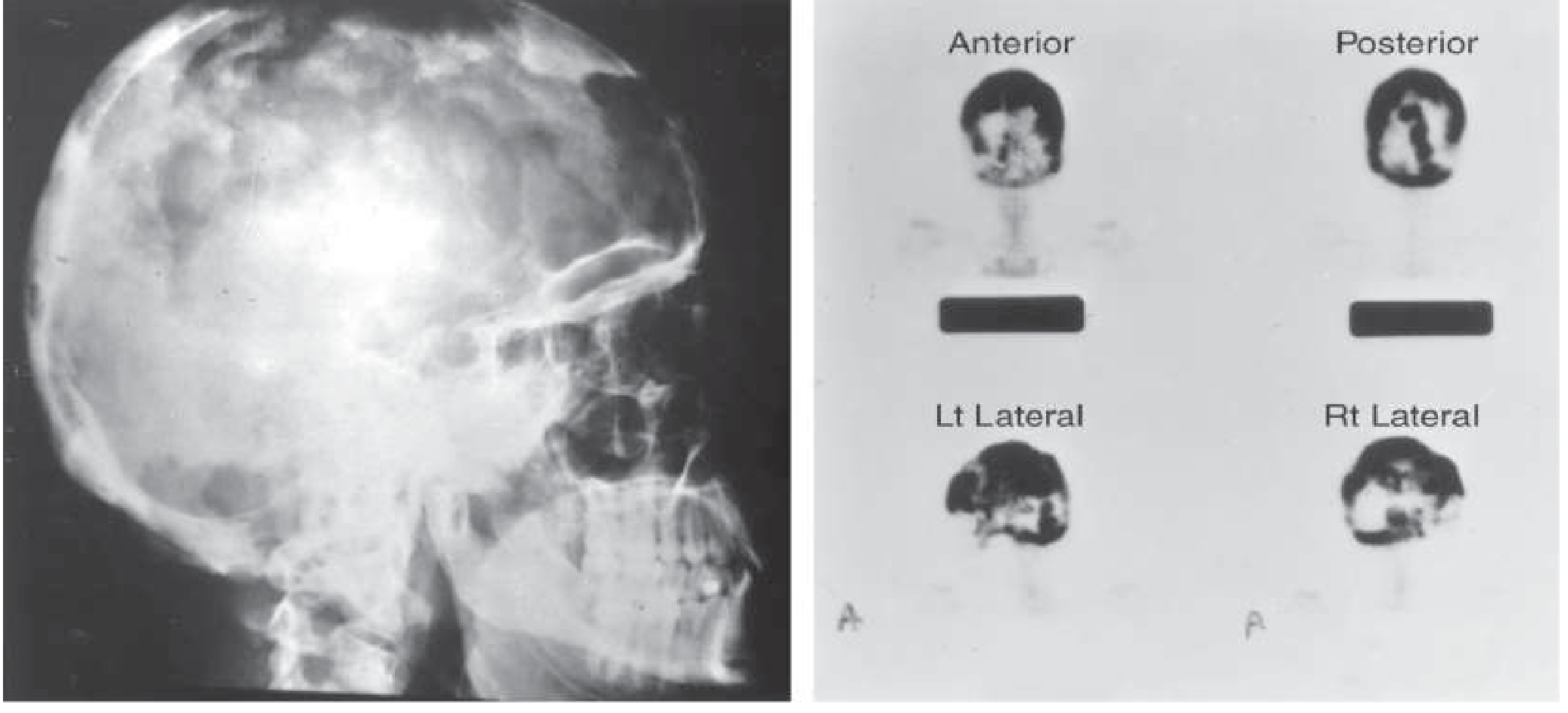
- "Cotton-wool" appearance: mixed lytic and sclerotic foci
- Osteoporosis circumscripta: well-defined lytic area (early/active phase) in frontal or occipital bone
- Thickening of diploic tables; enlargement and sclerosis of calvarium
- Bone scan: diffuse intense isotope uptake by frontal, parietal, occipital, and petrous bones
Spine
- "Picture-frame" vertebra: cortical thickening of superior and inferior endplates with central lucency
- "Ivory vertebra": diffuse radiodense enlargement of the entire vertebral body
Pelvis
- Disruption or fusion of sacroiliac joints
- Mixed porotic and radiodense lesions of ilium with coarse trabeculation (whorls)
- Brim sign: thickened and sclerotic iliopectineal line
- Softening with protrusio acetabuli; axial migration of hips
Long Bones
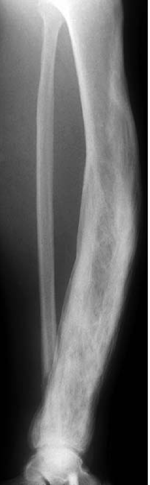
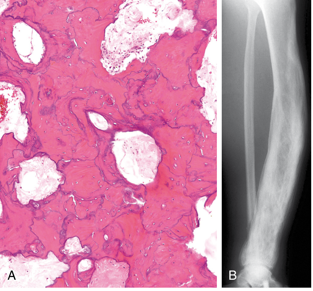
- Enlargement/expansion of the whole bone or involved segment
- Cortical thickening (both periosteal and endosteal)
- Coarsening of trabecular markings
- Bowing deformity (typically anterior in tibia/femur)
- Mixed lucency and sclerosis
- "Blade of grass" or "flame" sign: advancing lytic front at the edge of lesion in active phase
Other Imaging
- ⁹⁹ᵐTc bone scan: most sensitive modality for identifying active lesions and disease extent; less specific than plain films
- CT: useful for assessment of fracture, cortical detail, and nerve compression
- MRI: necessary when sarcomatous transformation, giant cell tumor, or metastatic disease is suspected in pagetic bone
Morphologic Features
Gross Pathology
- Affected bones are enlarged, thickened, and deformed
- Cortices are soft and porous - lack structural stability
- Coarsely thickened trabeculae
- Cut surface shows increased vascularity
Histology - Phase-Dependent
Phase 1: Osteolytic Phase
- Numerous abnormally large osteoclasts with up to 100 nuclei (normally ≤4)
- Prominent resorption pits (Howship's lacunae)
- Thin trabeculae being actively destroyed
- Some osteoclasts contain intranuclear inclusions (paramyxovirus-like)
Phase 2: Mixed Phase
- Both osteoclasts and osteoblasts active simultaneously
- Osteoblastic new bone formation alongside continued resorption
- Poorly mineralized woven bone being laid down
- Marrow replaced by loose fibrovascular connective tissue rich in osteoprogenitor cells and blood vessels
- Large osteoclasts visible close to areas of new bone formation; extensive marrow fibrosis
Phase 3: Osteosclerotic (Burned-out) Phase
- Hallmark: mosaic pattern of lamellar bone (jigsaw puzzle-like)
- Produced by unusually prominent, haphazardly oriented cement lines joining units of lamellar bone in all directions
- Cell activity decreases; fibrovascular tissue recedes and is replaced by normal marrow
- Coarsely thickened trabeculae; soft, porous cortices
Complications
1. Fractures
- "Chalk-stick" (transverse) fractures: characteristic of Paget disease; typically involve weight-bearing long bones (femur, tibia)
- Result from structurally inferior woven bone and cortical softening
- Vertebral compression fractures can cause kyphosis or spinal cord injury
2. Secondary Osteoarthritis
- Most common joint affected: hip (from femoral head deformity and protrusio acetabuli)
- Also knee and spine; results from bony deformity distorting joint mechanics
3. Neurologic Complications
- Sensorineural and conductive deafness: most common; temporal bone involvement affecting the ossicles and cochlea
- Spinal stenosis and radiculopathy from vertebral enlargement
- Cranial nerve palsies (VII, II)
- Platybasia with posterior fossa compression
- Spinal cord compression and myelopathy
4. Neoplastic Transformation (most feared)
- Secondary osteosarcoma: occurs in <1% of all Paget disease patients overall, but 5-10% of those with severe polyostotic disease; rapidly fatal with very poor prognosis
- Other tumors: giant cell tumor (especially in ZNF687 mutation carriers), fibrosarcoma, chondrosarcoma
- Suspect when a previously stable lesion rapidly enlarges or becomes increasingly painful
5. Cardiovascular Complications
- High-output cardiac failure: rare; seen in severe polyostotic disease where increased bone vascularity functions as an arteriovenous shunt
- May exacerbate underlying coronary artery disease
6. Hypercalcemia
- Typically only during immobilization (when osteoclastic resorption is uncoupled from osteoblastic deposition) - may precipitate nephrolithiasis
7. Systemic Proteinopathy (VCP mutations)
- Inclusion body myopathy + frontotemporal dementia + ALS in addition to bone disease
Biochemical Markers
| Marker | Significance |
|---|---|
| Serum ALP (total) | Test of choice - markedly elevated; reflects extent and severity; used for monitoring treatment response |
| Bone-specific ALP / PINP | Useful when total ALP normal but disease active at one site |
| Serum/urine N-telopeptide, C-telopeptide | Bone resorption markers; elevated in active disease; fall faster than ALP with treatment |
| Serum Ca²⁺, PO₄³⁻ | Usually normal (unless immobilized) |
| Osteocalcin | Not reliably elevated; not recommended |
Summary
Paget's disease of bone is a focal remodeling disorder driven primarily by pathologic osteoclast overactivation, most commonly linked to SQSTM1/NF-κB pathway dysregulation and possible paramyxoviral environmental triggers. It evolves through three histologic phases (lytic → mixed → sclerotic), producing the pathognomonic mosaic cement-line pattern on histology. Radiologically, it shows characteristic bone expansion, cortical thickening, mixed lysis/sclerosis, bowing deformities, and site-specific patterns (cotton-wool skull, picture-frame vertebra, brim sign in pelvis). Complications range from fractures and osteoarthritis to nerve compression and, most seriously, osteosarcomatous transformation in severe polyostotic disease.
Sources: Robbins, Cotran & Kumar Pathologic Basis of Disease (9780443264528, p. 1090-1091) | Harrison's Principles of Internal Medicine 22E (9781265977061, p. 3355-3358) | Goldman-Cecil Medicine (9780323930345, p. 2651-2653) | Firestein & Kelley's Textbook of Rheumatology (9780323935364) | Rheumatology 2-Volume Set, Elsevier (9780702081330, p. 572-691)
This is a shared conversation. Sign in to Orris to start your own chat.