
Lab diagnosis of MI
Reading File
Now let me search the medical library for lab diagnosis of MI in parallel:
Finding Sources
Finding Sources
Searching PubMed
"myocardial infarction" AND "biomarkers" AND "diagnosis"
Good - Henry's Clinical Diagnosis and Management by Laboratory Methods has a dedicated CARDIAC TROPONIN section. Let me read that plus search for broader MI lab markers:
Reading File
Finding Sources
Finding Sources
Excellent results. Let me now read the key sections from Harrison's 22E (2025) and Henry's Clinical Diagnosis - the most authoritative sources:
Reading File
Reading File
Reading File
I have the key data. Let me now fetch the biomarker kinetics graph (Fig 9.13) and also read the Henry's specific cardiac troponin section and Goldman-Cecil MI lab section:
Loading Image
Reading File
Reading File
I now have comprehensive data from multiple authoritative textbooks. Let me also quickly check for the LDH section and Washington Manual data:
Finding Sources
Reading File
I now have everything needed for a comprehensive answer. Let me compile the full response:
Laboratory Diagnosis of Myocardial Infarction (MI)
Principle
When myocardial cells are damaged, intracellular proteins leak through disrupted cell membranes into the bloodstream. The AMI (4th Universal Definition) requires a rise and/or fall in cardiac biomarker values with at least one value above the 99th percentile upper reference limit (URL), in the setting of symptoms, ECG changes, or imaging evidence of ischemia.
- Harrison's Principles of Internal Medicine 22E, p. 2159
Primary Biomarkers
1. Cardiac Troponins (cTnI and cTnT) - GOLD STANDARD
Troponin I and Troponin T are components of the cardiac contractile apparatus. Their amino acid sequences differ from skeletal muscle isoforms, allowing highly specific monoclonal antibody-based assays.
| Feature | Details |
|---|---|
| Onset of rise | 2-4 hours after MI (detectable within 1 hour with hs-assays) |
| Peak | ~24-48 hours (may peak earlier with reperfusion) |
| Duration of elevation | 7-10 days (up to 14 days in large infarcts) |
| Cut-off | 99th percentile of a reference population (URL) |
High-sensitivity (hs) troponin assays have detection thresholds of ~1-2 ng/L and a 99th percentile URL of ~10-30 ng/L. They enable:
- Rapid rule-out in 1-2 hours (serial testing)
- Negative predictive value >99% at presentation if patient is >2-3 hours from symptom onset
- Serial measurement at 0 h and 1-3 h (hs-assay) or 0 h and 3-6 h (conventional assay)
cTnI vs cTnT:
-
cTnI is entirely cardiac-specific; never expressed outside the heart under normal conditions
-
cTnT can be elevated in neuromuscular diseases (rare cases) without cardiac involvement - in such cases, cTnI is preferred
-
In renal failure, cTnT may be disproportionately elevated; cTnI is preferred
-
Henry's Clinical Diagnosis and Management by Laboratory Methods, p. 313; Goldman-Cecil Medicine, p. 657
Causes of troponin elevation other than MI (important for interpretation):
- Myocarditis, pericarditis
- Heart failure, pulmonary embolism
- Stroke, intracranial hemorrhage
- Septic shock, renal failure
- Cardiac contusion, cardioversion
2. Creatine Kinase-MB (CK-MB)
CK-MB is an isoenzyme of creatine kinase predominantly found in myocardium. It is no longer the recommended initial diagnostic marker but has specific utility.
| Feature | Details |
|---|---|
| Onset | 2-4 hours after MI |
| Peak | 24-48 hours |
| Return to normal | ~72 hours (3 days) |
| Relative index | CK-MB mass/CK activity ≥2.5 suggests myocardial source |
Remaining clinical role of CK-MB:
-
Diagnosis of early reinfarction - because CK-MB returns to normal faster than troponin (~72h vs 7-10 days), it is useful when re-MI is suspected in a patient already with an elevated troponin from the index event
-
CK-MB lacks specificity because it is present in both skeletal and cardiac muscle
-
Harrison's Principles of Internal Medicine 22E, p. 2159; Washington Manual of Medical Therapeutics
3. Myoglobin
| Feature | Details |
|---|---|
| Onset | Earliest marker - rises within 1-2 hours |
| Peak | 4-8 hours |
| Return to normal | 24 hours |
| Specificity | Low - present in skeletal muscle as well |
Myoglobin was historically useful for its early negativity (a normal myoglobin at 4-8 hours after symptom onset has good negative predictive value), but its poor specificity has limited its use. It has been largely superseded by hs-troponin assays.
4. Lactate Dehydrogenase (LDH)
| Feature | Details |
|---|---|
| Onset | 24-48 hours |
| Peak | 3-6 days |
| Duration | Up to 8-14 days |
| Isoenzyme | LDH1 > LDH2 ("flipped ratio") is cardiac-specific |
LDH1/LDH2 ratio >1 ("LDH flip") indicates myocardial damage. LDH is a late marker - historically useful for diagnosing MI when the patient presents late (>48-72h after symptom onset, after CK-MB has normalized). Now largely replaced by troponin's prolonged elevation.
Biomarker Kinetics Graph
This graph illustrates the temporal rise and fall of key biomarkers after MI:
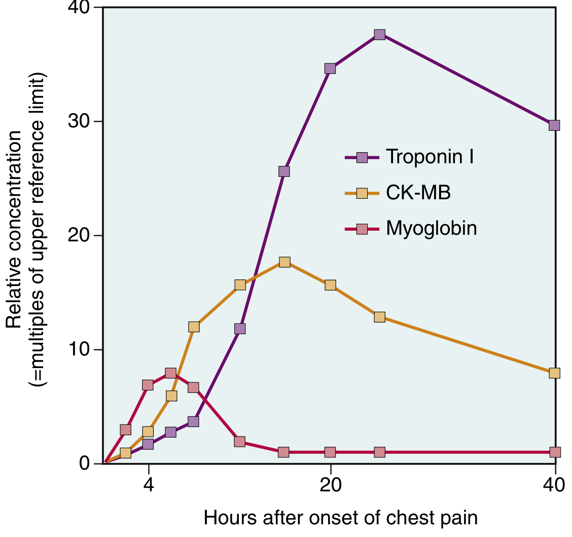
Fig. 9.13 from Robbins & Kumar Basic Pathology - Note how Troponin I rises highest and persists longest (7-10 days), CK-MB peaks at 24h and returns to normal by ~72h, and Myoglobin rises earliest but falls back quickly within 24h.
Summary Table: Biomarker Comparison
| Marker | Onset | Peak | Returns to Normal | Specificity | Current Role |
|---|---|---|---|---|---|
| cTnI / cTnT | 2-4 h (hs: <1h) | 24-48 h | 7-14 days | Very high | Primary marker - Gold standard |
| CK-MB | 2-4 h | 24-48 h | ~72 h | Moderate | Reinfarction detection |
| Myoglobin | 1-2 h | 4-8 h | 24 h | Low | Early rule-out (superseded) |
| LDH (LDH1>LDH2) | 24-48 h | 3-6 days | 8-14 days | Low-moderate | Late presentation (historical) |
Other Routine Laboratory Tests in MI
Along with cardiac biomarkers, the following are obtained in all patients:
- CBC - WBC leukocytosis (12,000-15,000/μL) peaks 2-4 days after MI (polymorphonuclear); provides baseline before antiplatelet/anticoagulant therapy
- Metabolic panel (BMP/CMP) - Electrolytes, glucose, renal function
- Fasting lipid panel - Baseline for statin therapy (note: lipids may fall in first 24-48h after MI)
- Coagulation tests (PT, aPTT) - Baseline before thrombolytic/anticoagulant therapy
- BNP/NT-proBNP - Elevated with ventricular wall stress; provides prognostic information; severe elevation suggests large infarction
- Blood glucose - Hyperglycemia is common; a marker of stress and poor prognosis
- Goldman-Cecil Medicine, pp. 657-658; Washington Manual of Medical Therapeutics, p. 133
Diagnostic Criteria (4th Universal Definition of MI)
The diagnosis of AMI requires acute myocardial injury (rising and/or falling troponin with ≥1 value above 99th percentile URL) plus at least one of:
- Symptoms of myocardial ischemia
- New ischemic ECG changes
- Development of pathological Q waves
- Imaging evidence of new loss of viable myocardium or wall motion abnormality in a pattern consistent with ischemic etiology
- Intracoronary thrombus identified on angiography or autopsy
The distinction between MI and other causes of myocardial injury (myocarditis, stress cardiomyopathy) requires clinical context - troponin elevation alone is not sufficient; a temporal rise-and-fall pattern must be demonstrated.
- Harrison's Principles of Internal Medicine 22E, p. 151
This is a shared conversation. Sign in to Orris to start your own chat.