
Orris
Hey! I'm Orris. How can I help you today?
Tell me about measles
Measles is a highly contagious viral infection caused by the Morbillivirus. It spreads via respiratory droplets and has a basic reproduction number (R0) of 12-18, making it one of the most contagious infectious diseases known. It primarily affects children but can infect anyone who is not immune. The classic presentation includes fever, cough, coryza, conjunctivitis, Koplik spots (pathognomonic white spots on the buccal mucosa), and a characteristic maculopapular rash spreading from the head downward. Complications can include pneumonia, encephalitis, and death. The MMR vaccine is highly effective at prevention.
Searching sources...
Reading File
Searching the Web
measles 2025 2026 outbreaks epidemiology updates
Now let me search the medical library for detailed textbook content on measles, in parallel with a PubMed search:
Finding Sources
Searching PubMed
"measles"[MeSH Terms] AND "vaccination"[MeSH Terms]
Reading File
Reading File
Reading File
Reading File
Loading Image
Here is a thorough overview of measles, drawing from authoritative medical textbooks and current epidemiological data.
Measles (Rubeola)
Causative Agent & Transmission
Measles is caused by the measles virus, a single-stranded RNA virus of the Paramyxoviridae family (the same family as mumps, RSV, parainfluenza, and human metapneumovirus). There is only one serotype worldwide.
It spreads primarily by the airborne route via aerosolized respiratory secretions - making it one of the most contagious pathogens known (R0 = 12-18). Transmission occurs from 4 days before to 4 days after the rash appears. The incubation period is 9-12 days.
Pathogenesis
The virus initially replicates in respiratory epithelium, then spreads to local lymphoid tissue, causing viremia. It then disseminates widely to the conjunctiva, skin, respiratory tract, urinary tract, small vessels, lymphatics, and CNS.
Three cell-surface receptors are involved:
- SLAMF1 (Signaling Lymphocytic Activation Molecule Family Member 1) - expressed on activated lymphocytes, dendritic cells, and monocytes; serves as the initial receptor
- Nectin-4 - on the basal surface of epithelial cells; important for respiratory entry and replication
T cell-mediated immunity controls infection and paradoxically produces the rash - this is why immunocompromised patients may have less obvious rash but more severe disease. Measles also causes transient but profound immunosuppression (lasting weeks to months), leaving patients vulnerable to secondary bacterial and viral infections, which account for much of measles-related mortality.
- Robbins, Cotran & Kumar Pathologic Basis of Disease
Clinical Features
Prodrome (days 1-4)
- High fever
- Malaise
- Conjunctivitis ("red eyes")
- Coryza (runny nose), sneezing
- Prominent cough
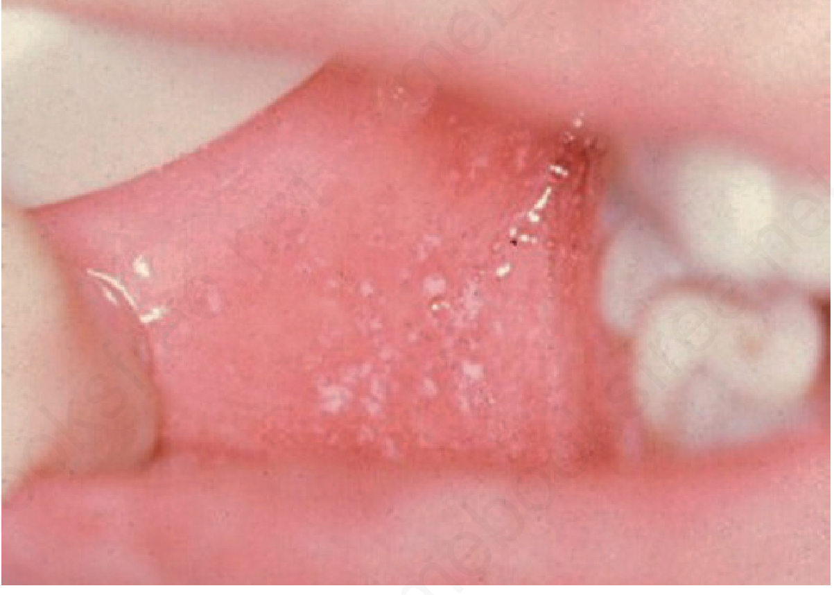
Koplik Spots (pathognomonic)
Appear during the prodrome as 1-mm white papules on an erythematous base, first on the buccal mucosa near the lower molars. They may spread to involve the wider buccal mucosa and pharynx. They precede the rash by 1-2 days and are pathognomonic for measles.
Exanthem (Rash)
The rash appears 1-7 days after the prodrome begins:
- Starts at the anterior scalp line and behind the ears
- Spreads over the face first, then over 2-3 days extends downward to trunk and extremities (cephalocaudal spread - this is slower than rubella)
- Lesions begin as discrete erythematous papules that coalesce
- Most prominent and confluent in earlier-affected areas; more discrete on extremities
- Purpura may be present on the extremities
- After 6-7 days, the rash clears with simultaneous fever resolution
Differential Diagnosis
Rubella, scarlet fever, secondary syphilis, enterovirus infections, drug eruptions
Pathology (Microscopy)
- Skin rash: dilated vessels, edema, mononuclear perivascular infiltrate
- Koplik spots: necrosis, neutrophilic exudate, neovascularization near Stensen ducts
- Lymphoid organs: marked follicular hyperplasia, large germinal centers, and Warthin-Finkeldey cells - pathognomonic multinucleate giant cells with eosinophilic nuclear and cytoplasmic inclusion bodies (also found in the lung and sputum)
Complications
| Complication | Notes |
|---|---|
| Otitis media | Most common complication |
| Pneumonia | Peribronchial/interstitial mononuclear infiltrate; major cause of death |
| Encephalitis | Rare (<1% of cases), but can be fatal |
| Thrombocytopenic purpura | Less common |
| "Black measles" | Rare DIC-like complication with purpuric/hemorrhagic rash |
| Fetal death | If infection occurs in pregnancy |
| Keratitis | Can lead to blindness; particularly in malnourished children |
| Subacute Sclerosing Panencephalitis (SSPE) | Rare, late complication (years later); defective virus accumulates mutations and spreads cell-to-cell; uniformly fatal |
| Measles Inclusion Body Encephalitis | In immunocompromised individuals |
Complications and fatalities are far more common in malnourished children and those with T-cell deficiencies (e.g. HIV). Children in low-resource countries are 10 to 1000 times more likely to die from measles than those in high-resource settings.
Diagnosis
- Clinical (Koplik spots + prodrome + cephalocaudal rash)
- Serology (IgM antibodies)
- PCR for viral RNA in respiratory secretions or urine
- Warthin-Finkeldey giant cells on histology (lung, lymph nodes)
Treatment
Mainly supportive. Vitamin A supplementation is recommended by WHO for all children with measles in resource-limited settings, as deficiency worsens outcomes significantly.
Vaccination (MMR)
-
Two doses of MMR (Measles-Mumps-Rubella) vaccine are required for full protection
-
Herd immunity threshold: ~95% coverage needed to achieve measles elimination
-
MMR is contraindicated in: pregnancy, severe immunocompromise (except non-severely immunocompromised HIV patients), and history of anaphylaxis to a prior dose
-
Common adverse effects: fever (<15%), rash (5%), transient lymphadenopathy (20%)
-
Persons born before 1957 are generally considered immune from natural infection
-
Goldman-Cecil Medicine International Edition
Current Epidemiology (2025-2026)
This is where the situation is especially urgent right now:
- The U.S. experienced its worst measles year since 1992 in 2025, with 3 deaths - more than in any year since measles elimination was declared in 2000
- In 2026, cases are already outpacing 2025. As of mid-2026, South Carolina alone has recorded over 600 confirmed cases, constituting the largest measles outbreak in U.S. history since elimination
- 94% of confirmed 2026 cases are in unvaccinated individuals
- The U.S. is at serious risk of losing its measles elimination status (Canada lost its status in November 2025 after 12+ months of continuous transmission)
- The CDC reports that falling vaccination coverage, increasing global measles activity, and travel-related importations are driving this surge
- Nationally, ~90% of Americans have received MMR, but in some regions coverage is below 60%, creating pockets of vulnerability
Recent Evidence
Recent systematic reviews confirm:
- MMR vaccine effectiveness remains high in African children (Endalamaw et al., 2024, PMID 39574018)
- Secondary vaccine failure can lead to onward transmission during outbreaks (Tranter et al., 2024, PMID 39173667)
- Adverse events following measles vaccines in infants <12 months are characterized and generally mild (Vittrup et al., 2025, PMID 39799850)
Bottom line: Measles is a vaccine-preventable, highly contagious viral disease that is resurgent globally due to declining vaccine coverage. Two doses of MMR remain the most effective preventive measure. The current U.S. outbreak situation (2025-2026) is a public health emergency that underscores the consequences of under-vaccination.
Diastasis recti vs ventral heenia
Diastasis recti is a separation of the rectus abdominis muscles along the midline (linea alba) without a true fascial defect - there is no hernia sac, so bowel/omentum cannot herniate and incarceration/strangulation do not occur. Ventral hernia involves a true defect in the abdominal wall fascia through which abdominal contents can protrude, creating a hernia sac with risks of incarceration and strangulation. They can coexist and are often confused clinically.
Searching sources...
Reading File
Searching the Web
diastasis recti vs ventral hernia diagnosis management 2025
Finding Sources
Finding Sources
Searching PubMed
"diastasis recti" AND "ventral hernia"
Reading File
Reading File
Finding Sources
Reading File
Reading File
Reading File
Here is a thorough comparison of diastasis recti vs. ventral hernia, drawn from major surgical textbooks.
Diastasis Recti vs. Ventral Hernia
These two conditions are frequently confused by patients and clinicians alike - they share a midline abdominal bulge but are fundamentally different in anatomy, risk profile, and management.
The Key Anatomical Distinction
| Feature | Diastasis Recti | Ventral Hernia |
|---|---|---|
| Definition | Separation of rectus abdominis muscles with widening/laxity of the linea alba | True defect (hole) in the abdominal wall fascia |
| Fascial defect? | No - fascia is intact, just stretched/thin | Yes - a discrete fascial ring/opening exists |
| Hernia sac? | No | Yes - peritoneal sac can contain bowel or omentum |
| Incarceration risk? | None | Yes |
| Strangulation risk? | None | Yes - can be life-threatening |
"This is not a true hernia as the midline fascia is intact, and as such incarceration and strangulation do not occur." - Schwartz's Principles of Surgery, 11th Ed.
"Patients often mistake diastasis recti for ventral hernia. Diastasis recti is a separation of the rectus abdominis muscles without a defect in the abdominal fascia... No fascial ring can be palpated." - Sleisenger & Fordtran's GI & Liver Disease
Diastasis Recti - In Detail
Definition: Abnormal separation of the rectus abdominis muscles and laxity at the linea alba. A midline separation >2 cm above the umbilicus is generally considered abnormal.
Epidemiology & Risk Factors
- Very common postpartum (up to 66% of women in the third trimester)
- Obesity
- Multiple pregnancies, older maternal age, cesarean sections
- Connective tissue disorders
- Congenital cases in newborns
- Males: less common but increasingly recognized (Axelsen et al., 2026 - PMID 41652086)
Clinical Features
- Fusiform midline bulge (long and wide, not focal)
- Bulge worsens with Valsalva or with rectus contraction (e.g., doing a sit-up)
- No palpable fascial ring edge
- May cause: back pain, abdominal wall weakness, difficulty engaging the core, cosmetic distress
- No bowel obstruction or acute abdomen
Diagnosis
- Often clinical (physical exam sufficient)
- Ultrasound: confirms diagnosis, measures inter-recti distance (IRD), rules out coexisting hernia
- CT scan: measures muscle separation precisely, useful when diagnosis is uncertain
Management
- Conservative (first-line):
- Weight loss
- Physical therapy - targeted core exercises (postpartum exercise reduces risk of progression)
- Note: standard sit-ups and crunches can worsen diastasis and should be avoided in early rehab
- Surgical (selected cases):
- Indications: disabling abdominal wall dysfunction, failure of conservative therapy, cosmesis
- Open or laparoscopic plication of the rectus sheath (midline fascial tightening)
- Mesh can be added but increases complication rates
- High long-term recurrence risk
- Also introduces a new risk of incisional hernia
Ventral Hernia - In Detail
Definition: A hernia through the anterior abdominal wall (excluding inguinal and femoral). Involves a true fascial defect through which abdominal contents can protrude.
Classification
| Type | Description |
|---|---|
| Umbilical | Through/near umbilical ring; common in infants and adults |
| Epigastric | Through linea alba above umbilicus; small defects, often contain preperitoneal fat |
| Spigelian | Through lateral edge of rectus sheath; can be occult on exam |
| Incisional | After prior abdominal surgery; up to 20% of laparotomies |
| Parastomal | Around a stoma; up to 50% of stoma patients |
| Trocar-site | After laparoscopic procedures (especially ports >10 mm) |
- Bailey and Love's Short Practice of Surgery, 28th Ed.
Risk Factors
- Prior abdominal surgery (incisional hernia)
- Obesity, advanced age, diabetes
- Wound infection after laparotomy
- Glucocorticoid use, immunosuppression
- Smoking, collagen vascular disease
- Vertical midline incisions (higher risk than transverse)
Clinical Features
- Focal bulge with a palpable fascial ring/neck
- Reducible (contents go back into abdomen) or irreducible
- Complications: incarceration (contents trapped, bowel obstruction) and strangulation (vascular compromise - surgical emergency)
Diagnosis
- Clinical exam (palpable defect edges)
- CT scan (gold standard): defines defect size, contents, and anatomy for surgical planning
- Ultrasound: useful for small or occult defects
Management
- Watchful waiting: acceptable for small, asymptomatic hernias containing only fat (low strangulation risk)
- Elective repair: indicated for symptomatic hernias causing pain or obstructive symptoms
- Urgent repair: incarcerated hernia not reducible
- Emergency repair: strangulated hernia
- Mesh-based repair recommended for defects >2 cm and all incisional hernias
- Primary suture repair acceptable only for small primary hernias <2 cm
- Approaches: open, laparoscopic (lower wound complications), or robotic
Coexistence & Clinical Importance
These two conditions frequently coexist:
- In a series of small umbilical/epigastric hernias, concomitant diastasis recti was found in 45% of patients
- Diastasis is a risk factor for hernia recurrence after suture repair - mesh should be strongly considered when diastasis is present alongside a small primary hernia
- Hernia repair alone without midline reinforcement in a patient with diastasis significantly increases the recurrence rate (31.2% in one series)
- Patients undergoing complex abdominal wall reconstruction should have their diastasis addressed at the same time
"Management of diastasis recti is a trending topic without consensus... Diastasis recti has been shown to be a risk factor for recurrence after suture repair of a primary VH." - Sabiston Textbook of Surgery
Quick Comparison Summary
| Diastasis Recti | Ventral Hernia | |
|---|---|---|
| Fascial defect | No | Yes |
| Hernia sac | No | Yes |
| Palpable ring | No | Yes |
| Strangulation risk | None | Present |
| Bulge character | Wide, fusiform | Focal, discrete |
| Worsens with Valsalva | Yes | Yes |
| Reducible | N/A | Often yes |
| First-line treatment | Conservative (PT, exercise) | Observation or surgery |
| Surgery urgency | Elective only (cosmesis/function) | Can be urgent/emergency |
| Imaging to distinguish | Ultrasound or CT | CT (surgical planning) |
Recent Evidence
A 2025 systematic review and meta-analysis on minimally invasive endoscopic onlay repair found good short-term outcomes when both diastasis recti and ventral hernia are repaired together in a single procedure (Brucchi et al., 2025 - PMID 39920372), reflecting the growing trend toward combined repair when both pathologies coexist.
This is a shared conversation. Sign in to Orris to start your own chat.