
cVC spleen
spleen venous drainage splenic vein portal system anatomy
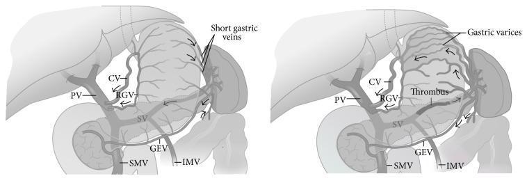
This medical illustration consists of two side-by-side comparison diagrams depicting the pathophysiology of sinistral (left-sided) portal hypertension due to splenic vein thrombosis. The diagrams show the hepatobiliary and gastrointestinal venous anatomy, including the liver, stomach, spleen, and pancreas. The left diagram represents normal venous anatomy where the splenic vein (SV), portal vein (PV), superior mesenteric vein (SMV), and inferior mesenteric vein (IMV) are patent with physiological blood flow. The right diagram illustrates the effects of a thrombus within the splenic vein. This obstruction leads to retrograde flow and increased pressure in the collateral venous pathways, specifically causing the development of prominent gastric varices, dilatation of the short gastric veins, coronary vein (CV), and gastroepiploic vein (GEV). Notably, in both the normal and pathological states, the portal vein, SMV, and IMV remain patent, highlighting that the hypertension is localized to the splenic drainage system. Arrows indicate the direction of venous blood flow, shifting toward the gastric wall in the presence of the thrombus.
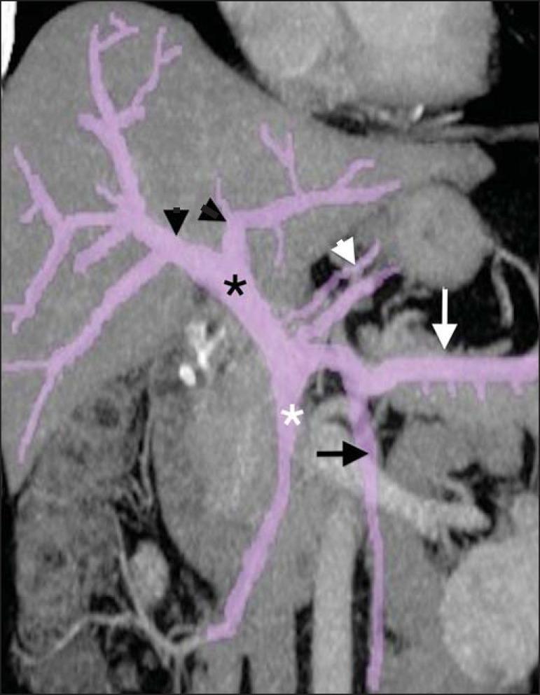
This reformatted coronal CT scan highlights the vascular anatomy of the human portal venous system, with vessels enhanced in purple for educational clarity. The image demonstrates the formation of the main portal vein (black asterisk) at the confluence of the superior mesenteric vein (white asterisk) and the splenic vein (white arrow). The splenic vein is shown receiving drainage from the gastroepiploic vein and pancreatic branches (white arrowhead). The inferior mesenteric vein (black arrow) is visible draining into the splenic vein. Distally, the main portal vein enters the liver hilum and bifurcates into its primary right and left intrahepatic portal branches (black arrowheads). This diagnostic image is essential for understanding the hepatoportal circulation, identifying the origins of portal hypertension, and mapping pre-surgical anatomy for liver or pancreatic interventions. The visualization clearly depicts the relationship between mesenteric drainage and the hepatic inflow tract.
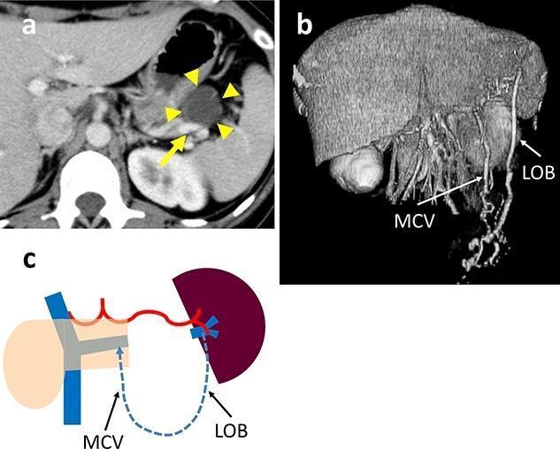
This composite educational figure illustrates the clinical imaging and venous anatomy associated with a pancreatic tail tumor. (a) Axial contrast-enhanced CT scan demonstrates a well-circumscribed, 35-mm hypodense cystic mass (yellow arrowheads) in the pancreatic tail. The lesion shows direct contact with the splenic vein (SpV, yellow arrow), which is a critical consideration for spleen-preserving distal pancreatectomy. (b) 3D vascular reconstruction highlights postoperative or compensatory collateral circulation, identifying a prominent arcade formed by a dilated left omental branch (LOB) and the middle colic vein (MCV). These vessels represent alternative venous drainage pathways from the spleen. (c) Pathophysiology diagram (schema) summarizes the venous return logic, showing blood flow (dashed blue line) from the spleen via the LOB and MCV into the portal system, bypassing the original splenic vein route. The content is designed for advanced surgical and radiological education, focusing on oncological staging and the preservation of splenic perfusion in pancreatic surgery.
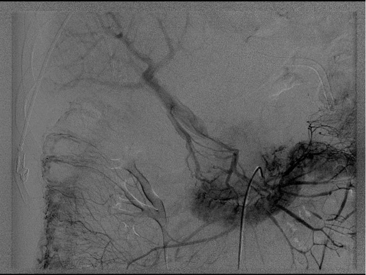
This diagnostic image is a splenoportogram (venous phase) illustrating acute portal vein thrombosis. The radiograph reveals a total lack of contrast enhancement in the main portal vein and the confluence of the superior mesenteric and splenic veins, indicating a complete vascular occlusion at this anatomical site. Significant findings include the absence of physiological venous return to the liver through the primary portal system. Instead, the image demonstrates extensive compensatory mechanisms, characterized by the visualization of numerous tortuous collateral vessels and small collateral veins providing alternative routes for venous drainage. The study highlights the diagnostic features of acute venous obstruction in the hepatobiliary system, which in this clinical context led to mesenteric ischemia. The image is a critical educational resource for understanding mesenteric vascular anatomy, pathological thrombus localization, and the secondary development of collateral circulation in response to portal hypertension or acute occlusion.
Venous Circulation (cVC) of the Spleen
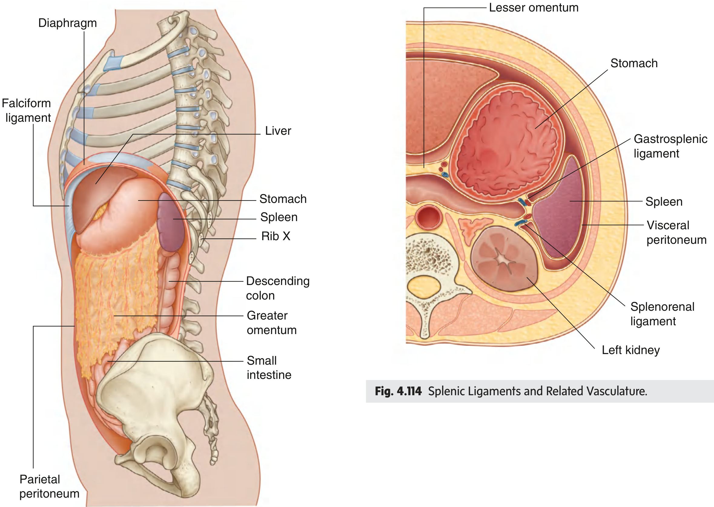
Position and Ligamentous Context
- Gastrosplenic ligament - connects spleen to the greater curvature of the stomach; carries the short gastric veins and left gastro-omental vein
- Splenorenal ligament - connects spleen to the left kidney; carries the splenic vessels (artery and vein)
Venous Drainage: The Splenic Vein
| Feature | Detail |
|---|---|
| Origin | Exits the splenic hilum on the medial surface of the spleen |
| Course | Runs along the upper border of the pancreas, accompanying the splenic artery |
| Tributaries received | Pancreatic veins, short gastric veins, left gastro-omental vein |
| Inferior mesenteric vein | Joins the splenic vein behind the body of the pancreas |
| Termination | Unites with the superior mesenteric vein (SMV) behind the head of the pancreas to form the portal vein |
The Portal Venous System: Overview
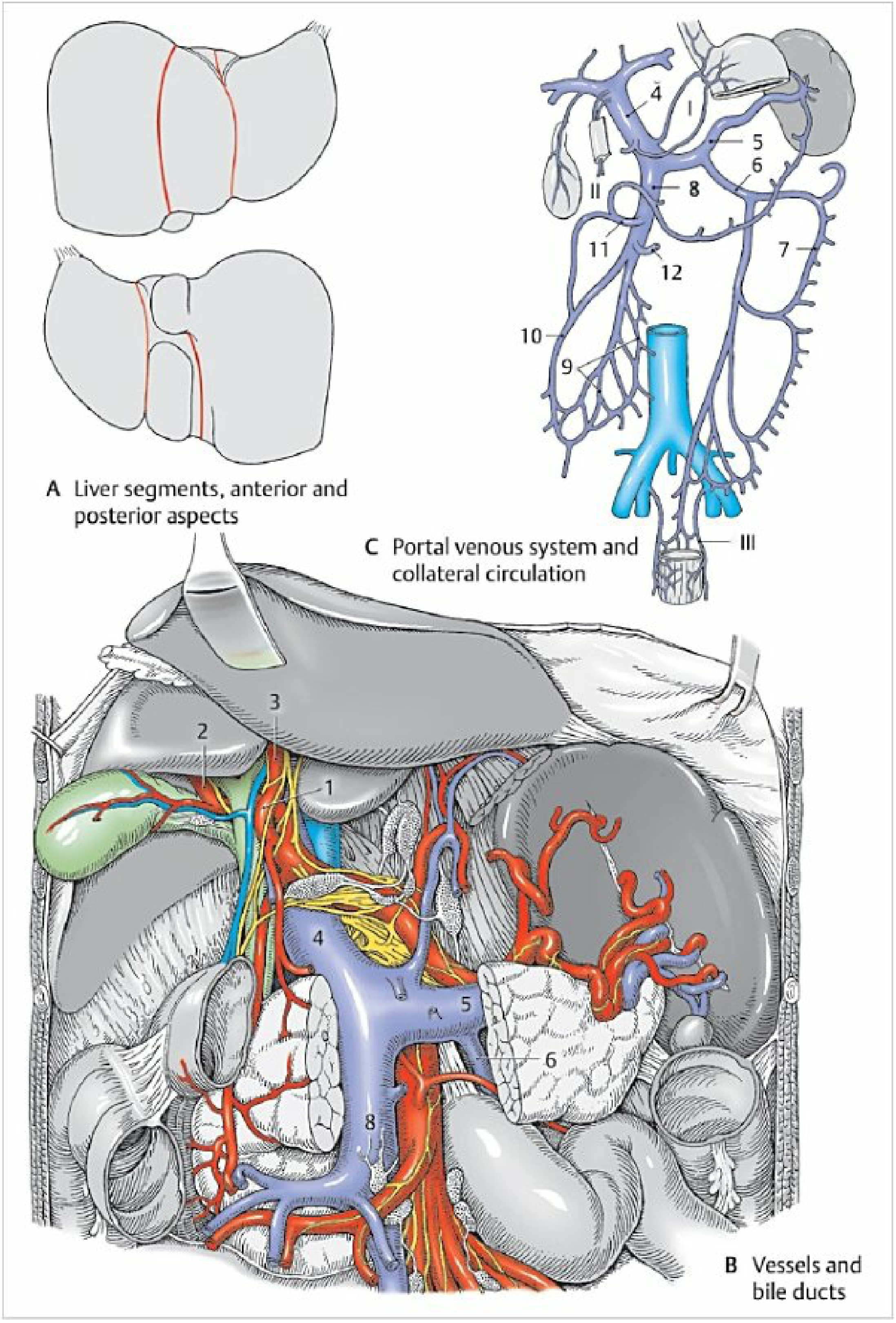
- Splenic vein - from the spleen, pancreas, short gastric veins, left gastro-omental vein
- Superior mesenteric vein (SMV) - from jejunum, ileum, right colon, pancreas
- Inferior mesenteric vein (IMV) - drains into the splenic vein (opens behind pancreatic body); receives left colic vein, sigmoid veins, superior rectal vein
Portal-Caval (cVC) Anastomoses
| Site | Portal side | Caval side | Clinical result (if hypertension) |
|---|---|---|---|
| Esophagus (I) | Left gastric (coronary) vein | Esophageal veins → azygos → SVC | Esophageal varices (most dangerous; ~60% fatal if they bleed) |
| Umbilicus/abdominal wall (II) | Paraumbilical veins (via ligamentum teres) | Thoracoepigastric veins → SVC | Caput medusae |
| Rectum (III) | Superior rectal vein → IMV | Middle and inferior rectal veins → IVC | Rectal/anorectal varices |
| Retroperitoneum | Colic/mesenteric veins (veins of Retzius) | Lumbar/renal/phrenic veins → IVC | Retroperitoneal varices |
Clinical Relevance: Splenic Vein Thrombosis
- Portal vein, SMV, and IMV remain patent
- Blood backs up through short gastric veins and coronary vein into the stomach wall
- Result: isolated gastric varices (without esophageal varices) - a surgically correctable condition (splenectomy is curative)
Summary Flow
Spleen
└─► Splenic vein (runs along upper border of pancreas)
├── receives: short gastric veins, left gastro-omental vein
├── receives: IMV (behind pancreatic body)
└─► Joins SMV (behind pancreatic head)
└─► Portal vein (5-8 cm)
└─► Liver (right & left lobes)
└─► Hepatic veins → IVC → Right heart