
Amelogenesis kuhs university bds first year essay
Reading File
Amelogenesis is the process of enamel formation, one of the most tested topics in BDS first year (Oral Histology). Here is a quick overview while I pull detailed content from the textbooks:
- Amelogenesis occurs in two main stages: secretory stage and maturation stage
- Ameloblasts are the key cells, derived from inner enamel epithelium
- Enamel matrix proteins (amelogenin, enamelin, ameloblastin) are secreted and later removed
- The process results in the hardest tissue in the body with ~96% inorganic content
Searching sources...
Finding Sources
Finding Sources
Finding Sources
Finding Sources
Reading File
Finding Sources
Reading File
Reading File
Reading File
Reading File
Now I have comprehensive content. Let me also fetch the key diagram images.
Loading Image
Loading Image
Excellent - I now have everything needed for a comprehensive KUHS BDS 1st year essay on Amelogenesis. Here is the full essay:
AMELOGENESIS
(Process of Enamel Formation)
INTRODUCTION
Amelogenesis is the process by which enamel, the hardest and most mineralized tissue in the human body, is formed. Enamel covers the anatomic crown of the tooth and consists of 96-98% calcium hydroxyapatite with only 2-3% organic material and water. Unlike other mineralized tissues, enamel is derived from epithelium (specifically from the inner enamel epithelium of the enamel organ), not from connective tissue. Once fully formed and after tooth eruption, enamel cannot be replaced because the cells responsible for its formation - the ameloblasts - are lost.
ORIGIN OF AMELOBLASTS
Ameloblasts develop from the ectodermal lining of the embryonic oral cavity. During tooth development, the inner enamel epithelium (IEE) cells of the enamel organ differentiate into pre-ameloblasts and then into secretory-stage ameloblasts. This differentiation is triggered by signaling from the adjacent odontoblasts and the dental papilla.
The enamel organ consists of four cell layers:
- Outer enamel epithelium (OEE) - outermost single layer
- Stellate reticulum - star-shaped cells, provides structural support and nutrients
- Stratum intermedium - flat cells adjacent to ameloblasts, important during secretory stage
- Inner enamel epithelium (IEE) - differentiates into ameloblasts
STAGES OF AMELOGENESIS
Amelogenesis is classically divided into two main stages:
1. SECRETORY STAGE (Matrix Production Stage)
Sequence of events:
-
Dentin is formed first - Odontoblasts differentiate from the dental papilla and secrete predentin, which mineralizes to form the first layer of dentin. This inductive signal then triggers the IEE cells to differentiate into ameloblasts.
-
Differentiation of ameloblasts - IEE cells elongate, become tall columnar cells, and their organelles reorganize. The nucleus shifts to the proximal (basal) end, and the synthetic organelles (rER, Golgi apparatus) occupy the main body of the cell.
-
Secretion of enamel matrix - Secretory-stage ameloblasts secrete an organic proteinaceous matrix directly onto the surface of the previously formed dentin. This matrix is rich in specific enamel proteins and undergoes rapid, partial mineralization immediately upon secretion.
Structure of the secretory ameloblast:
The secretory ameloblast is a tall, polarized columnar cell. At its apical (distal) pole is a unique conical cytoplasmic extension called the Tomes' process. This process is surrounded by developing enamel and contains numerous secretory granules carrying enamel matrix proteins. Key ultrastructural features include:
- Nucleus at the proximal end
- rER and Golgi in the mid-body for protein synthesis
- Tomes' process at the apical end for matrix secretion
- Junctional complexes at both apical and basal poles to maintain cell integrity and polarity
- Actin filaments that aid in moving the ameloblast away from the DEJ
As each ameloblast retreats away from the dentinoenamel junction (DEJ), it leaves behind an enamel rod (prism). Therefore, each enamel rod is the product of one ameloblast, and the direction of the rod records the path taken by that ameloblast.
Enamel rods (prisms):
- 4 µm wide, 8 µm high in cross-section
- Keyhole (or "paddlelock") shape in cross-section - head oriented coronally, tail toward root
- Span the full thickness of enamel from DEJ to surface (~2-2.5 mm at cusps)
- Hydroxyapatite crystals run parallel to long axis in the head; oblique in the tail
The secretory stage continues until the full thickness of future enamel is laid down.
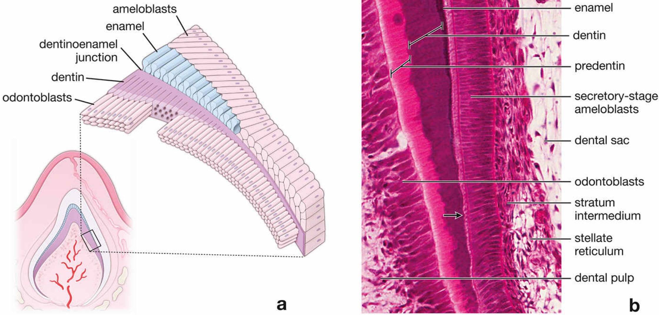
Fig: Secretory stage of amelogenesis. (a) Diagram showing ameloblasts laying enamel on dentin. (b) H&E photomicrograph showing secretory-stage ameloblasts adjacent to developing enamel, stratum intermedium, stellate reticulum, and dental pulp.
2. MATURATION STAGE
After the full thickness of enamel matrix is deposited, the matrix must mature to achieve its final hardness. This involves:
- Removal of organic matrix (amelogenins and other proteins are degraded and reabsorbed)
- Continued influx of calcium (Ca²⁺) and phosphate into the enamel
- Net result: enamel mineral content increases from ~30% to ~96-98%
Maturation-stage ameloblasts:
These cells differentiate from secretory-stage ameloblasts and function primarily as a transporting epithelium - moving substances both into and out of the maturing enamel.
The hallmark feature of maturation-stage ameloblasts is the presence of a striated (ruffled) border on their apical surface.
Modulation cycle - Maturation-stage ameloblasts undergo cyclical changes:
- ~70% of cells at any time display a striated (ruffled) border: these secrete bicarbonate ions (HCO₃⁻) and contain plasma membrane Ca²⁺-ATPases (PMCA) that pump Ca²⁺ into the maturing enamel
- ~30% display a smooth border: these secrete proteolytic enzymes (matrix metalloproteinases, kallikrein-4) to degrade and remove the organic matrix
During maturation, the stratum intermedium, stellate reticulum, and outer enamel epithelium collapse on each other and cannot be distinguished individually. Blood vessels invaginate into this reorganized layer to form the papillary layer (stellate papillary cells) adjacent to maturation ameloblasts. Both maturation ameloblasts and papillary cells are rich in mitochondria, reflecting their high energy demands as transport epithelium.
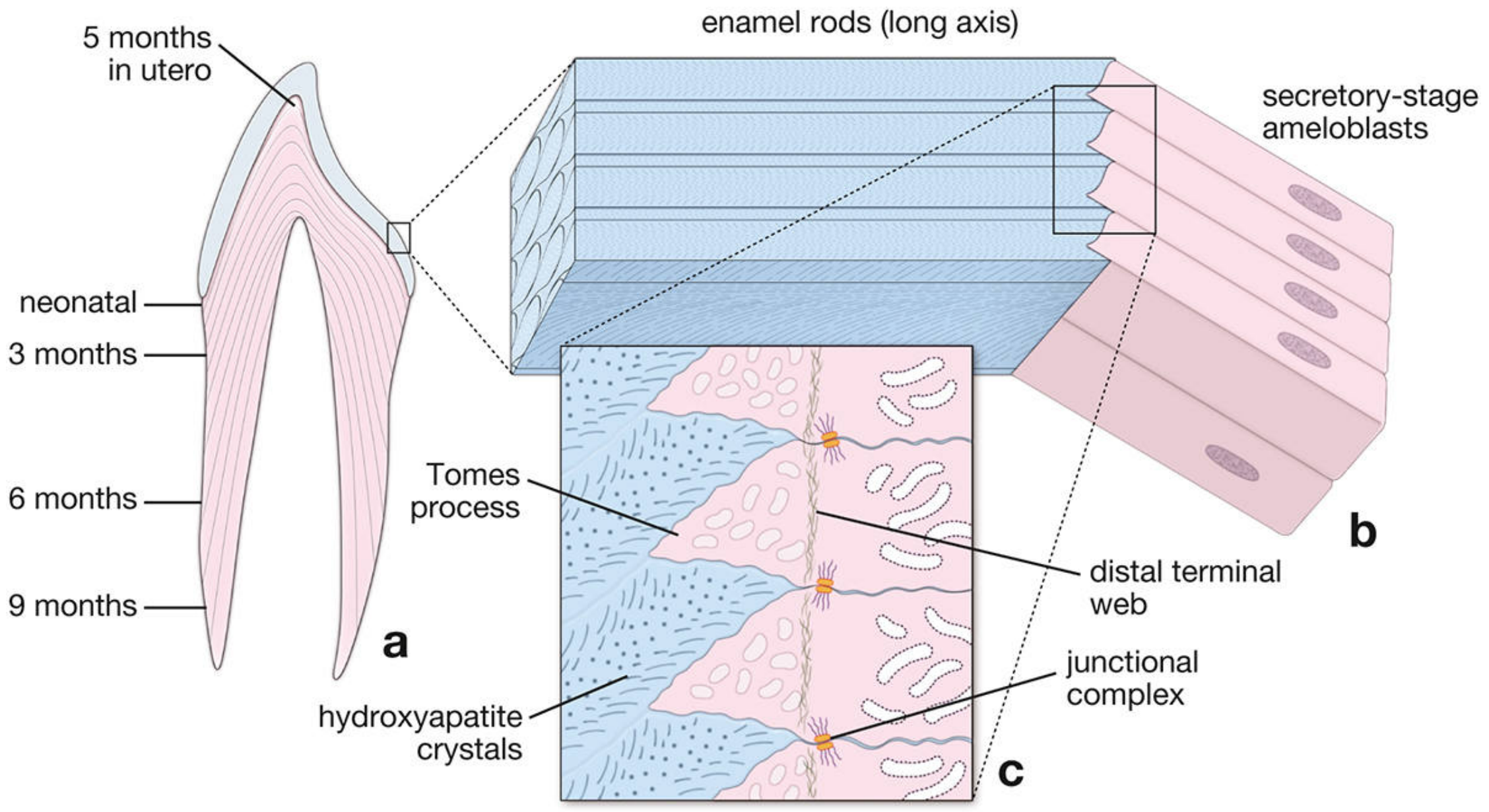
Fig: Detailed schematic of amelogenesis. (a) Enamel rods with incremental lines at different developmental stages. (b) Secretory-stage ameloblasts in relation to enamel rods. (c) Close-up of Tomes' process, hydroxyapatite crystals, distal terminal web, and junctional complexes.
ENAMEL MATRIX PROTEINS
The extracellular matrix of developing enamel contains several unique proteins, all of which are eventually removed during maturation:
| Protein | Function |
|---|---|
| Amelogenin (~90% of matrix) | Main structural protein; initiates and guides hydroxyapatite crystal growth; maintains spacing between enamel rods |
| Enamelin | Distributed throughout enamel layer; undergoes proteolytic cleavage as enamel matures; fragments remain on crystal surfaces |
| Ameloblastin (Amelin/Sheathlin) | Produced from early secretory to late maturation stages; controls crystal elongation; forms junctional complexes between crystals |
| Tuftelin | Earliest detected protein; located near DEJ; acidic and insoluble - aids in nucleation of enamel crystals; accounts for enamel tufts (areas of hypomineralization) |
FATE OF AMELOBLASTS
After enamel maturation is complete and just before tooth eruption, the ameloblasts, along with the reduced enamel epithelium (fused remnant of the enamel organ), form the junctional epithelium and oral epithelium over the crown. The ameloblasts are then lost - no cells capable of re-forming enamel remain, which is why enamel cannot regenerate after damage.
INCREMENTAL LINES IN ENAMEL
The rhythmic, cyclical nature of enamel secretion results in characteristic incremental lines:
- Striae of Retzius (Contour lines of Retzius) - Brown bands visible in ground sections; represent rhythmic growth periods (approximately every 8 days); run obliquely across enamel rods; appear as surface ridges called perikymata on the enamel surface
- Neonatal line - A wider, prominent line of hypomineralization seen in deciduous teeth and the first permanent molar; marks the metabolic disturbance at birth (transition from prenatal to postnatal nutrition); clinically useful as a reference point
FLUORIDE AND ENAMEL
Fluoride ions (F⁻) can be incorporated into hydroxyapatite crystals during enamel development, converting them to fluorapatite. Fluorapatite is more resistant to acidic dissolution by oral microorganisms, which is the scientific basis for fluoridation of water and toothpaste in caries prevention.
AMELOGENESIS IMPERFECTA
Amelogenesis imperfecta (AI) is a group of hereditary conditions in which a mutation in the genes encoding enamel matrix proteins (most commonly amelogenin gene, AMELX, on the X chromosome) or the proteins involved in enamel maturation results in defective enamel formation. Clinically, affected teeth show:
- Hypoplastic AI: thin, pitted enamel (defect in secretory stage)
- Hypomaturation AI: enamel of normal thickness but soft and opaque (defect in maturation stage)
- Hypocalcified AI: enamel easily chips from dentin
SUMMARY TABLE
| Feature | Secretory Stage | Maturation Stage |
|---|---|---|
| Main cell | Secretory ameloblast | Maturation ameloblast |
| Cell process | Tomes' process | Striated/Ruffled border |
| Main function | Lay down organic matrix | Remove organic matrix; mineralize |
| Mineral content | ~30% | ~96-98% |
| Stratum intermedium | Present | Absent (collapses) |
| Papillary layer | Absent | Present |
CONCLUSION
Amelogenesis is a tightly regulated, two-stage process of enamel formation. The secretory stage involves highly specialized ameloblasts that deposit a protein-rich matrix via their Tomes' processes. The maturation stage converts this matrix into the hardest biologic tissue through mineral influx and organic matrix removal. Key proteins - amelogenin, enamelin, ameloblastin, and tuftelin - orchestrate crystal growth and rod organization. Because ameloblasts are lost before eruption, enamel is permanent and irreplaceable, making the process clinically relevant to understanding caries, developmental defects, and restorative dentistry.
Sources:
- Histology: A Text and Atlas with Correlated Cell and Molecular Biology (Ross & Pawlina), Chapter 16 - Oral Cavity and Associated Structures
- Junqueira's Basic Histology: Text and Atlas, 17e, Chapter 15 - Oral Cavity
This is a shared conversation. Sign in to Orris to start your own chat.