
I have my university exam of 1 st year of subject anatomy These are the important questions give me diagrams of each topic of the hard and neck for anatomy exam"Cavernous sinus, Parotid gland, Tongue, Facial nerve, Palatine tonsil, Face, Facial artery, Nasal septum, Maxillary air sinus, Vocal cord, Ansa cervicalis, Development of tongue" and remeber I don't want any theory I just want diagrams
cavernous sinus anatomy diagram labeled cross section
development of tongue embryology diagram labeled
vocal cord larynx anatomy diagram labeled true false vocal folds
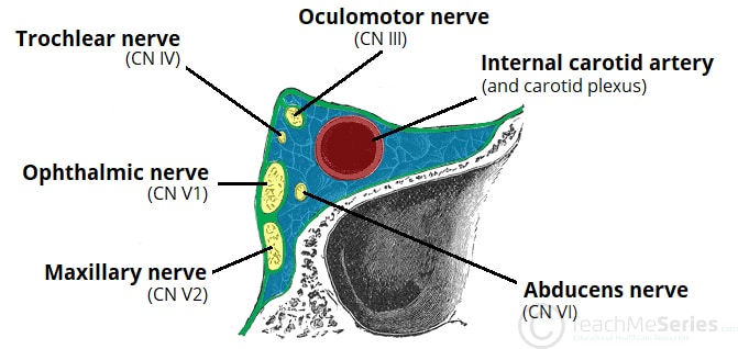
1. Cavernous Sinus
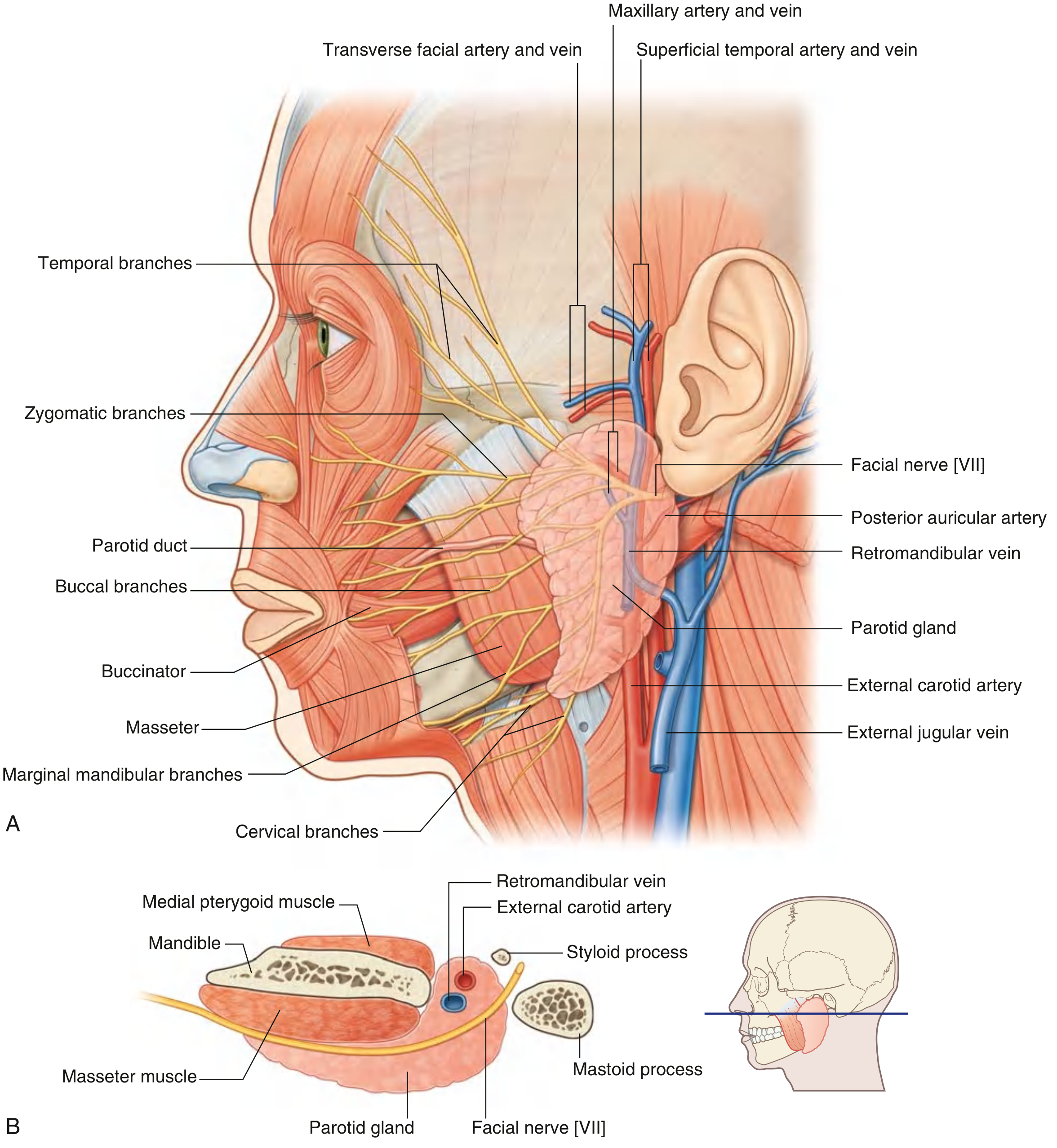
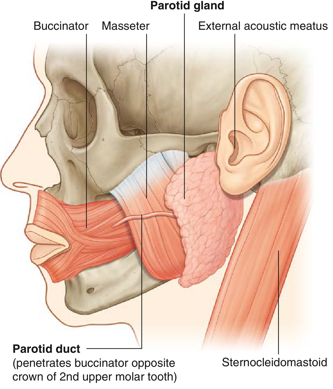
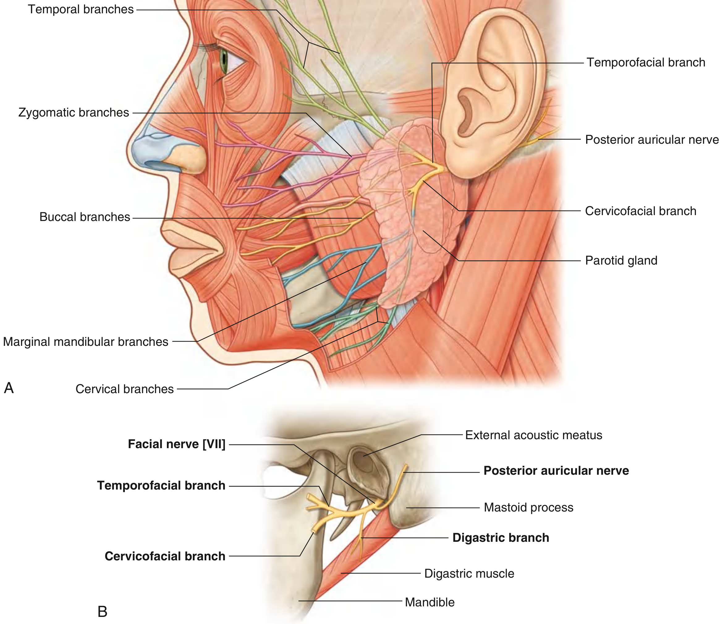
2. Parotid Gland
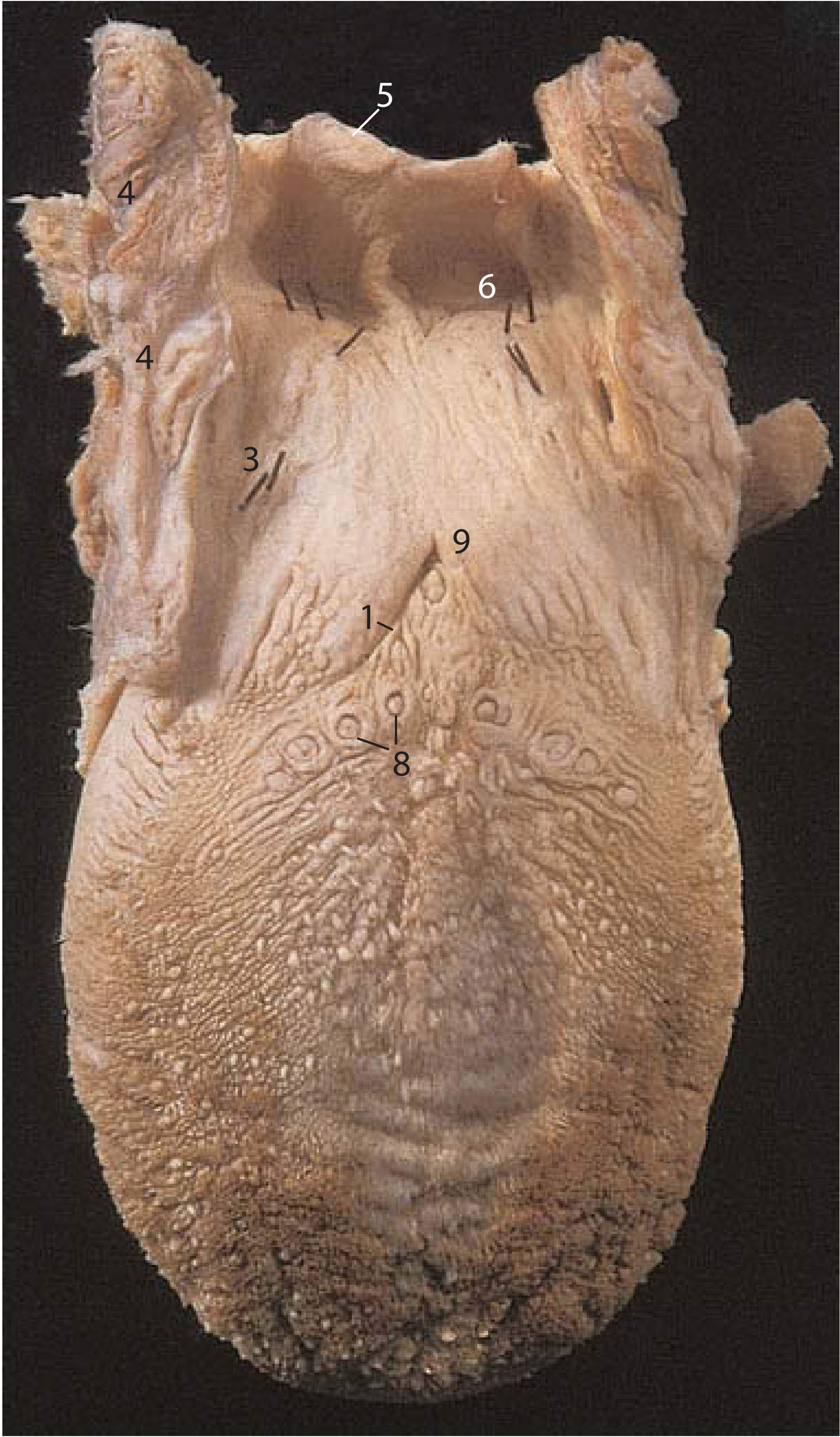
3. Tongue
4. Facial Nerve
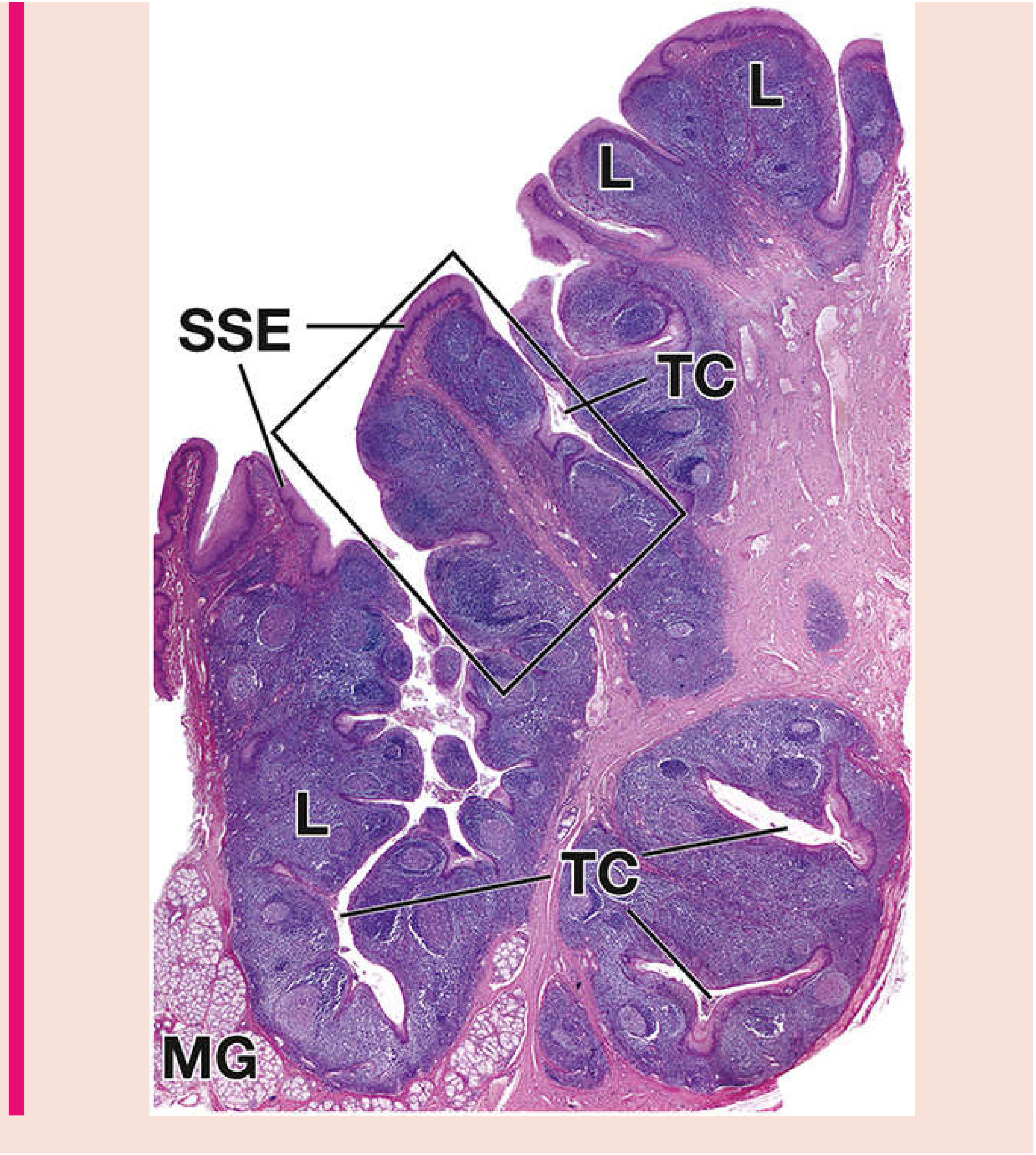
5. Palatine Tonsil
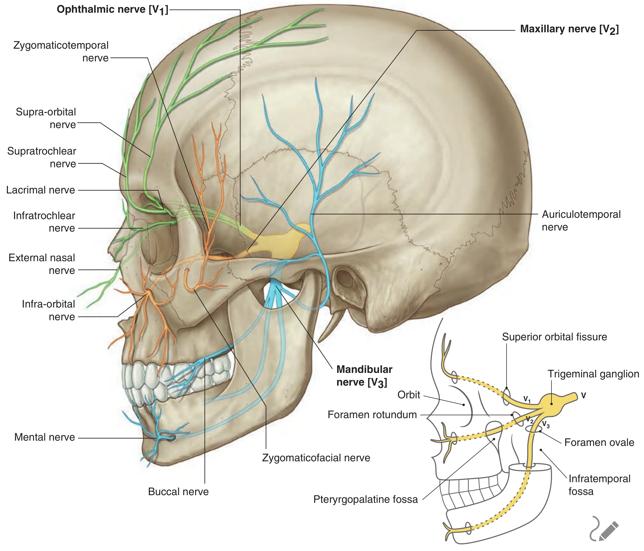
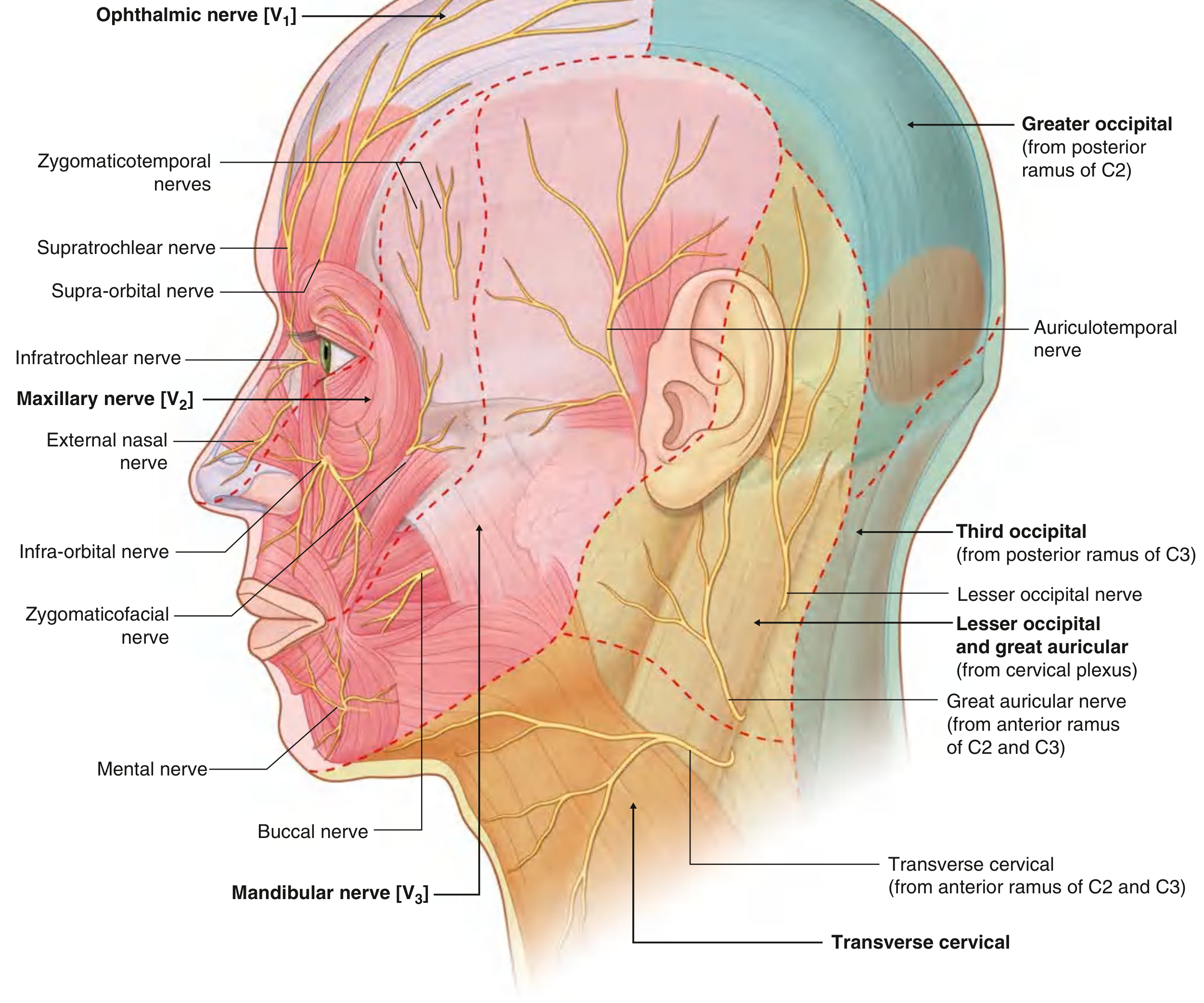
6. Face - Sensory Innervation (Trigeminal Nerve)
7. Facial Artery
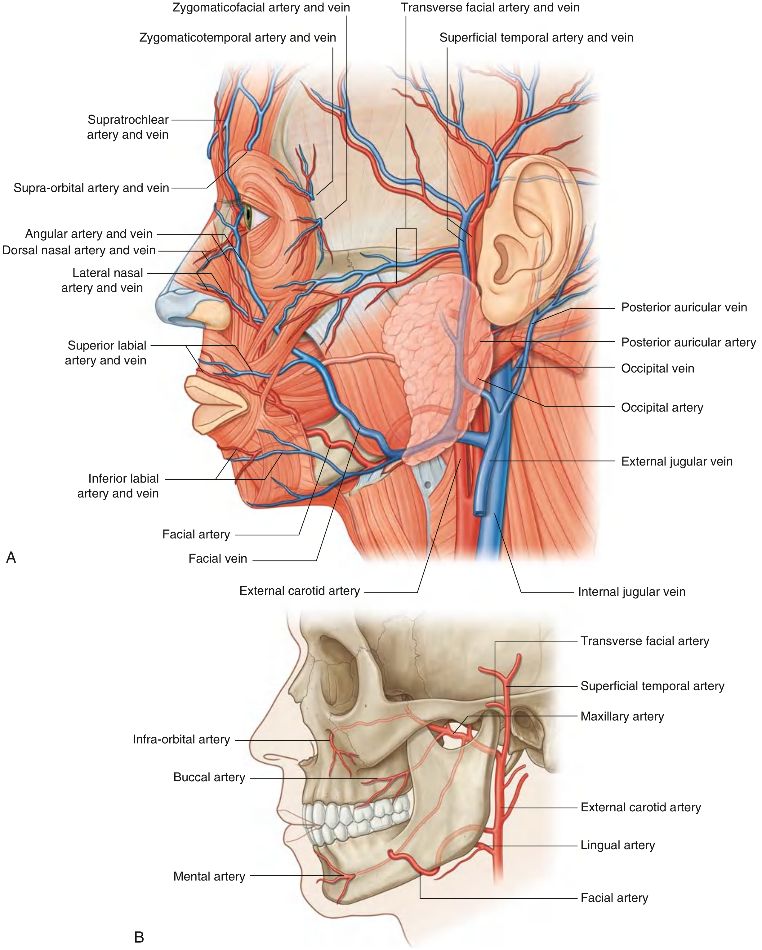
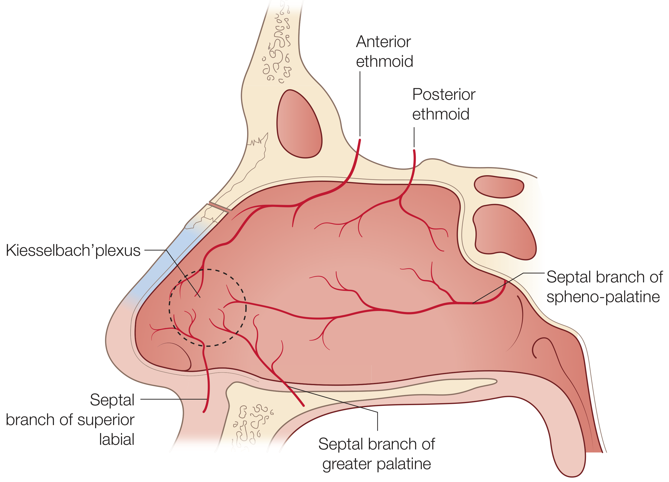
8. Nasal Septum

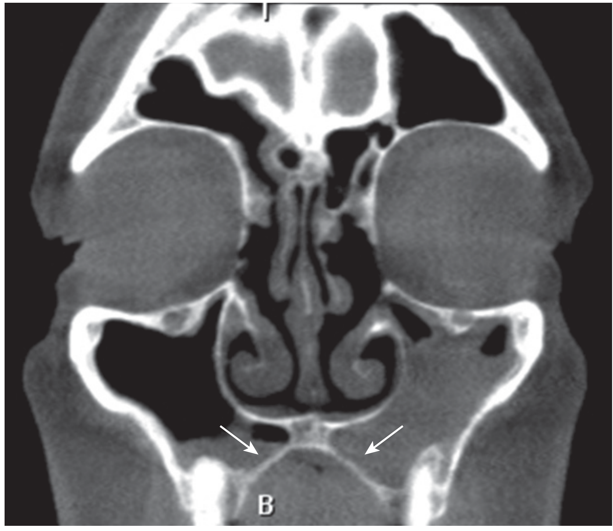
9. Maxillary Air Sinus
10. Vocal Cord (Larynx)
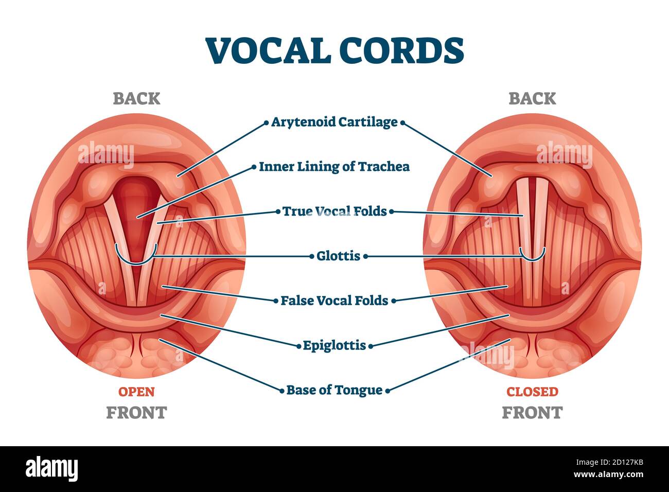
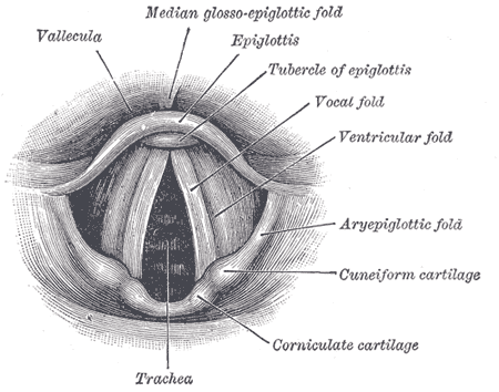
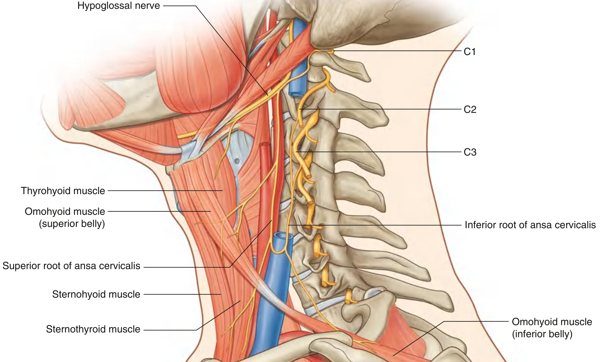
11. Ansa Cervicalis
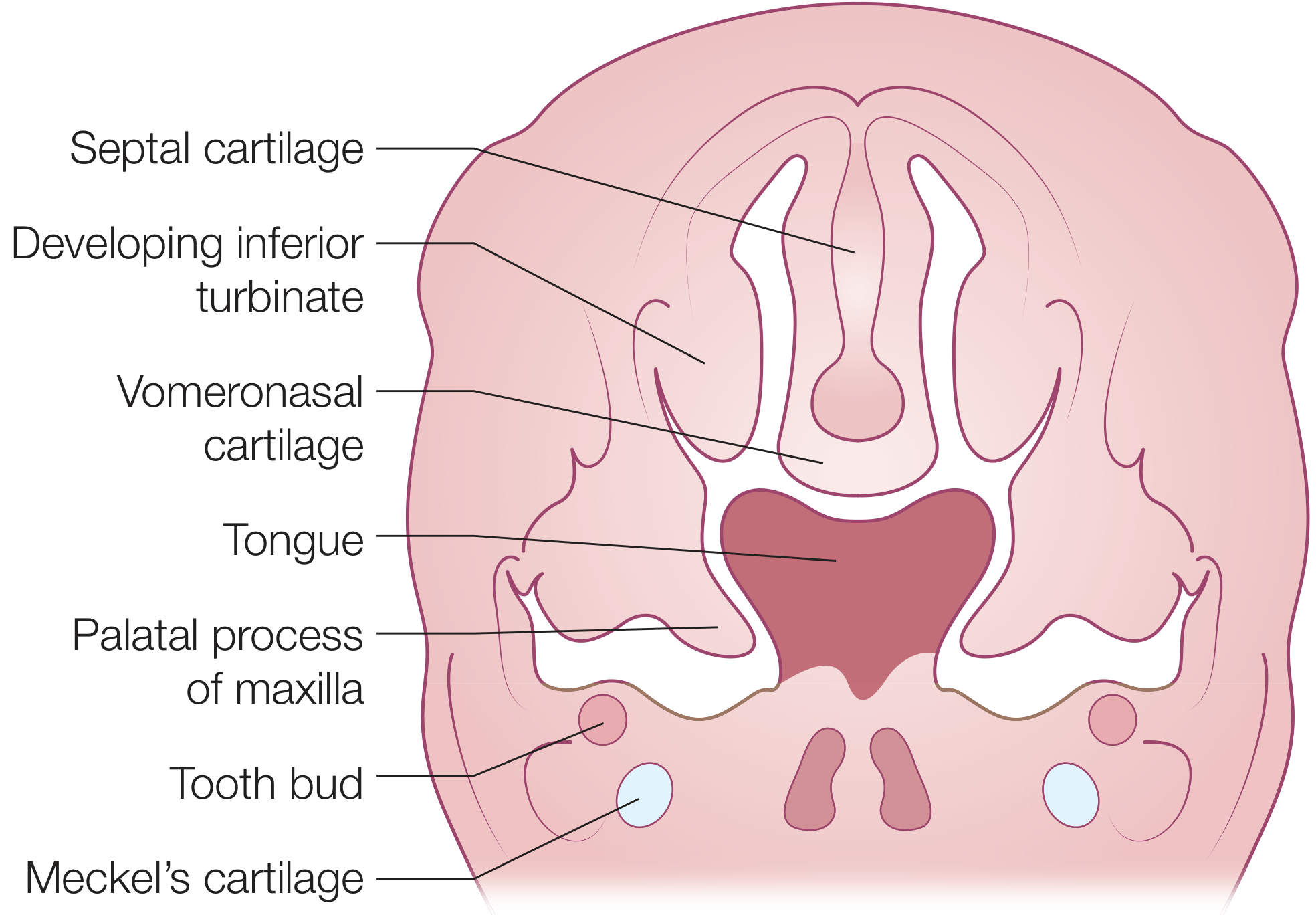
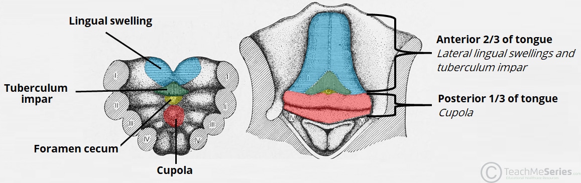
12. Development of Tongue
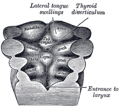
Sources: Gray's Anatomy for Students (9780323934237), Scott-Brown's Otorhinolaryngology Head & Neck Surgery (9781138094642, 9781138094611), Histology: A Text and Atlas (9781975181512), TeachMeAnatomy, Wikipedia/Gray's public domain. All images are from standard anatomical atlases used in 1st year anatomy curricula.
These diagrams are good for learning but I need easy diagrams to draw in exam
How would you like the easy exam diagrams? · Which topics do you want first? (I'll do all 12, just prioritizing)
Simple hand-drawn style anatomy exam diagram of the Cavernous Sinus, coronal cross-section. Clean pencil sketch style with clear black labels. Show: the sinus cavity in the center, Internal Carotid Artery (ICA) inside with label, CN VI (Abducens) inside with label, CN III (Oculomotor) in lateral wall, CN IV (Trochlear) in lateral wall, CN V1 (Ophthalmic) in lateral wall, CN V2 (Maxillary) in lateral wall. Label Pituitary gland medially, Sphenoid bone at bottom, Diaphragma sellae at top. Add a small legend box. Title at top: "CAVERNOUS SINUS - Coronal Section". Simple clean lines, easy to copy in a university anatomy exam.
Simple hand-drawn style anatomy exam diagram of the Parotid Gland, lateral view of the face. Clean pencil sketch with black labels easy to copy in exam. Show: outline of face/jaw from side, Parotid gland shaded lightly between ear and masseter, Facial nerve VII entering gland at stylomastoid foramen and branching into 5 branches labeled: Temporal, Zygomatic, Buccal, Marginal Mandibular, Cervical. Show Parotid duct crossing masseter opening near 2nd upper molar. Label: External carotid artery passing through gland, Retromandibular vein, Masseter muscle, Sternocleidomastoid. Title: "PAROTID GLAND". Simple clean lines easy to reproduce.
Simple hand-drawn style anatomy exam diagram of the Facial Nerve (CN VII) extracranial course and branches. Clean pencil sketch with black labels. Show: Stylomastoid foramen at top where nerve exits skull, nerve entering Parotid gland, then dividing into 2 trunks (Temporofacial and Cervicofacial), then 5 terminal branches with clear labels and arrows showing their distribution on the face outline: 1-Temporal branch (to forehead/frontalis), 2-Zygomatic branch (to orbicularis oculi), 3-Buccal branch (to buccinator, upper lip), 4-Marginal Mandibular branch (to lower lip/chin), 5-Cervical branch (to platysma). Title: "FACIAL NERVE [VII] - Extracranial Branches". Simple lines easy to copy.
Simple hand-drawn anatomy exam diagram of the Facial Artery course on the face. Clean pencil sketch style with clear black labels. Show: side profile of face outline, External Carotid Artery origin in neck labeled, Facial artery looping over mandible (label: "winds around lower border of mandible"), tortuous course up face, labeling branches with arrows: Submental artery, Inferior labial artery, Superior labial artery (with Septal branch), Lateral nasal artery, Angular artery (terminal branch at medial canthus). Mark the facial vein running parallel. Mark danger area of face with dotted circle. Title: "FACIAL ARTERY". Simple clean lines for exam.
Simple hand-drawn anatomy exam diagram of Sensory Nerve Supply of the Face. Clean pencil sketch with black labels easy to copy. Show: front-facing face outline divided into 3 zones with dotted lines. Label each zone clearly: Zone 1 (upper - forehead/upper eyelid) = CN V1 Ophthalmic with branches Supraorbital, Supratrochlear, Lacrimal. Zone 2 (middle - cheek/lower eyelid/upper lip) = CN V2 Maxillary with branches Infraorbital, Zygomaticofacial, Zygomaticotemporal. Zone 3 (lower - lower lip/chin/jaw) = CN V3 Mandibular with branches Mental, Buccal, Auriculotemporal. Title: "SENSORY NERVE SUPPLY OF FACE (CN V)". Very simple, easy to draw in exam.
Is it done
Simple hand-drawn style anatomy exam diagram of the Cavernous Sinus coronal cross-section. Clean pencil sketch with clear black labels. Show the sinus cavity, Internal Carotid Artery (ICA) inside, CN VI (Abducens) inside the sinus, CN III (Oculomotor) in lateral wall, CN IV (Trochlear) in lateral wall, CN V1 (Ophthalmic) in lateral wall, CN V2 (Maxillary) in lateral wall. Label Pituitary gland medially, Sphenoid bone at bottom. Title at top: CAVERNOUS SINUS - Coronal Section. Simple clean lines easy to copy in a university anatomy exam.
Simple hand-drawn anatomy exam diagram of Parotid Gland lateral view of the face. Clean pencil sketch with black labels. Show outline of face and jaw from the side, Parotid gland lightly shaded between ear and masseter, Facial nerve VII entering gland at stylomastoid foramen branching into 5 labeled branches: Temporal, Zygomatic, Buccal, Marginal Mandibular, Cervical. Show Parotid duct crossing masseter opening near 2nd upper molar. Label External carotid artery, Retromandibular vein, Masseter muscle. Title: PAROTID GLAND. Simple and easy to reproduce in exam.
Simple hand-drawn anatomy exam diagram of Facial Nerve CN VII extracranial branches. Clean pencil sketch. Show stylomastoid foramen at top where nerve exits, nerve trunk entering Parotid gland, dividing into upper Temporofacial trunk and lower Cervicofacial trunk, then 5 terminal branches labeled with arrows on face outline: Temporal branch to forehead, Zygomatic branch to eye, Buccal branch to cheek and upper lip, Marginal Mandibular branch to lower lip and chin, Cervical branch to platysma. Title: FACIAL NERVE VII - Branches. Easy to draw in exam.
Simple hand-drawn anatomy exam diagram of Facial Artery course on the face. Clean pencil sketch with clear black labels. Show side profile of face, External Carotid Artery in neck, Facial artery looping over lower border of mandible, tortuous course up the face, branches labeled with arrows: Submental artery, Inferior labial artery, Superior labial artery with Septal branch, Lateral nasal artery, Angular artery at medial corner of eye as terminal branch. Title: FACIAL ARTERY. Simple and easy to copy in exam.
400, message: Got more than 4194304 bytes when reading: b'data: {"candidates": [{"content": {"role": "model","parts": [{"inlineData": {"mimeType": "image/png"...'.
We encountered an error while processing your request. Please try again or reach out to us for help.