
Male reproductive organs from neet level (UG) Give me the points as such that I can teach them directly to class 12th students Ncert baded too
🔬 Male Reproductive Organs - Class 12 / NEET UG Teaching Notes
📌 OVERVIEW: Male Reproductive System
| Internal Genitalia | External Genitalia |
|---|---|
| Testes, Epididymis, Vas deferens, Prostate, Seminal vesicles, Bulbourethral glands | Penis, Scrotum |
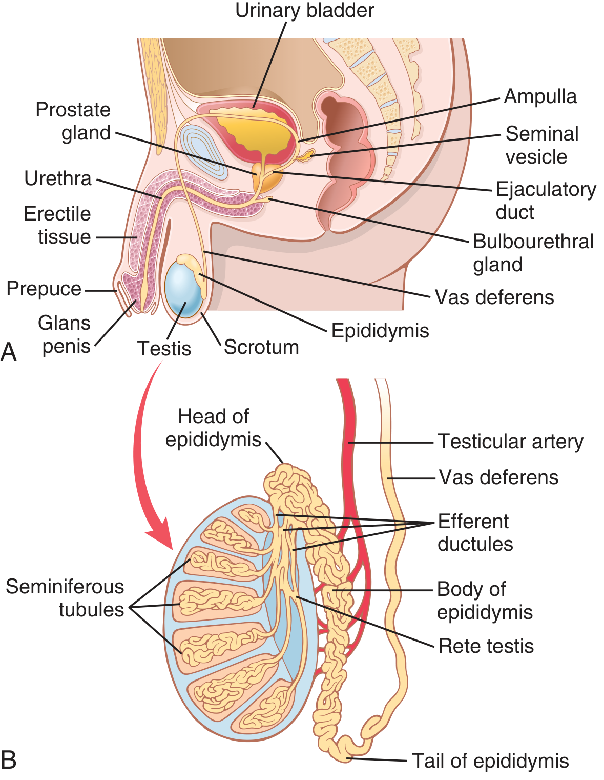
1. TESTES (Primary Male Reproductive Organs)
- Paired organs located inside the scrotum, outside the body cavity
- Why outside? Temperature must be 2-2.5°C lower than body temperature (~35°C) for normal spermatogenesis
- Maintained by a countercurrent heat exchange mechanism between testicular arteries and veins
- Two main functions:
- Spermatogenesis - production of sperm
- Steroidogenesis - secretion of testosterone (male sex hormone)
Internal Structure of Testis:
- Each testis has ~250 lobules
- Each lobule contains 1-3 seminiferous tubules
- 80% of testis = Seminiferous tubules (site of sperm production)
- 20% of testis = Connective tissue + Leydig cells (interstitial cells)
Cells in Seminiferous Tubules:
| Cell | Function |
|---|---|
| Spermatogonia | Stem cells; undergo mitosis to generate spermatocytes |
| Sertoli cells | "Nurse cells" - support developing sperm |
| Leydig cells | Located in interstitium; secrete testosterone |
Sertoli Cell Functions (4 key points for NEET):
- Nutrition - provide nutrients to developing sperm (sperm are isolated from bloodstream)
- Blood-testis barrier - form tight junctions with each other, protect sperm from the immune system
- Fluid secretion - secrete aqueous fluid to transport sperm toward epididymis
- Androgen-binding protein (ABP) - keeps local testosterone levels high to support spermatogenesis
2. SPERMATOGENESIS (Most Important - High NEET Weightage)
- Begins at puberty (~13-14 years) and continues throughout life
- Occurs in the seminiferous tubules
- Takes about 64-74 days to complete one full cycle
- ~120 million sperm produced per testis per day
Stages of Spermatogenesis:
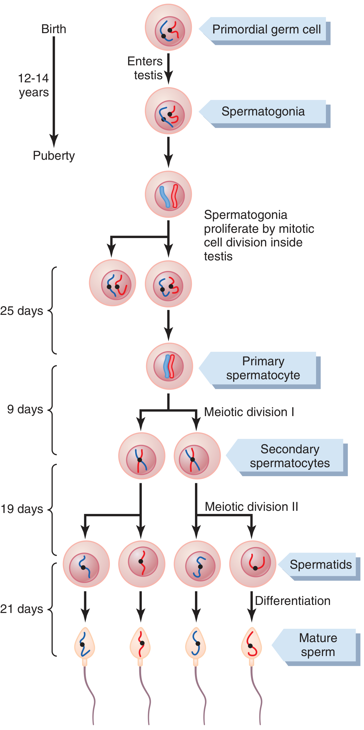
| Stage | Cell | Ploidy | Division |
|---|---|---|---|
| Spermatogonia (2n) | Type A & B | Diploid (46 chr) | Mitosis |
| Primary spermatocyte | Large cell | Diploid (46 chr) | Before Meiosis I |
| Secondary spermatocyte | Smaller | Haploid (23 chr) | After Meiosis I |
| Spermatids | Round cells | Haploid (23 chr) | After Meiosis II |
| Spermatozoa | Mature sperm | Haploid (23 chr) | Spermiogenesis |
Spermiogenesis:
- Spermatid → Spermatozoon (no more division, just differentiation/morphological changes)
- Loss of cytoplasm, development of flagellum, acrosome formation
3. STRUCTURE OF SPERMATOZOON (Mature Sperm)
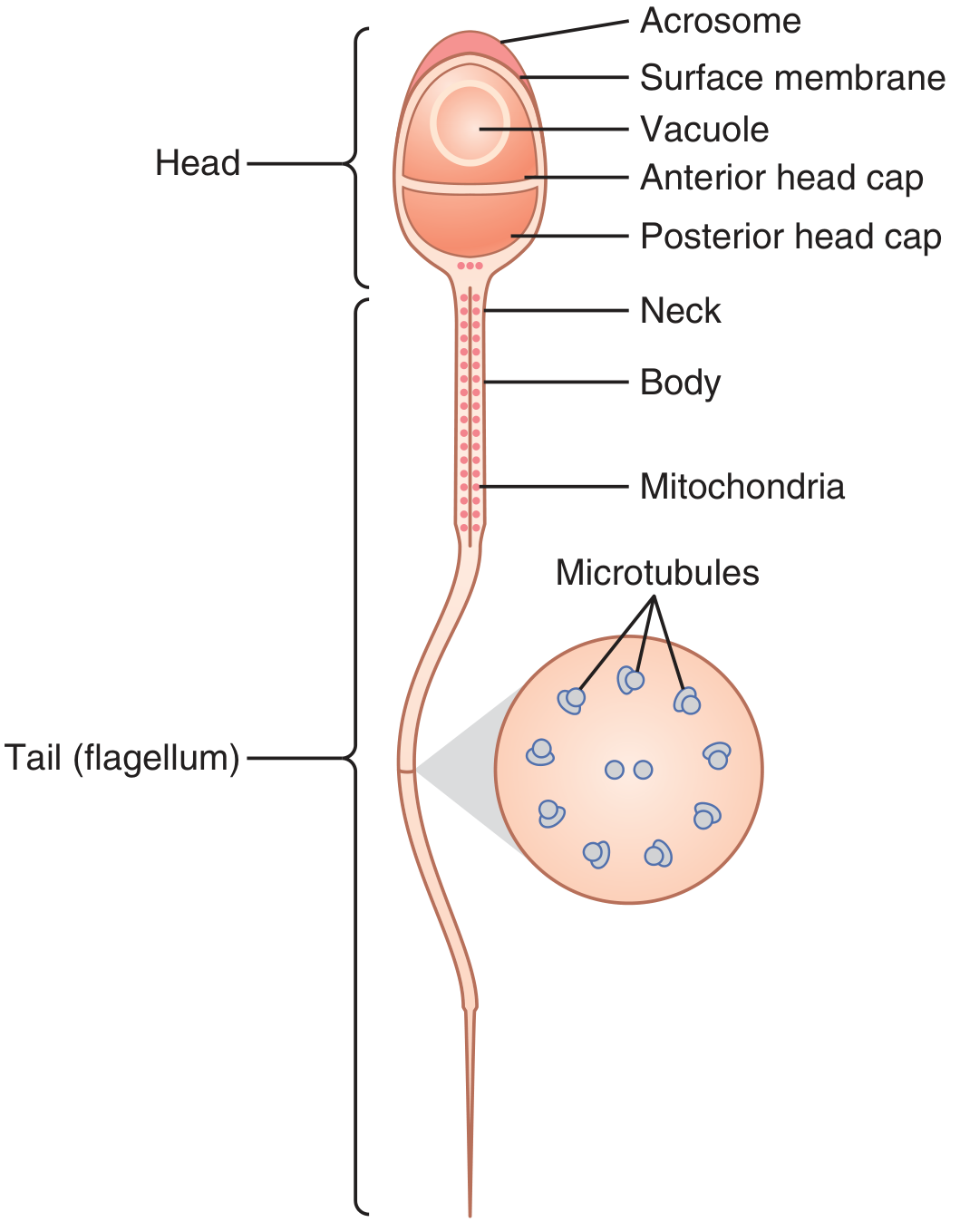
| Part | Details |
|---|---|
| Head | Contains condensed nucleus (haploid DNA) |
| Acrosome (anterior 2/3 of head) | Cap-like structure from Golgi apparatus; contains enzymes - hyaluronidase and proteases (for penetrating egg) |
| Neck | Short connecting region |
| Middle piece (Body) | Contains mitochondria - powerhouse for sperm motility (ATP production) |
| Tail (Flagellum) | Contains axoneme (9+2 microtubule arrangement); provides motility |
4. ACCESSORY DUCTS
(a) Epididymis
- Highly coiled tube (~6 m long) lying on posterior surface of testis
- 3 parts: Head → Body → Tail
- Functions:
- Maturation of sperm (takes 18-24 hours for sperm to gain motility)
- Storage of sperm (for up to several months)
- Sperm are non-motile and non-fertilizable when they leave the testis
- Become motile after passing through the epididymis
(b) Vas Deferens (Ductus Deferens)
- Muscular tube that carries sperm from epididymis to the ejaculatory duct
- Passes through the inguinal canal into the pelvic cavity
- Vasectomy = surgical cutting of vas deferens = male contraception
- Ampulla = dilated terminal part of vas deferens, acts as a storage reservoir
(c) Ejaculatory Duct
- Formed by the junction of vas deferens and duct of seminal vesicle
- Opens into the urethra
- Short duct (~2 cm)
(d) Urethra
- Common passage for urine and semen (not simultaneously)
- Passes through the prostate and penis
5. ACCESSORY GLANDS
| Gland | Location | Secretion / Function |
|---|---|---|
| Seminal vesicles (2) | Behind urinary bladder | ~60% of semen volume; secretes fructose (energy for sperm), prostaglandins, fibrinogen |
| Prostate gland (1) | Below urinary bladder, surrounds urethra | ~30% of semen volume; secretes citric acid, enzymes, PSA (prostate-specific antigen); slightly alkaline - neutralizes vaginal acidity |
| Bulbourethral glands / Cowper's glands (2) | Below prostate, lateral to urethra | Pre-ejaculatory fluid; lubricates urethra, neutralizes residual urine acidity before sperm passage |
6. PENIS (External Organ)
- Organ of copulation and urination
- Contains erectile tissue - 2 corpora cavernosa + 1 corpus spongiosum
- Glans penis = tip of penis; covered by prepuce (foreskin)
- Corpus spongiosum surrounds the urethra
7. SCROTUM
- External sac of skin that houses the testes
- Maintains testicular temperature 2-2.5°C below body temperature
- Contains dartos muscle (wrinkles the scrotal skin) and cremaster muscle (raises/lowers testes to regulate temperature)
8. HORMONAL CONTROL OF MALE REPRODUCTION
| Hormone | Source | Function |
|---|---|---|
| GnRH | Hypothalamus | Stimulates anterior pituitary to release LH and FSH |
| LH (ICSH) | Anterior pituitary | Stimulates Leydig cells → testosterone secretion |
| FSH | Anterior pituitary | Stimulates Sertoli cells → supports spermiogenesis; stimulates ABP secretion |
| Testosterone | Leydig cells | Spermatogenesis, secondary sexual characters, libido |
| Inhibin | Sertoli cells | Negative feedback on FSH (inhibits FSH release) |
9. SPERM PATHWAY (Sperm Route - Must Memorize)
10. QUICK NEET FACTS TO REMEMBER
| Fact | Value |
|---|---|
| Duration of spermatogenesis | 64-74 days |
| Sperm produced per day | ~120 million per testis |
| Sperm motility acquired in | Epididymis (18-24 hrs) |
| Epididymis length | ~6 metres |
| Largest accessory gland | Prostate |
| Largest contributor to semen volume | Seminal vesicles (~60%) |
| Acrosome enzyme | Hyaluronidase, Acrosin |
| Energy for sperm motility | Fructose (from seminal vesicles) + mitochondria in middle piece |
| Sperm viability in female tract | 48-72 hours |
| Temperature for spermatogenesis | 2-2.5°C below body temp |
COMMON NEET MCQ TRAPS
- Leydig cells vs Sertoli cells - Leydig = testosterone; Sertoli = ABP + inhibin + nurse function
- Spermiogenesis = spermatid → sperm (morphological change only, NO division)
- Spermiation = release of sperm from Sertoli cells into the lumen
- Acrosome is derived from the Golgi apparatus
- Middle piece of sperm has mitochondria - NOT the head
- Inhibin - from Sertoli cells - inhibits FSH (not LH)
- Vasectomy cuts vas deferens; sperm production continues but is reabsorbed
- Sperm are haploid (n = 23) after meiosis
Where is mullerian and wolfian ducts present and their function Neet legel only
🧬 Müllerian & Wolffian Ducts - NEET UG Notes (Class 12)
📌 KEY CONCEPT FIRST
📍 Where Are They Present?
- Both ducts arise from the embryonic mesonephros (a temporary embryonic kidney)
- Present in the pelvic/abdominal region of the embryo during early foetal development
- Both ducts run parallel to each other in the embryo from about Week 4-7 of gestation
- By Week 10, only ONE duct system survives - depending on whether the embryo is male or female
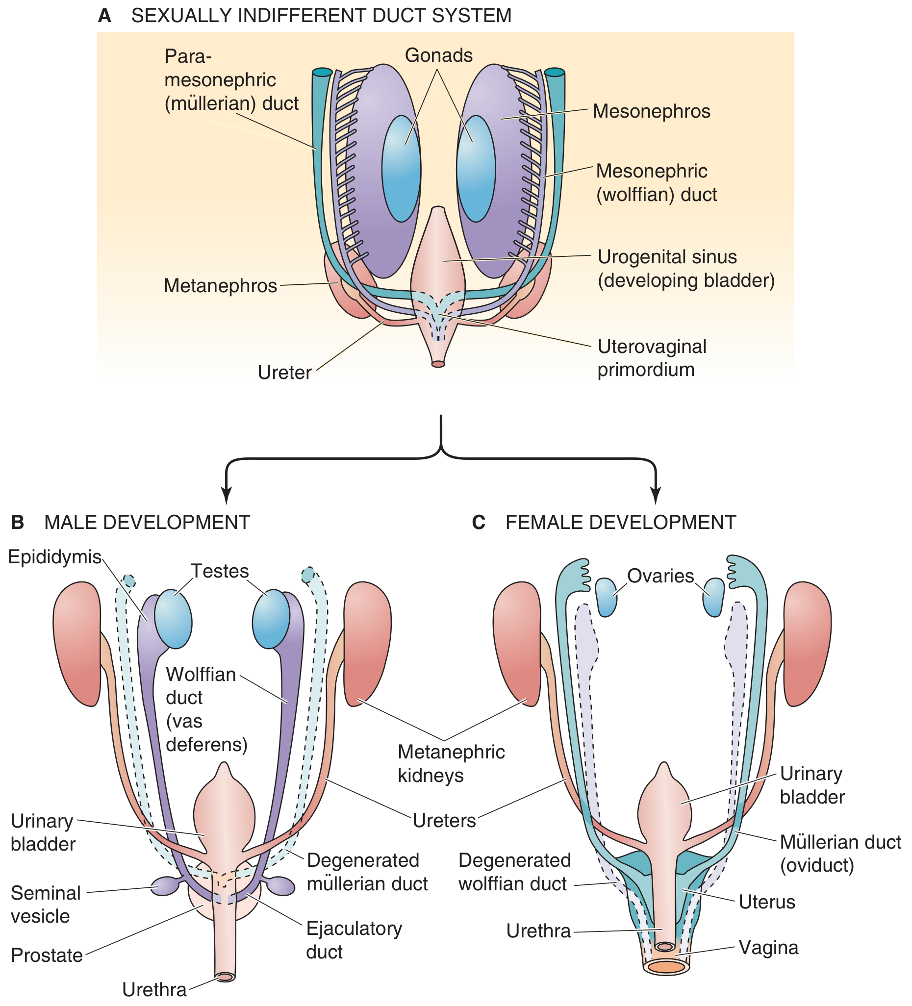
1. WOLFFIAN DUCT (Mesonephric Duct)
| Feature | Detail |
|---|---|
| Other name | Mesonephric duct |
| Embryonic origin | Mesonephros (transient embryonic kidney) |
| Present in | All embryos initially (both sexes) |
| Stimulated by | Testosterone (from Leydig cells of testis) |
Fate in MALES (survives):
Wolffian duct → Epididymis + Vas deferens + Seminal vesicle + Ejaculatory duct
- Proximal part → Epididymis (head, body, tail)
- Middle part → Vas deferens
- Lateral outgrowth at distal end → Seminal vesicle
- Terminal part (before urethra) → Ejaculatory duct
Fate in FEMALES (degenerates):
- In the absence of testosterone, the Wolffian duct involutes (degenerates)
- No male internal structures are formed
2. MÜLLERIAN DUCT (Paramesonephric Duct)
| Feature | Detail |
|---|---|
| Other name | Paramesonephric duct |
| Embryonic origin | Invagination of coelomic epithelium, lateral to the Wolffian duct |
| Present in | All embryos initially (both sexes) |
| Inhibited by | AMH / MIS (Anti-Müllerian Hormone / Müllerian Inhibiting Substance) from Sertoli cells |
Fate in FEMALES (survives):
Müllerian duct → Fallopian tubes + Uterus + Cervix + Upper 1/3 of vagina
- Upper part (unfused) → Fallopian tubes (oviducts)
- Lower part (two ducts fuse at midline) → Uterus + Cervix
- Lowest fused part → Upper 1/3 of vagina
Fate in MALES (degenerates):
- Sertoli cells secrete AMH (Anti-Müllerian Hormone) → causes regression/degeneration of Müllerian ducts
- No female internal structures are formed
🔁 SUMMARY TABLE (Most Important for NEET)
| Feature | Wolffian Duct | Müllerian Duct |
|---|---|---|
| Other name | Mesonephric duct | Paramesonephric duct |
| Present in | Both sexes (embryo) | Both sexes (embryo) |
| Develops in | Male | Female |
| Degenerates in | Female | Male |
| Hormone needed to develop | Testosterone | No hormone needed (default) |
| Hormone causing degeneration | None in female (absence of testosterone) | AMH / MIS (in males) |
| Source of hormone | Leydig cells of testis | Sertoli cells of testis |
| Develops into (in active sex) | Epididymis, Vas deferens, Seminal vesicle, Ejaculatory duct | Fallopian tubes, Uterus, Cervix, Upper vagina (1/3) |
🔑 HORMONAL CONTROL (The "Jost Experiment" Logic)
| Condition | Result |
|---|---|
| Testis present → secretes testosterone | Wolffian duct develops (male internal organs form) |
| Testis present → secretes AMH | Müllerian duct regresses (no female organs) |
| No testis (female / castrated male embryo) | Wolffian duct degenerates (no testosterone) |
| No testis (female / castrated male embryo) | Müllerian duct develops (no AMH present) = default pathway |
| Ovary present or absent | Does NOT matter - Müllerian development happens with or without ovary |
⚡ NEET MCQ TRAPS
- Wolffian duct = Mesonephric duct (same thing, different names - both used in MCQs)
- Müllerian duct = Paramesonephric duct (same thing)
- AMH is secreted by Sertoli cells (NOT Leydig cells) - very common trap
- Testosterone causes Wolffian to develop (NOT Müllerian)
- Vagina is NOT entirely from Müllerian duct - only upper 1/3; lower 2/3 from urogenital sinus
- Ovary is NOT needed for Müllerian duct development (proven by Jost experiment)
- In a castrated male embryo → female anatomy develops (no testosterone = no Wolffian; no AMH = Müllerian survives)
- Both ducts are present in embryo up to ~7 weeks; divergence happens around week 8-10
🧠 MNEMONIC
"Males LOSE Müllerian (by AMH), KEEP Wolffian (by Testosterone)" "Females KEEP Müllerian (default), LOSE Wolffian (no testosterone)"
Testis components and some details about them that might be relevant to neet ug level
🔬 Testis - Components & Details | NEET UG Class 12
📌 OVERVIEW
| Compartment | Contents | Function |
|---|---|---|
| Tubular compartment | Seminiferous tubules | Spermatogenesis (sperm production) |
| Interstitial compartment | Leydig cells + connective tissue + blood vessels | Steroidogenesis (testosterone production) |
COMPONENT 1: SEMINIFEROUS TUBULES
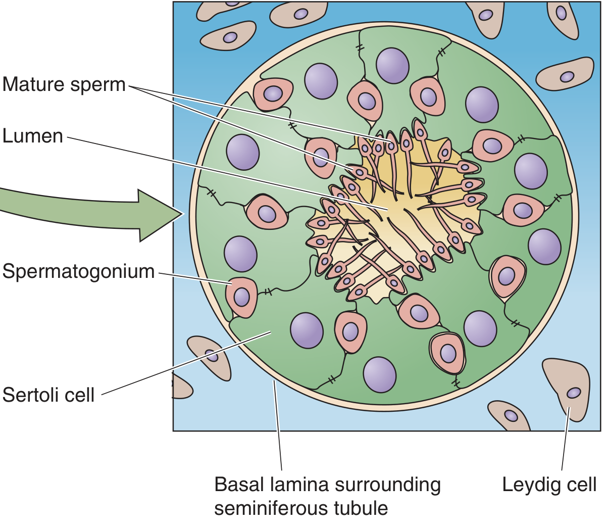
- Make up ~80% of total testis volume
- Highly coiled/convoluted loops, arranged in ~250 lobules
- Each lobule has 1-3 seminiferous tubules
- Total length of all tubules in both testes = ~250 metres
- The lumen of the tubule opens into the rete testis, which connects to the epididymis via efferent ductules
Cells INSIDE the Seminiferous Tubule:
🔴 A. Spermatogonia (Germ Cells / Stem Cells)
- Located at the periphery (outer wall / basal layer) of the tubule
- They are diploid (2n) stem cells
- Divide by mitosis to produce primary spermatocytes
- As cells mature, they move inward toward the lumen
- The most immature cells = near the periphery; most mature = near the lumen
Spermatogonia → Primary spermatocytes → Secondary spermatocytes → Spermatids → Spermatozoa (at lumen)
🟡 B. Sertoli Cells (Supporting / Nurse Cells)
- Also called "sustentacular cells"
- Large, tall columnar cells that extend from the basal lamina to the lumen of the tubule
- Spermatogenic cells are embedded within them
- Do NOT divide after puberty
- Stimulated by FSH from anterior pituitary
Functions of Sertoli Cells (5 key functions - all NEET relevant):
| # | Function | Detail |
|---|---|---|
| 1 | Nutrition | Provide nutrients (glucose, lactate, amino acids) to developing sperm - sperm are isolated from blood |
| 2 | Blood-Testis Barrier (BTB) | Form tight junctions with adjacent Sertoli cells → protect developing sperm from immune system (sperm antigens are foreign) |
| 3 | Fluid secretion | Secrete luminal fluid that transports sperm toward epididymis |
| 4 | Androgen-Binding Protein (ABP) | Secreted into tubule lumen; binds testosterone and keeps local testosterone concentration HIGH to support spermatogenesis |
| 5 | Hormone secretion | Secrete Inhibin (inhibits FSH) and AMH (in foetal life - causes Müllerian duct regression) |
| 6 | Phagocytosis | Engulf residual cytoplasm shed by spermatids during spermiogenesis |
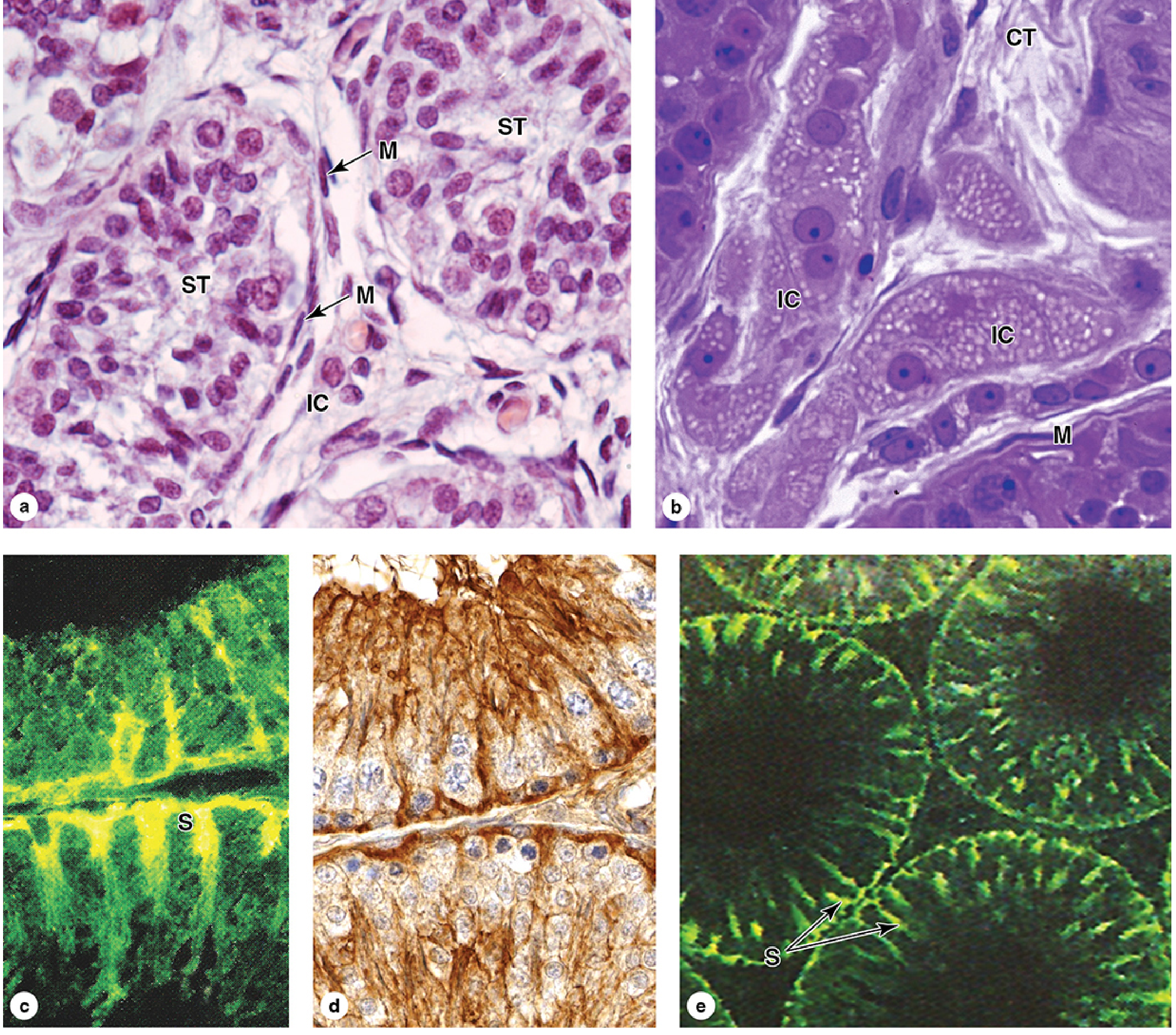
COMPONENT 2: LEYDIG CELLS (Interstitial Cells)
- Located in the connective tissue BETWEEN the seminiferous tubules (not inside the tubule)
- Also called "interstitial cells of Leydig"
- Make up the ~20% of testis that is NOT tubular tissue
- Contain lipid droplets in cytoplasm (typical of steroid-secreting cells)
- Stimulated by LH (= ICSH in males) from anterior pituitary
Functions of Leydig Cells:
- Primary function = Synthesise and secrete TESTOSTERONE
- Also produce small amounts of estrogens and DHEA
- Testosterone has both:
- Local (paracrine) effect - diffuses into Sertoli cells to support spermatogenesis
- Endocrine effect - enters bloodstream → secondary sexual characteristics, muscle growth, libido, etc.
LH → Leydig cells → Testosterone → (1) spermatogenesis support + (2) secondary sex characters
COMPONENT 3: MYOID CELLS (Peritubular Cells)
- Flat, smooth-muscle-like cells that surround each seminiferous tubule on the outside (just outside the basal lamina)
- Their rhythmic contractions help propel sperm and fluid through the tubule toward the rete testis
- Often tested in histology-based MCQs as "cells with elongated nuclei around the tubule"
COMPONENT 4: RETE TESTIS
- A network of anastomosing channels at the hilum (mediastinum) of the testis
- Collects sperm from the seminiferous tubules
- Connects to the efferent ductules, which carry sperm to the epididymis
- Part of the sperm pathway: Seminiferous tubule → Rete testis → Efferent ductules → Epididymis
COMPONENT 5: COVERING LAYERS OF TESTIS
| Layer | Details |
|---|---|
| Tunica vaginalis | Outermost serous layer; derived from peritoneum; has visceral + parietal layers |
| Tunica albuginea | Thick fibrous white capsule of dense connective tissue; gives shape and protection; sends septa inward to divide testis into lobules |
| Tunica vasculosa | Innermost layer; contains blood vessels and loose connective tissue |
Tunica albuginea is the most commonly asked covering in NEET MCQs.
📊 COMPLETE SUMMARY TABLE
| Component | Location | Stimulated by | Key Product / Function |
|---|---|---|---|
| Spermatogonia | Inside tubule, periphery | Testosterone (mitosis) | Stem cells → spermatozoa |
| Sertoli cells | Inside tubule wall (full height) | FSH | ABP, Inhibin, AMH, BTB, nutrition |
| Leydig cells | Between tubules (interstitium) | LH | Testosterone |
| Myoid cells | Around tubule (outside) | - | Contraction → sperm movement |
| Rete testis | Hilum / mediastinum testis | - | Collects + channels sperm out |
| Tunica albuginea | Capsule of testis | - | Protection, sends septa → lobules |
⚡ HIGH-YIELD NEET POINTS
- Leydig cells → BETWEEN tubules | Sertoli cells → INSIDE tubule wall
- LH → Leydig | FSH → Sertoli (the most tested distinction)
- Inhibin → from Sertoli cells → inhibits FSH (negative feedback)
- ABP is secreted by Sertoli cells, NOT Leydig cells
- AMH is secreted by Sertoli cells during foetal life only
- Blood-testis barrier = tight junctions between adjacent Sertoli cells (NOT Leydig cells)
- Spermatogonia are at the periphery; mature sperm are at the lumen - this gradient is important
- Testosterone produced by Leydig cells can be converted to estrogen by aromatase within Sertoli cells
- ~120 million sperm/testis/day produced; spermatogenesis takes 64-74 days
- Testis temperature = 2-2.5°C below body temp (maintained by countercurrent heat exchange between pampiniform plexus of veins and testicular artery)
🧠 QUICK MNEMONICS
- "Leydig = LH = testosterone" → "3 L's together"
- "Sertoli = FSH = Feeds, Supports, nourishes" (FSH → Sertoli → feeds sperm)
- "Sertoli makes ABP, Inhibin, AMH" = "AIA" or "Sertoli does AIA work"
Now from your understanding, give me a teacher's overview of the next topic relevant to neet UG only
I think we should learn about the ducts, secretions, glands, male accesory organs etc, in a sequence before jumping to female
🧫 Male Accessory Ducts, Glands & Semen | NEET UG Class 12
📍 THE SPERM PATHWAY - Learn This First
Seminiferous tubule → Rete testis → Efferent ductules → Epididymis → Vas deferens → Ejaculatory duct → Urethra → Outside
🅰️ ACCESSORY DUCTS (4 in sequence)
1. RETE TESTIS
- A network of tubular channels inside the testis (at the mediastinum/hilum)
- Receives sperm from the seminiferous tubules
- Connects to the efferent ductules (15-20 in number)
- Does NOT add any secretion - purely a conducting channel
2. EFFERENT DUCTULES (Vasa efferentia)
- 15-20 small tubes connecting rete testis to the head of epididymis
- Lined with alternating tall ciliated and short non-ciliated cells (gives a "scalloped" look histologically)
- Cilia beat toward epididymis to help move sperm
- Often asked as "first connection between testis and epididymis"
3. EPIDIDYMIS ⭐ (Most Important Duct - High NEET Weightage)
- A single, highly coiled tube (~6 metres long) lying on the posterior surface of testis
- Has 3 parts: Head (caput) → Body (corpus) → Tail (cauda)
| Part | Location | Function |
|---|---|---|
| Head | Receives from efferent ductules | Initial sperm entry |
| Body | Middle portion | Maturation continues |
| Tail | Lowermost | Main storage site; continues as vas deferens |
Functions of Epididymis:
| # | Function | Key Point |
|---|---|---|
| 1 | Sperm maturation | Sperm gain motility and fertilizing ability here (takes 18-24 hours) |
| 2 | Sperm storage | Stores sperm for up to several months; mainly in the tail |
| 3 | Concentration | Water is reabsorbed; sperm become concentrated |
| 4 | Nutrition | Epididymal fluid provides carnitine and glycerylphosphorylcholine as energy substrates |
Critical NEET point: Sperm leaving the testis are non-motile and non-fertilizable. They become motile ONLY after passing through the epididymis.
- Lined by pseudostratified columnar epithelium with stereocilia (long, non-motile microvilli - NOT true cilia)
- Surrounded by smooth muscle that gradually thickens toward the tail
- Androgen (testosterone)-dependent - epididymal function fails without testosterone
4. VAS DEFERENS (Ductus Deferens)
- Direct continuation of the epididymis tail
- A thick-walled, muscular tube (~45 cm long) with a very narrow lumen
- Passes through the inguinal canal → enters pelvic cavity
- Dilates near the bladder to form the Ampulla of vas deferens (storage reservoir)
- Joins with the duct of the seminal vesicle to form the ejaculatory duct
Functions:
- Rapid transport of sperm during ejaculation (powerful smooth muscle contractions)
- Storage of sperm in the ampulla
Important clinical fact:
- Vasectomy = surgical cutting/ligation of vas deferens = male sterilisation
- After vasectomy: sperm still produced but reabsorbed; testosterone production continues normally (Leydig cells unaffected)
- ~50% develop anti-sperm antibodies after vasectomy
5. EJACULATORY DUCT
- Formed by the union of ampulla of vas deferens + duct of seminal vesicle
- Short (~2 cm), passes through the prostate gland
- Opens into the prostatic urethra
- Carries both sperm AND seminal vesicle secretion
6. URETHRA
- Common passage for both urine and semen (NOT simultaneously - sphincters prevent mixing)
- Three parts:
- Prostatic urethra (passes through prostate)
- Membranous urethra (short, passes through urogenital diaphragm - Cowper's glands open here)
- Penile/Spongy urethra (runs through penis to outside)
🅱️ ACCESSORY GLANDS (3 glands - All contribute to semen)
1. SEMINAL VESICLES ⭐⭐ (Seminal Glands)
| Feature | Detail |
|---|---|
| Number | 2 (paired) |
| Location | Behind the urinary bladder, above the prostate |
| Duct | Opens into the ampulla of vas deferens → forms ejaculatory duct |
| Contribution to semen volume | ~60% (largest contributor) |
Secretion contains:
| Substance | Function |
|---|---|
| Fructose ⭐ | Primary energy source for sperm motility (sperm use fructose, not glucose) |
| Prostaglandins | (1) Make cervical mucus more receptive to sperm; (2) stimulate reverse peristalsis in uterus to push sperm toward fallopian tubes |
| Fibrinogen | Causes semen to coagulate (clot) temporarily after ejaculation |
| Citric acid | Antioxidant support |
| Vitamin C (ascorbic acid) | Antioxidant |
NEET trap: Seminal vesicles are NOT storage sites for sperm (sperm are stored in the epididymis and ampulla of vas deferens)
2. PROSTATE GLAND ⭐⭐
| Feature | Detail |
|---|---|
| Number | 1 (single, unpaired) |
| Location | Just below the urinary bladder, surrounds the prostatic urethra |
| Largest male accessory gland | Yes - single largest gland |
| Contribution to semen volume | ~30% |
Secretion contains:
| Substance | Function |
|---|---|
| Prostatic fluid (thin, milky) | Gives semen its characteristic appearance |
| Citric acid | Provides nutrition, helps pH buffering |
| Calcium & phosphate | Buffering |
| Clotting enzyme (Fibrinogenase) | Clots fibrinogen from seminal vesicles → temporary semen coagulum |
| Fibrinolysin (PSP94/Profibrinolysin) | Dissolves the coagulum within 15-30 min → allows sperm to become free and motile |
| PSA (Prostate-Specific Antigen) | Liquefies semen; used as a clinical marker for prostate cancer |
| PAP (Prostatic Acid Phosphatase) | Enzyme; also a clinical/forensic marker |
| Zinc | Antibacterial; stabilises DNA in sperm head |
Why is prostatic fluid important for fertility?
- Vas deferens fluid is slightly acidic (inhibits sperm)
- Vaginal secretions are acidic (pH 3.5-4.0) (kills sperm)
- Sperm need pH 6.0-6.5 to be optimally motile
- Prostatic fluid is slightly alkaline → neutralises the acidity → sperm become fully motile
3. BULBOURETHRAL GLANDS (Cowper's Glands)
| Feature | Detail |
|---|---|
| Number | 2 (paired) |
| Location | Below the prostate, within the urogenital diaphragm, on either side of the urethra |
| Opens into | Membranous/Penile urethra (directly) |
| Contribution to semen volume | Small (~1-2%) |
Secretion:
| Substance | Function |
|---|---|
| Pre-ejaculatory fluid (clear, mucoid) | Secreted during arousal BEFORE ejaculation |
| Lubricant | Lubricates the urethra to protect sperm during ejaculation |
| Neutralises residual urine | Urine is acidic; Cowper's fluid neutralises the urethra before sperm pass through |
NEET note: Cowper's gland fluid is secreted before ejaculation, not during. It can contain a small number of sperm, which is why pre-ejaculatory fluid can sometimes cause pregnancy.
🧬 SEMEN - Composition & Facts
| Parameter | Value |
|---|---|
| Volume per ejaculate | 2.5-5 mL |
| pH | 7.35-7.5 (slightly alkaline) |
| Colour | White/opalescent (from prostate) |
| Consistency | Mucoid (from seminal vesicles) |
| Sperm count | ~100 million/mL average; < 20% abnormal forms |
| Speed of sperm in female tract | ~3 mm/min |
| Sperm viability in female tract | 1-2 days (up to 48-72 hours) |
Semen Volume Breakdown:
| Source | % of semen volume |
|---|---|
| Seminal vesicles | ~60% |
| Prostate gland | ~30% |
| Vas deferens + sperm | ~10% |
| Bulbourethral + urethral glands | Small (~1-2%) |
Semen Coagulation-Liquefaction Sequence (NEET MCQ):
- At ejaculation → clotting enzyme (prostate) acts on fibrinogen (seminal vesicles) → semen forms a temporary coagulum (holds semen near cervix)
- Within 15-30 minutes → fibrinolysin/PSA (prostate) dissolves the clot → sperm are released and become free to swim
⚡ NEET MCQ RAPID FIRE
| Question angle | Answer |
|---|---|
| Which gland contributes maximum to semen volume? | Seminal vesicles (~60%) |
| Energy source for sperm motility? | Fructose (from seminal vesicles) |
| Where do sperm gain motility? | Epididymis |
| Where are sperm stored? | Epididymis (mainly tail) + ampulla of vas deferens |
| Which gland surrounds the urethra? | Prostate |
| PSA is marker for which gland? | Prostate |
| Pre-ejaculatory fluid is from? | Bulbourethral (Cowper's) glands |
| Vasectomy cuts which structure? | Vas deferens |
| Why is prostate fluid alkaline? | To neutralise acidic vaginal environment and activate sperm motility |
| Semen pH? | 7.35-7.5 |
| Prostaglandins in semen - source? | Seminal vesicles |
| Cowper's glands open into? | Membranous/penile urethra (NOT prostatic urethra) |
| Ejaculatory duct is formed by? | Ampulla of vas deferens + duct of seminal vesicle |
| Ejaculatory duct opens into? | Prostatic urethra |
🧠 SUMMARY MNEMONIC
Seminal vesicles = Fructose + Prostaglandins + Fibrinogen (FPF) Prostate = Alkaline + Clotting enzyme + PSA + Zinc (ACZ) Cowper's = Lubrication before ejaculation
Cremasteric reflex, just wjat they need Only that much
Cremasteric Reflex - NEET Level Only
What is it?
- A reflex that moves the testis up or down to regulate scrotal temperature
- Controlled by the cremaster muscle (skeletal muscle in the spermatic cord)
How it works:
| Stimulus | Response | Purpose |
|---|---|---|
| Cold temperature | Cremaster contracts → testis pulled UP (closer to body) | To warm the testis |
| High temperature | Cremaster relaxes → testis hangs DOWN (away from body) | To cool the testis |
Why does it matter?
- Maintains testicular temperature at 2-2.5°C below body temperature
- This is essential for normal spermatogenesis
- If testes stay at body temperature (as in cryptorchidism = undescended testis) → spermatogenesis fails → infertility
Key Points for NEET:
- Cremaster muscle = skeletal muscle (NOT smooth muscle) - controlled by somatic nervous system
- The dartos muscle (in scrotal skin) also helps - it is smooth muscle, wrinkles the scrotal skin in cold
- Both cremaster + dartos work together for temperature regulation
- Reflex arc: stroke inner thigh → L1-L2 spinal nerve → cremaster contracts → testis rises (this is the classic clinical reflex test - not asked in NEET UG but good context)
One-line distinction (exam favourite):
| Muscle | Type | Action |
|---|---|---|
| Cremaster | Skeletal muscle | Raises/lowers testis |
| Dartos | Smooth muscle | Wrinkles scrotal skin |
Spermatogenesis
🔬 Spermatogenesis | NEET UG Class 12
📌 DEFINITION
- Starts at puberty (~13-14 years)
- Continues throughout life (decreases in old age)
- Takes 64-74 days for one full cycle
- ~120 million sperm produced per testis per day
- Regulated by testosterone (acts via Sertoli cells)
📍 WHERE it happens - Inside the Seminiferous Tubule
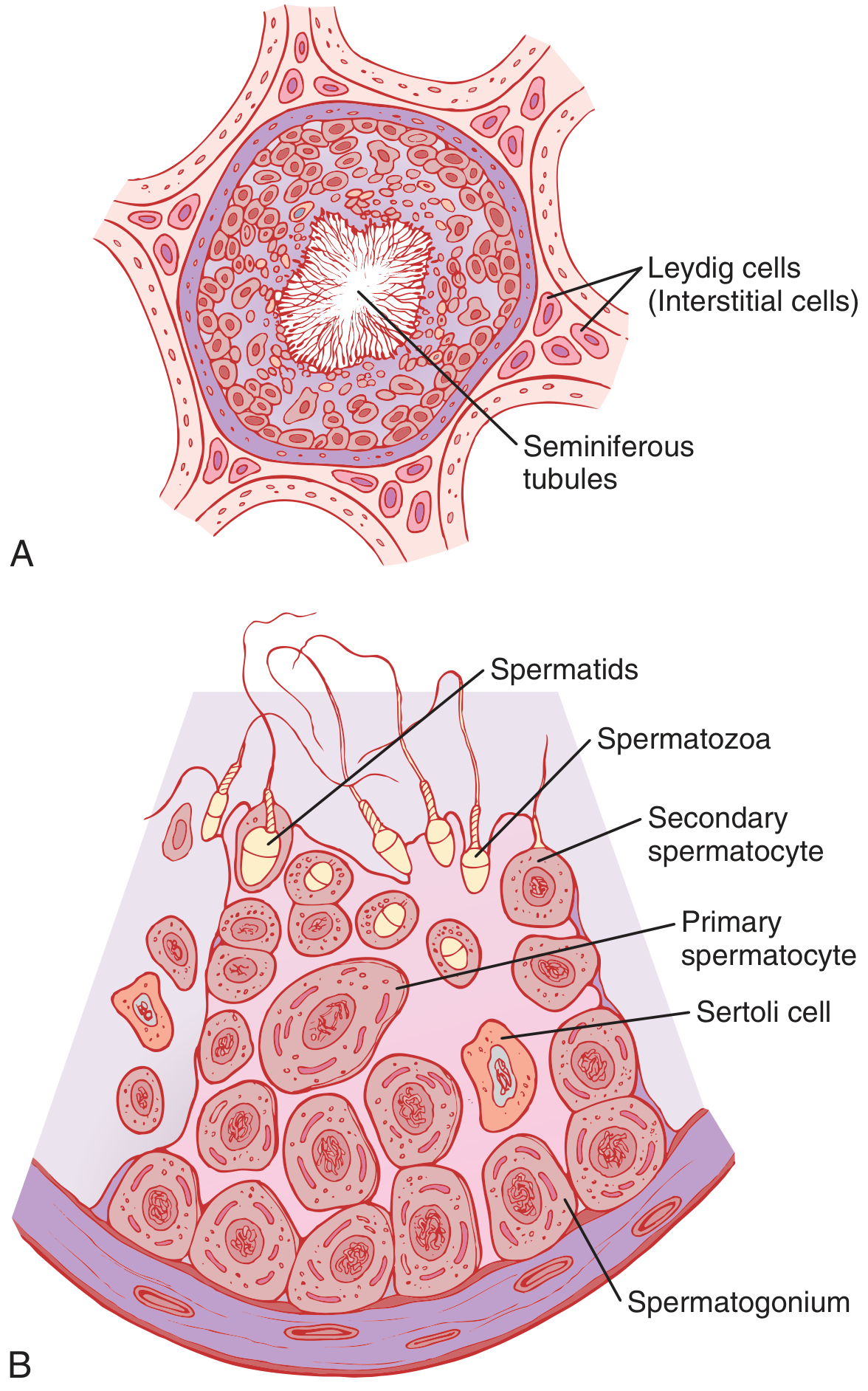
🔢 THREE PHASES OF SPERMATOGENESIS
Phase 1: MULTIPLICATIVE PHASE → Mitosis of spermatogonia
Phase 2: MEIOTIC PHASE → Meiosis I + II
Phase 3: SPERMIOGENESIS → Spermatid → Sperm (no division)
PHASE 1 - MULTIPLICATIVE PHASE (Mitosis)
- Spermatogonia (2n, 46 chromosomes) are the diploid stem cells at the periphery
- They divide by mitosis to produce more spermatogonia
- Two types:
- Type A spermatogonia → remain as stem cells (self-renewal) - ensures lifelong sperm production
- Type B spermatogonia → committed cells that go on to become primary spermatocytes
Key point: Mitosis here is for multiplication only - chromosome number does NOT change (still 2n = 46)
PHASE 2 - MEIOTIC PHASE
Meiosis I (Reductional division):
- Type B spermatogonium grows and becomes a Primary spermatocyte (2n = 46)
- Primary spermatocyte is the LARGEST germ cell in the seminiferous tubule
- It undergoes Meiosis I → produces 2 Secondary spermatocytes
- Each secondary spermatocyte = haploid (n = 23)
- This is where crossing over occurs (genetic recombination)
Meiosis II (Equational division):
- Each Secondary spermatocyte (n = 23) undergoes Meiosis II
- Produces 2 Spermatids each
- Total from one primary spermatocyte = 4 spermatids
- Each spermatid = haploid (n = 23)
COMPLETE FLOW CHART
| Cell | Ploidy | Chromosome # | Division | No. of cells |
|---|---|---|---|---|
| Spermatogonium | Diploid | 2n = 46 | Mitosis | 1 → many |
| Primary spermatocyte | Diploid | 2n = 46 | Before Meiosis I | 1 |
| Secondary spermatocyte | Haploid | n = 23 | After Meiosis I | 2 |
| Spermatid | Haploid | n = 23 | After Meiosis II | 4 |
| Spermatozoon | Haploid | n = 23 | After spermiogenesis | 4 |
NEET rule: 1 primary spermatocyte → 4 spermatozoa (all functional)
PHASE 3 - SPERMIOGENESIS ⭐
- No cell division occurs in spermiogenesis - only morphological differentiation
- Spermatid → Sperm through the following changes:
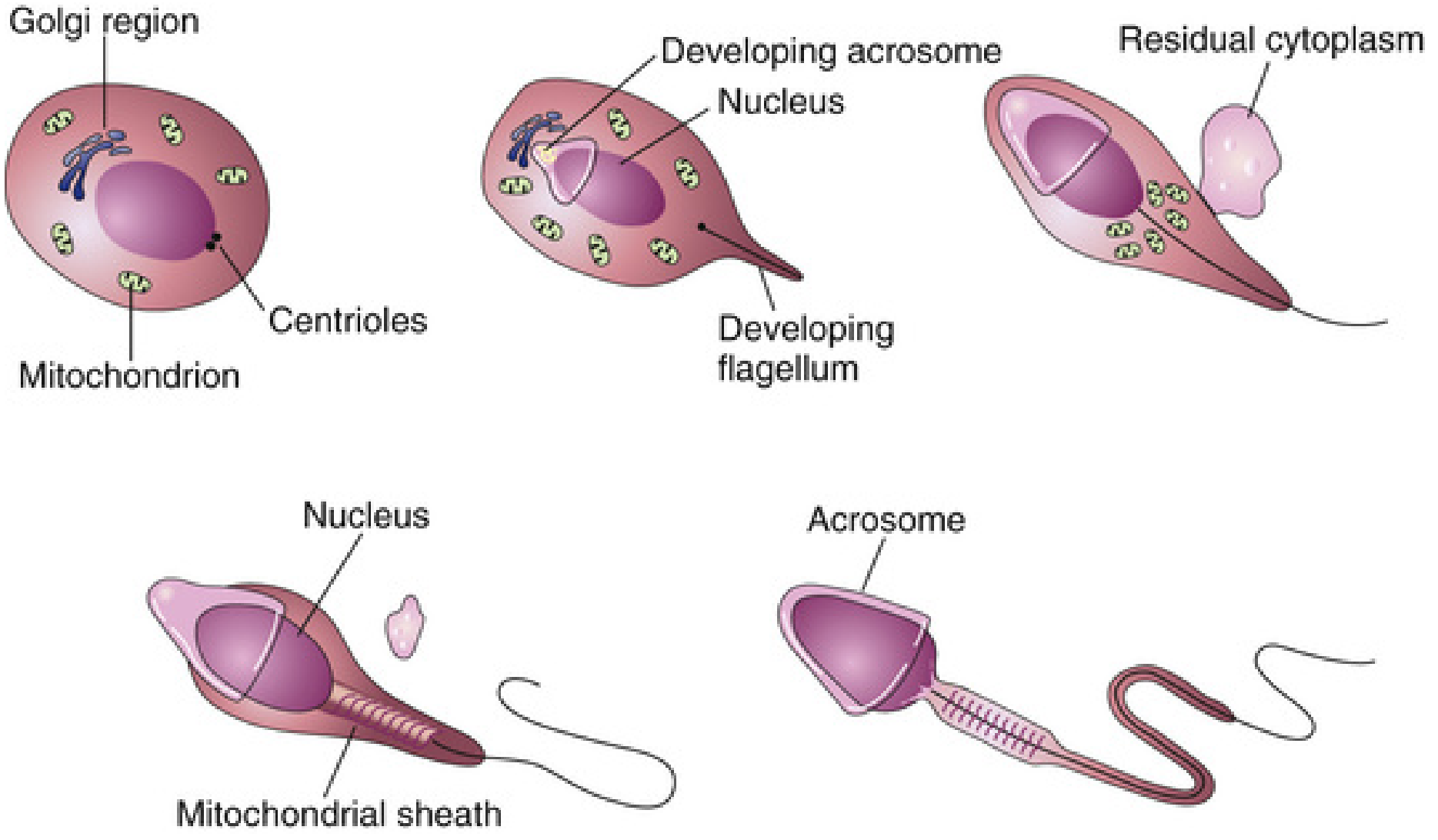
Changes during Spermiogenesis:
| Change | Detail |
|---|---|
| Acrosome formation | Golgi apparatus forms the acrosome cap over the anterior 2/3 of nucleus |
| Nucleus condenses | Chromatin becomes tightly packed; cell becomes smaller |
| Flagellum develops | Centrioles form the axoneme (9+2 microtubule arrangement) → tail grows |
| Mitochondria | Migrate and arrange around the axoneme in the middle piece to form mitochondrial sheath |
| Cytoplasm shed | Residual cytoplasm is discarded as "residual body" → phagocytosed by Sertoli cells |
Final event - SPERMIATION:
- Mature spermatozoa are released from Sertoli cells into the lumen of the seminiferous tubule
- This release is called Spermiation
STRUCTURE OF MATURE SPERMATOZOON
| Part | Structure | Function |
|---|---|---|
| Head | Condensed nucleus (haploid DNA) | Carries genetic material |
| Acrosome | Cap over anterior 2/3 of head; from Golgi | Contains hyaluronidase + acrosin (enzymes to penetrate egg) |
| Neck | Short connecting piece | Junction of head and tail |
| Middle piece (Body) | Axoneme + mitochondrial sheath | ATP production for motility |
| Tail (Flagellum) | 9+2 axoneme of microtubules | Motility - whip-like movement |
HORMONAL CONTROL OF SPERMATOGENESIS
| Hormone | Source | Role in Spermatogenesis |
|---|---|---|
| GnRH | Hypothalamus | Stimulates LH + FSH release |
| LH | Anterior pituitary | Stimulates Leydig cells → testosterone |
| FSH | Anterior pituitary | Stimulates Sertoli cells → ABP secretion → supports spermiogenesis |
| Testosterone | Leydig cells | Essential for all stages; acts via Sertoli cells |
| Estrogen | Sertoli cells (from testosterone by aromatase) | Needed for spermiogenesis |
| Growth hormone | Anterior pituitary | Promotes early division of spermatogonia |
| Inhibin | Sertoli cells | Negative feedback → inhibits FSH |
SPERMATOGENESIS vs SPERMIOGENESIS - Most Common NEET Confusion
| Spermatogenesis | Spermiogenesis | |
|---|---|---|
| Meaning | Entire process (spermatogonia → sperm) | Only last phase (spermatid → sperm) |
| Includes | Mitosis + Meiosis + Spermiogenesis | Only morphological differentiation |
| Cell division? | Yes (mitosis + meiosis) | NO cell division |
| Duration | 64-74 days total | Part of that 74 days |
⚡ NEET RAPID FIRE - NUMBERS
| Fact | Value |
|---|---|
| Duration of spermatogenesis | 64-74 days |
| Sperm produced per testis/day | ~120 million |
| Chromosome number of spermatogonium | 2n = 46 |
| Chromosome number of spermatozoon | n = 23 |
| Spermatozoa from 1 primary spermatocyte | 4 |
| Largest cell in seminiferous tubule | Primary spermatocyte |
| Acrosome is derived from | Golgi apparatus |
| Energy for sperm motility | ATP from mitochondria (middle piece) |
| Acrosome enzymes | Hyaluronidase + Acrosin |
| Residual body phagocytosed by | Sertoli cells |
| Motility acquired in | Epididymis (sperm non-motile when leaving testis) |
🧠 MNEMONICS
"Some Pretty Sexy Sperms" Spermatogonia → Primary spermatocyte → Secondary spermatocyte → Spermatid → Spermatozoa
"ANCE" = Acrosome forms, Nucleus condenses, Cytoplasm shed, Elongation + flagellum
"FSH = Feeds Sertoli Here" (FSH → Sertoli) "LH = Loves Leydig" (LH → Leydig → testosterone)
Create a spermatogenesis flowchart PDF for classroom display
mkdir -p /tmp/workspace/spermatogenesis && ls /tmp/workspace/
~/spermatogenesis/create_flowchart.py
from reportlab.lib.pagesizes import A3, landscape
from reportlab.lib import colors
from reportlab.lib.units import cm, mm
from reportlab.pdfgen import canvas
from reportlab.lib.colors import HexColor, white, black
# ── colour palette ──────────────────────────────────────────────
BG = HexColor("#F0F4FF") # page background
TITLE_BG = HexColor("#1A237E") # dark navy
TITLE_FG = white
BOX_STEM = HexColor("#4A90D9") # spermatogonia blue
BOX_PRIMARY = HexColor("#7B1FA2") # primary spermatocyte purple
BOX_SECOND = HexColor("#00796B") # secondary teal
BOX_SPTID = HexColor("#E65100") # spermatid orange
BOX_SPERM = HexColor("#1B5E20") # spermatozoon dark green
BOX_FG = white
ARROW_CLR = HexColor("#37474F") # dark grey arrows
PHASE_BG = HexColor("#E8EAF6") # phase band background
PHASE_FG = HexColor("#1A237E")
LABEL_CLR = HexColor("#BF360C") # red-orange for labels
NUM_CLR = HexColor("#0D47A1") # blue numbers
SIDE_BG = HexColor("#FFF8E1") # note card yellow
SIDE_BORDER = HexColor("#F9A825")
FOOTER_CLR = HexColor("#546E7A")
# ── page setup ───────────────────────────────────────────────────
W, H = landscape(A3) # 420 × 297 mm
c = canvas.Canvas("/tmp/workspace/spermatogenesis/Spermatogenesis_Flowchart.pdf",
pagesize=(W, H))
def filled_rect(x, y, w, h, fill, stroke=None, radius=6):
c.setFillColor(fill)
if stroke:
c.setStrokeColor(stroke)
c.setLineWidth(1.5)
else:
c.setStrokeColor(fill)
c.setLineWidth(0)
c.roundRect(x, y, w, h, radius, fill=1, stroke=1 if stroke else 0)
def text_block(text, x, y, w, h, fg=white, size=11, bold=False, center=True):
c.setFillColor(fg)
c.setFont("Helvetica-Bold" if bold else "Helvetica", size)
if center:
c.drawCentredString(x + w/2, y + h/2 - size*0.35, text)
else:
c.drawString(x + 6, y + h/2 - size*0.35, text)
def draw_arrow(x1, y1, x2, y2, label="", label2=""):
"""Draws a vertical downward arrow with optional label on the right."""
c.setStrokeColor(ARROW_CLR)
c.setFillColor(ARROW_CLR)
c.setLineWidth(2)
c.line(x1, y1, x2, y2 + 8)
# arrowhead
p = c.beginPath()
p.moveTo(x2, y2)
p.lineTo(x2 - 7, y2 + 12)
p.lineTo(x2 + 7, y2 + 12)
p.close()
c.drawPath(p, fill=1, stroke=0)
if label:
c.setFillColor(LABEL_CLR)
c.setFont("Helvetica-Bold", 9)
c.drawString(x2 + 14, (y1 + y2) / 2 + 4, label)
if label2:
c.setFillColor(HexColor("#555555"))
c.setFont("Helvetica", 8)
c.drawString(x2 + 14, (y1 + y2) / 2 - 8, label2)
def multi_line(text_lines, x, y, w, h, fg=white, sizes=None, bold_first=True):
"""Draw multiple lines centred in a box."""
n = len(text_lines)
line_h = h / (n + 0.5)
for i, line in enumerate(text_lines):
sz = sizes[i] if sizes else 10
c.setFillColor(fg)
c.setFont("Helvetica-Bold" if (bold_first and i == 0) else "Helvetica", sz)
c.drawCentredString(x + w/2, y + h - (i + 1) * line_h + line_h * 0.2, line)
# ════════════════════════════════════════════════════════════════
# BACKGROUND
# ════════════════════════════════════════════════════════════════
c.setFillColor(BG)
c.rect(0, 0, W, H, fill=1, stroke=0)
# ════════════════════════════════════════════════════════════════
# TITLE BANNER
# ════════════════════════════════════════════════════════════════
filled_rect(0, H - 2.2*cm, W, 2.2*cm, TITLE_BG, radius=0)
c.setFillColor(TITLE_FG)
c.setFont("Helvetica-Bold", 22)
c.drawCentredString(W/2, H - 1.5*cm, "SPERMATOGENESIS — Complete Flow Chart")
c.setFont("Helvetica", 11)
c.drawCentredString(W/2, H - 2.0*cm, "NEET UG | Class 12 Biology · Chapter: Human Reproduction")
# ════════════════════════════════════════════════════════════════
# PHASE BANDS (background strips)
# ════════════════════════════════════════════════════════════════
# We'll lay out 5 main boxes top → bottom in the centre column
# plus side note cards on right
TOP_Y = H - 2.4*cm # just below title
BOT_Y = 1.2*cm # footer area
USABLE = TOP_Y - BOT_Y # usable height
# centre column
CX = W * 0.18 # left edge of centre column
CW = W * 0.44 # width of centre column
BOX_H = 2.3*cm
BOX_W = CW
# vertical positions for the 5 main cells (plus header)
# we space them evenly
TOTAL_BOXES = 5
GAP = (USABLE - TOTAL_BOXES * BOX_H) / (TOTAL_BOXES + 1)
def box_y(i):
"""i=0 is topmost box"""
return TOP_Y - GAP*(i+1) - BOX_H*(i+1)
# Phase band spans
phase_data = [
# (label, top_box_i, bot_box_i, colour)
("PHASE 1\nMULTIPLICATIVE\n(Mitosis)", 0, 0, HexColor("#E3F2FD")),
("PHASE 2\nMEIOTIC\n(Meiosis I & II)", 1, 2, HexColor("#F3E5F5")),
("PHASE 3\nSPERMIOGENESIS\n(Differentiation)", 3, 3, HexColor("#FFF3E0")),
("EPIDIDYMIS\nMaturation", 4, 4, HexColor("#E8F5E9")),
]
BAND_X = CX - 3.5*cm
BAND_W = CW + 7.0*cm # full width behind everything
for label, ti, bi, col in phase_data:
py_top = box_y(ti) + BOX_H + GAP*0.5
py_bot = box_y(bi) - GAP*0.5
ph = py_top - py_bot
filled_rect(BAND_X, py_bot, BAND_W, ph, col, stroke=HexColor("#CCCCCC"), radius=8)
# phase label on left
c.setFillColor(PHASE_FG)
lines = label.split("\n")
lx = BAND_X + 5
lw = 2.4*cm
mid_y = (py_top + py_bot) / 2
c.setFont("Helvetica-Bold", 8)
c.drawCentredString(lx + lw/2, mid_y + 8, lines[0])
c.setFont("Helvetica-Bold", 8)
c.drawCentredString(lx + lw/2, mid_y - 2, lines[1] if len(lines) > 1 else "")
c.setFont("Helvetica", 7.5)
c.drawCentredString(lx + lw/2, mid_y - 13, lines[2] if len(lines) > 2 else "")
# ════════════════════════════════════════════════════════════════
# MAIN BOXES
# ════════════════════════════════════════════════════════════════
boxes = [
# (colour, lines, sizes)
(BOX_STEM, ["SPERMATOGONIUM", "Diploid | 2n = 46 chromosomes", "Stem cell — lies at periphery of seminiferous tubule"],
[13, 10, 9]),
(BOX_PRIMARY, ["PRIMARY SPERMATOCYTE", "Diploid | 2n = 46 chromosomes", "Largest germ cell in the seminiferous tubule"],
[13, 10, 9]),
(BOX_SECOND, ["SECONDARY SPERMATOCYTE (×2)", "Haploid | n = 23 chromosomes", "Each is ~half the size of primary spermatocyte"],
[13, 10, 9]),
(BOX_SPTID, ["SPERMATID (×4)", "Haploid | n = 23 chromosomes", "Round cell — no tail yet; undergoes spermiogenesis"],
[13, 10, 9]),
(BOX_SPERM, ["MATURE SPERMATOZOON (×4)", "Haploid | n = 23 chromosomes", "Motility acquired in epididymis (18–24 hrs)"],
[13, 10, 9]),
]
for i, (col, lines, szs) in enumerate(boxes):
by = box_y(i)
filled_rect(CX, by, BOX_W, BOX_H, col, stroke=white, radius=10)
multi_line(lines, CX, by, BOX_W, BOX_H, fg=white, sizes=szs, bold_first=True)
# ════════════════════════════════════════════════════════════════
# ARROWS between boxes with division labels
# ════════════════════════════════════════════════════════════════
arrow_data = [
# (from_box_i, label_main, label_sub)
(0, "MITOSIS", "Type B → Primary spermatocyte"),
(1, "MEIOSIS I", "Reductional division · Crossing over occurs"),
(2, "MEIOSIS II", "Equational division · 4 spermatids total"),
(3, "SPERMIOGENESIS", "No division · Morphological change only"),
]
ax = CX + BOX_W / 2
for from_i, lbl, sub in arrow_data:
y_start = box_y(from_i)
y_end = box_y(from_i + 1) + BOX_H
draw_arrow(ax, y_start, ax, y_end, lbl, sub)
# ════════════════════════════════════════════════════════════════
# RIGHT SIDE — NOTE CARDS
# ════════════════════════════════════════════════════════════════
NX = CX + CW + 1.0*cm
NW = W - NX - 0.8*cm
def note_card(title, lines, y, h, title_col=HexColor("#1A237E")):
filled_rect(NX, y, NW, h, SIDE_BG, stroke=SIDE_BORDER, radius=8)
# title strip
filled_rect(NX, y + h - 0.7*cm, NW, 0.7*cm, title_col, radius=6)
c.setFillColor(white)
c.setFont("Helvetica-Bold", 9)
c.drawCentredString(NX + NW/2, y + h - 0.46*cm, title)
# body lines
c.setFont("Helvetica", 8.5)
ly = y + h - 0.7*cm - 0.3*cm
for line in lines:
c.setFillColor(HexColor("#212121"))
bullet = line.startswith("•")
indent = 10 if bullet else 6
c.drawString(NX + indent, ly, line)
ly -= 0.42*cm
# Card 1 — Key Numbers
note_card("⏱ KEY NUMBERS (NEET Favourites)",
["• Duration: 64–74 days total",
"• Sperm produced: ~120 million / testis / day",
"• 1 Primary spermatocyte → 4 spermatozoa",
"• Epididymis: 6 m long; 18–24 hrs for maturity",
"• Temperature: 2–2.5°C below body temp"],
y=box_y(0) - 0.1*cm, h=3.2*cm,
title_col=HexColor("#0D47A1"))
# Card 2 — Spermiogenesis changes
note_card("🔬 SPERMIOGENESIS CHANGES",
["• Acrosome forms (from Golgi apparatus)",
"• Nucleus condenses (DNA tightly packed)",
"• Flagellum grows (from centrioles)",
"• Mitochondria → mitochondrial sheath (middle piece)",
"• Residual cytoplasm shed → phagocytosed by Sertoli cells",
"• Spermiation = release from Sertoli into lumen"],
y=box_y(1) + 0.2*cm, h=3.6*cm,
title_col=HexColor("#6A1B9A"))
# Card 3 — Hormones
note_card("💊 HORMONAL CONTROL",
["• LH → Leydig cells → Testosterone",
"• FSH → Sertoli cells → ABP secretion",
"• Testosterone → drives all stages",
"• Inhibin (Sertoli) → inhibits FSH (–ve feedback)",
"• GH → promotes spermatogonia division"],
y=box_y(2) + 0.0*cm, h=3.0*cm,
title_col=HexColor("#004D40"))
# Card 4 — Sperm structure
note_card("🔵 SPERM STRUCTURE",
["Head: nucleus + acrosome (anterior 2/3)",
"Acrosome: hyaluronidase + acrosin",
"Middle piece: mitochondria → ATP",
"Tail (flagellum): 9+2 axoneme microtubules",
"Speed: 1–4 mm/min in fluid"],
y=box_y(3) + 0.0*cm, h=3.0*cm,
title_col=HexColor("#BF360C"))
# Card 5 — NEET trap
note_card("⚠ COMMON NEET TRAPS",
["Spermiogenesis ≠ Spermatogenesis (no division!)",
"Spermiation = release from Sertoli cell",
"Sperm motile only AFTER epididymis",
"Primary spermatocyte = LARGEST germ cell",
"Acrosome from Golgi (NOT from nucleus)",
"Inhibin inhibits FSH (NOT LH)"],
y=box_y(4) + 0.0*cm, h=3.2*cm,
title_col=HexColor("#B71C1C"))
# ════════════════════════════════════════════════════════════════
# CHROMOSOME COUNT INDICATORS (left of main boxes)
# ════════════════════════════════════════════════════════════════
chr_data = [
(0, "2n=46", NUM_CLR),
(1, "2n=46", NUM_CLR),
(2, "n=23", HexColor("#006064")),
(3, "n=23", HexColor("#006064")),
(4, "n=23", HexColor("#1B5E20")),
]
for bi, txt, col in chr_data:
by = box_y(bi) + BOX_H/2 - 0.25*cm
filled_rect(CX - 2.2*cm, by, 1.9*cm, 0.5*cm, col, radius=4)
c.setFillColor(white)
c.setFont("Helvetica-Bold", 9)
c.drawCentredString(CX - 1.25*cm, by + 0.14*cm, txt)
# cell count badges (right side of boxes, inside)
count_data = [
(0, "×∞"),
(1, "×1"),
(2, "×2"),
(3, "×4"),
(4, "×4"),
]
for bi, cnt in count_data:
by = box_y(bi) + BOX_H - 0.65*cm
bx = CX + BOX_W - 1.3*cm
filled_rect(bx, by, 1.1*cm, 0.52*cm, HexColor("#FFFFFF44"), radius=4)
c.setFillColor(white)
c.setFont("Helvetica-Bold", 10)
c.drawCentredString(bx + 0.55*cm, by + 0.15*cm, cnt)
# ════════════════════════════════════════════════════════════════
# FOOTER
# ════════════════════════════════════════════════════════════════
filled_rect(0, 0, W, 1.1*cm, HexColor("#CFD8DC"), radius=0)
c.setFillColor(FOOTER_CLR)
c.setFont("Helvetica", 8.5)
c.drawString(1*cm, 0.4*cm, "NEET UG | Class 12 Biology | Unit: Human Reproduction")
c.drawCentredString(W/2, 0.4*cm,
"Spermatogonia → Primary S/C → Secondary S/C → Spermatid → Spermatozoon")
c.setFont("Helvetica-Bold", 8.5)
c.drawRightString(W - 1*cm, 0.4*cm, "All 4 spermatozoa are functional ✓")
c.save()
print("PDF created successfully.")
cd /tmp/workspace/spermatogenesis && python create_flowchart.py
ls -lh /tmp/workspace/spermatogenesis/
Spermatogenesis Flowchart
PDF Document · PDF
- Central flowchart column - 5 colour-coded boxes flowing top to bottom:
- 🔵 Spermatogonium → 🟣 Primary spermatocyte → 🟢 Secondary spermatocyte → 🟠 Spermatid → 🟩 Spermatozoon
- Arrows with division labels between each box (Mitosis / Meiosis I / Meiosis II / Spermiogenesis)
- Phase bands on the left margin clearly marking Phase 1 (Multiplicative), Phase 2 (Meiotic), Phase 3 (Spermiogenesis)
- Chromosome count badges (2n=46 / n=23) beside every box
- Cell count indicators (×1, ×2, ×4) inside each box
- 5 colour-coded note cards on the right:
- Key numbers (NEET favourites)
- Spermiogenesis changes
- Hormonal control
- Sperm structure
- Common NEET traps
- Clean footer with the full sequence as a one-liner
Complete spermatogenesis important definitions and everything for neet UG for better understanding pf concepts First teach the mitosis and meiosis frok scratch to a level that's required to understand gametogenesis
🧬 Mitosis, Meiosis & Spermatogenesis | NEET UG - Complete Notes
PART 1: ESSENTIAL BACKGROUND - KEY TERMS AND DEFINITIONS
📖 CRITICAL DEFINITIONS
| Term | Definition |
|---|---|
| Chromosome | Condensed, coiled structure of DNA + proteins (histones). Visible during cell division. |
| Chromatid | Each of the two identical copies of a chromosome after DNA replication, joined at the centromere |
| Sister chromatids | The two identical copies joined together after DNA replication - they are ONE chromosome |
| Homologous chromosomes (homologs) | Pairs of chromosomes - one from mother, one from father - same size, same gene loci, but possibly different alleles |
| Centromere | The constricted region of a chromosome where sister chromatids are held together; where spindle fibres attach |
| Kinetochore | Protein complex at the centromere where spindle microtubules attach |
| Spindle fibres | Microtubule structures that pull chromosomes to opposite poles during division |
| Diploid (2n) | Cell with TWO sets of chromosomes (one from each parent). In humans: 2n = 46 |
| Haploid (n) | Cell with ONE set of chromosomes. In humans: n = 23 |
| Ploidy | The number of chromosome sets in a cell |
| DNA replication | Copying of DNA; occurs during S phase of interphase, BEFORE any cell division |
| Bivalent (Tetrad) | A pair of homologous chromosomes (each made of 2 sister chromatids) joined together = 4 chromatids total |
| Crossing over | Exchange of segments between non-sister chromatids of homologous chromosomes during Prophase I of meiosis |
| Chiasma (pl. chiasmata) | The visible X-shaped point where crossing over has occurred |
| Synapsis | Precise alignment and pairing of homologous chromosomes during Prophase I of meiosis |
| Synaptonemal complex | Protein scaffold that holds homologous chromosomes together during synapsis |
| Nondisjunction | Failure of chromosomes to separate properly during meiosis → abnormal chromosome number in gametes |
HUMAN CHROMOSOME NUMBERS - Must Know
| Cell type | Chromosome count | Ploidy |
|---|---|---|
| Body (somatic) cells | 46 (23 pairs) | Diploid (2n) |
| Spermatogonia | 46 | Diploid (2n) |
| Primary spermatocyte | 46 | Diploid (2n) |
| Secondary spermatocyte | 23 | Haploid (n) |
| Spermatid | 23 | Haploid (n) |
| Spermatozoon | 23 | Haploid (n) |
| Ovum | 23 | Haploid (n) |
| Zygote (fertilised egg) | 46 | Diploid (2n) |
PART 2: CELL CYCLE - The Setup Before Division
INTERPHASE (cell grows + replicates DNA)
├── G1 phase: Cell grows; organelles duplicated; protein synthesis
├── S phase: DNA replication (2n → 4n DNA content; still 46 chromosomes but each now has 2 chromatids)
└── G2 phase: Cell prepares for division; more growth; spindle proteins made
MITOTIC PHASE (M phase) = actual division
Key point for NEET: DNA replication happens in S phase of interphase, BEFORE division starts. So by the time Prophase begins, every chromosome already has 2 sister chromatids (but the cell is still considered 2n = 46 chromosomes).
PART 3: MITOSIS - "Equal Division for Growth"
Stages of Mitosis
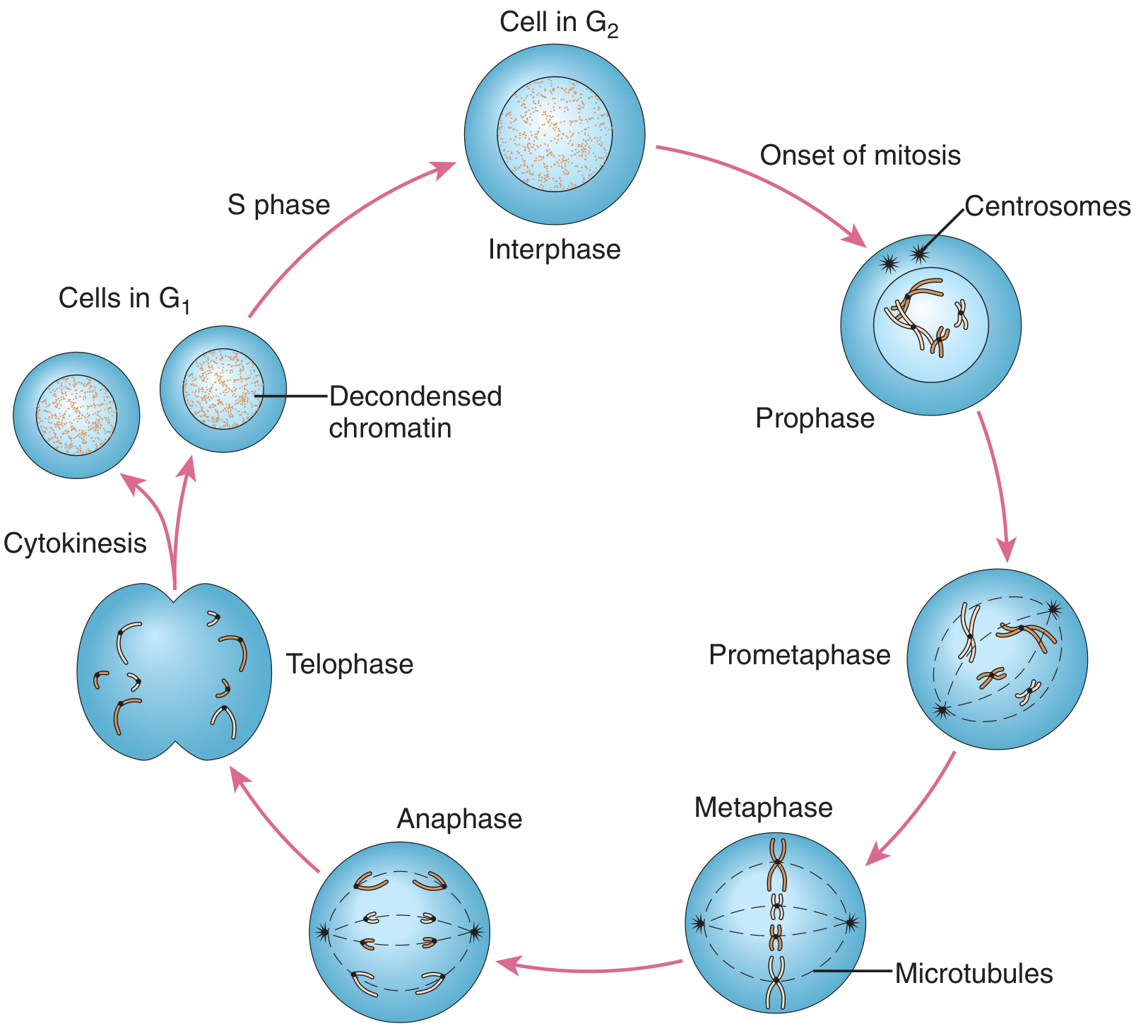
Mnemonic: "PMAT" = Prophase → Metaphase → Anaphase → Telophase (+ Prometaphase between P and M)
Stage 1: PROPHASE
- Chromosomes condense (become visible under microscope)
- Centrosomes move to opposite poles; spindle fibre formation begins
- Nucleolus disappears
- Nuclear envelope still present at start
Stage 2: PROMETAPHASE
- Nuclear envelope breaks down (dissolves)
- Spindle fibres attach to chromosomes at kinetochores
- Chromosomes begin moving toward the centre
Stage 3: METAPHASE ⭐ (Most important stage for cytology)
- Chromosomes maximally condensed (best stage to count and study chromosomes - karyotype made from this stage)
- All chromosomes align at the equatorial plate / metaphase plate (centre of the cell)
- Each chromosome = 2 sister chromatids held at centromere
- Each centromere connected to BOTH poles by spindle fibres
Stage 4: ANAPHASE
- Centromeres split
- Sister chromatids separate and are pulled to opposite poles by spindle fibres
- Each chromatid is now considered a separate chromosome
- At this point the cell momentarily has 92 chromosomes (46 at each pole)
Stage 5: TELOPHASE
- Chromosomes arrive at poles and begin to decondense
- Nuclear envelope reforms around each set of chromosomes
- Nucleolus reappears
- Two new nuclei formed
CYTOKINESIS (not part of mitosis but follows it)
- Division of the cytoplasm
- In animal cells: cleavage furrow forms and pinches the cell in two
- Result: 2 daughter cells, each 2n = 46
Summary of Mitosis:
| Feature | Value |
|---|---|
| Number of divisions | 1 |
| Cells produced | 2 |
| Ploidy of daughter cells | Diploid (2n = 46) |
| Are daughters identical? | Yes (genetically identical to parent) |
| Crossing over? | No |
| Where in spermatogenesis? | Multiplication of spermatogonia (Phase 1) |
PART 4: MEIOSIS - "Halving Division for Reproduction"
MEIOSIS I - "Reductional Division" (Most Complex, Most Important)
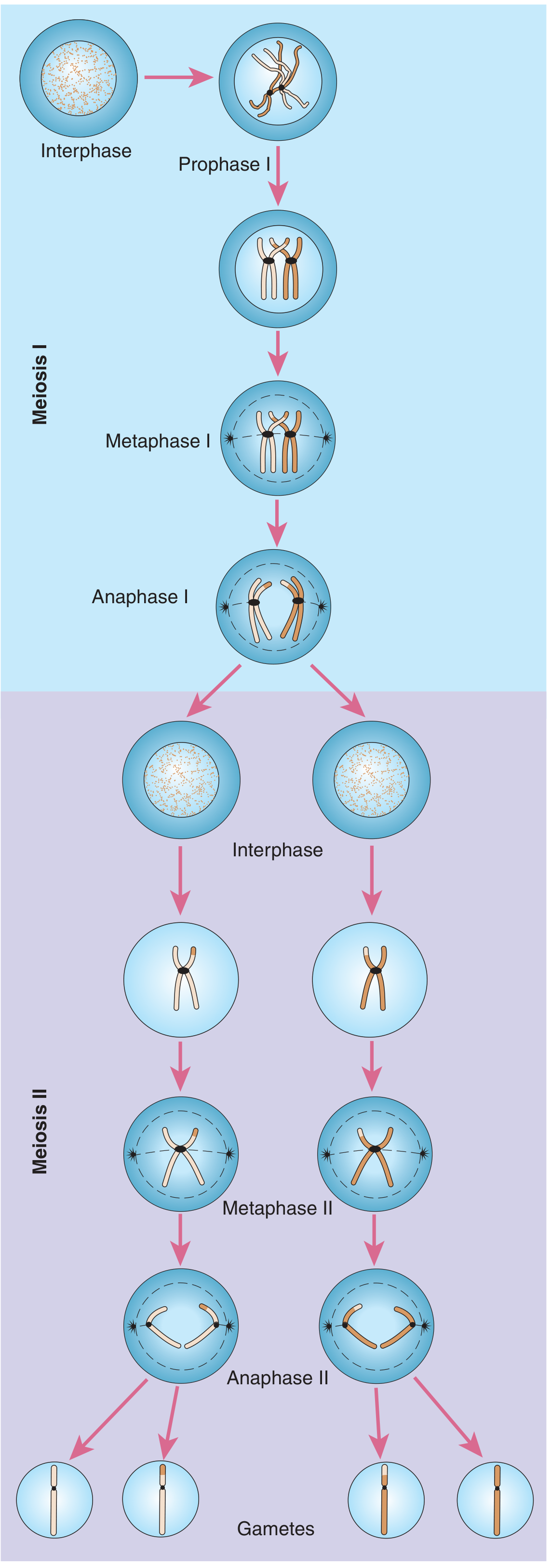
PROPHASE I ⭐⭐ (Most Important and Longest Stage of Entire Meiosis)
Leptotene → Zygotene → Pachytene → Diplotene → Diakinesis
| Substage | Key Events |
|---|---|
| Leptotene | Chromosomes begin to condense and become visible; thin thread-like appearance |
| Zygotene | Homologous chromosomes begin to pair (synapse) - synaptonemal complex forms; paired unit = bivalent |
| Pachytene ⭐ | Synapsis complete; CROSSING OVER occurs between non-sister chromatids of homologs; chiasmata form; genetic recombination happens here |
| Diplotene | Synaptonemal complex dissolves; homologs begin to separate slightly but remain connected at chiasmata; chromosomes clearly double-stranded visible |
| Diakinesis | Maximum condensation; chiasmata move toward ends (terminalization); nuclear envelope breaks down; bivalents move to equatorial plate |
NEET key: Crossing over happens in Pachytene. Chiasmata are visible in Diplotene.
METAPHASE I
- Bivalents (homologous pairs) align at the equatorial plate (not individual chromosomes like in mitosis)
- Centromeres of homologs face opposite poles
- This is where independent assortment occurs (each pair can face either pole randomly)
ANAPHASE I ⭐ (Key difference from mitosis)
- Homologous chromosomes (not sister chromatids) are pulled to opposite poles
- Each chromosome still has 2 sister chromatids joined at centromere
- Chromosome number halved at this point
- This is called disjunction
TELOPHASE I
- Nuclear envelope may or may not reform (varies)
- Cytokinesis → 2 haploid cells (n = 23), but each chromosome still has 2 chromatids
Between Meiosis I and Meiosis II: No DNA replication occurs (no S phase)
MEIOSIS II - "Equational Division"
PROPHASE II
- Chromosomes condense again
- Spindle formation
- Nuclear envelope breaks down
METAPHASE II
- Individual chromosomes (each with 2 sister chromatids) align at equatorial plate
ANAPHASE II ⭐
- Sister chromatids separate and move to opposite poles
- (This is the same event as mitotic anaphase)
TELOPHASE II
- Nuclear envelopes reform
- Cytokinesis → 4 haploid cells total
Summary Comparison: Mitosis vs Meiosis
| Feature | Mitosis | Meiosis |
|---|---|---|
| Occurs in | Somatic (body) cells | Germ cells only (gonads) |
| Rounds of division | 1 | 2 (Meiosis I + II) |
| DNA replication | Once (before division) | Once (before Meiosis I only) |
| Cells produced | 2 | 4 |
| Chromosome number in products | 2n = 46 (diploid) | n = 23 (haploid) |
| Are daughter cells identical? | Yes | No (genetically unique) |
| Crossing over / recombination? | No | Yes (Pachytene of Prophase I) |
| Synapsis of homologs? | No | Yes (Prophase I) |
| Purpose | Growth, repair | Gamete formation |
| In spermatogenesis | Phase 1 (spermatogonia multiply) | Phase 2 (spermatocyte → spermatids) |
What makes each gamete UNIQUE? (2 reasons)
- Independent assortment - During Metaphase I, each pair of homologs can face either pole randomly → 2²³ = over 8 million possible combinations
- Crossing over - During Pachytene, segments exchanged between non-sister chromatids → shuffles genes within chromosomes → enormous additional variation
PART 5: HOW MEIOSIS CONNECTS TO SPERMATOGENESIS
SPERMATOGONIUM (2n = 46)
↓ MITOSIS (Phase 1 - multiplication)
SPERMATOGONIUM (2n = 46) [some self-renew, some proceed]
↓ grows into...
PRIMARY SPERMATOCYTE (2n = 46)
↓ MEIOSIS I (Reductional division)
• Prophase I: crossing over, synapsis
• Chromosome number halved
2 × SECONDARY SPERMATOCYTE (n = 23) [each still has 2 chromatids per chromosome]
↓ MEIOSIS II (Equational division) [NO DNA replication between]
• Sister chromatids separate
4 × SPERMATID (n = 23) [round cells, no tail]
↓ SPERMIOGENESIS (Phase 3 - differentiation, NO division)
4 × SPERMATOZOON (n = 23) [mature sperm with head + tail]
PART 6: SPERMATOGENESIS - COMPLETE DEFINITIONS
| Term | Definition |
|---|---|
| Spermatogenesis | The entire process of formation of mature spermatozoa from spermatogonia; includes mitosis, meiosis, and spermiogenesis |
| Spermatogonium | Diploid (2n) stem cell at the periphery of the seminiferous tubule; the starting cell of spermatogenesis |
| Type A spermatogonium | Self-renewing stem cell that maintains the pool of spermatogonia for lifelong sperm production |
| Type B spermatogonium | Committed cell that proceeds to become a primary spermatocyte |
| Primary spermatocyte | Diploid (2n=46) cell that undergoes Meiosis I; largest germ cell in the tubule |
| Secondary spermatocyte | Haploid (n=23) cell produced after Meiosis I; undergoes Meiosis II |
| Spermatid | Haploid (n=23) round cell produced after Meiosis II; no tail yet; undergoes spermiogenesis |
| Spermiogenesis | Transformation of spermatid into mature spermatozoon; involves morphological differentiation only - NO cell division |
| Spermatozoon | Mature haploid (n=23) sperm cell with head, neck, middle piece, and tail |
| Spermiation | Release of mature spermatozoa from Sertoli cells into the lumen of the seminiferous tubule |
| Capacitation | Physiological changes sperm undergo in the female reproductive tract that enable them to fertilise an egg |
| Acrosome reaction | Release of acrosomal enzymes (hyaluronidase, acrosin) when sperm contacts the zona pellucida of the egg |
PART 7: SPERMIOGENESIS - Step-by-Step Changes
| Change | Detail | NEET point |
|---|---|---|
| Acrosome formation | Golgi apparatus forms vesicles → fuse to form the acrosome cap over anterior 2/3 of nucleus | Acrosome = from Golgi |
| Nuclear condensation | Nucleus shrinks, DNA tightly packed, histone proteins replaced by protamines | Makes sperm head compact |
| Flagellum development | Centrioles (9+2 arrangement) form the axoneme backbone of the tail | Tail = axoneme = 9+2 microtubules |
| Mitochondrial sheath | Mitochondria migrate and wrap around the axoneme in the middle piece | Middle piece = energy for motility |
| Cytoplasm shedding | Excess cytoplasm shed as residual body → phagocytosed by Sertoli cells | Sertoli cells clean up debris |
| Cell elongation | Round spermatid becomes elongated spermatozoon | Final shape achieved |
| Spermiation | Mature sperm released from Sertoli cell grip into tubule lumen | Last step of spermiogenesis |
PART 8: SPERM STRUCTURE - Every Part Explained
| Part | What's in it | Function |
|---|---|---|
| Head | Condensed nucleus (haploid DNA) + acrosome cap | Carries genetic info; acrosome penetrates egg |
| Acrosome | Hyaluronidase + Acrosin (serine protease) | Digests corona radiata and zona pellucida |
| Neck | Centrioles | Connects head to tail |
| Middle piece | Axoneme + mitochondrial sheath | ATP synthesis → powers tail movement |
| Principal piece (tail) | Axoneme (9+2 microtubules) + fibrous sheath | Main propulsion |
| End piece | Axoneme only | Terminal segment |
PART 9: NONDISJUNCTION - Important Concept
- n+1 (24 chromosomes) = disomic gamete
- n-1 (22 chromosomes) = nullisomic gamete
- Trisomy 21 (Down syndrome) - extra chromosome 21
- Klinefelter syndrome (XXY) - extra X in male
- Monosomy X (Turner syndrome, 45, X)
PART 10: RAPID FIRE NEET NUMBERS
| Fact | Value |
|---|---|
| Duration of spermatogenesis | 64-74 days |
| Sperm produced per day (per testis) | ~120 million |
| 1 primary spermatocyte → how many sperm? | 4 |
| Chromosome no. of primary spermatocyte | 2n = 46 |
| Chromosome no. of spermatozoon | n = 23 |
| Largest germ cell in seminiferous tubule | Primary spermatocyte |
| Stage with crossing over | Pachytene (Prophase I) |
| Stage where chiasmata are visible | Diplotene (Prophase I) |
| Acrosome derived from | Golgi apparatus |
| Tail motility powered by | Mitochondria (ATP) in middle piece |
| Axoneme structure | 9+2 arrangement of microtubules |
| Sperm motility acquired in | Epididymis |
| Sperm viability in female tract | 48-72 hours |
| Sperm speed in female tract | ~3 mm/min |
PART 11: MASTER COMPARISON TABLE - ALL STAGES
| Cell | Division used | Ploidy | Chr. no. | # Cells | Largest? |
|---|---|---|---|---|---|
| Spermatogonium | Mitosis | 2n | 46 | Many | No |
| Primary spermatocyte | - (after Meiosis I) | 2n | 46 | 1 | Yes - largest |
| Secondary spermatocyte | Meiosis II | n | 23 | 2 | No |
| Spermatid | - (after Meiosis II) | n | 23 | 4 | No |
| Spermatozoon | - (spermiogenesis) | n | 23 | 4 | No |
Spermatid to sperm What happens during maturation And structure of spermatid and mature sperm too For ug neet