
Immunotherapy in ovarian cancer
"ovarian cancer" AND immunotherapy
PMID: 41763143
PMID: 39764856
"ovarian cancer" AND "CAR-T" OR "adoptive cell therapy"
PMID: 40650991
"ovarian cancer" AND "pembrolizumab" OR "durvalumab" OR "atezolizumab"
"ovarian cancer" AND ("avelumab" OR "JAVELIN" OR "PAOLA" OR "IMagyn050")
PMID: 37625235
"ovarian cancer" AND "durvalumab" AND "olaparib"
immunotherapy ovarian cancer 2025 2026 new approvals trials update
PMID: 41380962
https://www.oncologynewscentral.com/category/ovarian-cancer
Immunotherapy in Ovarian Cancer
1. Immunological Basis
- Regulatory T cells (Tregs) that secrete IL-10 and TGF-beta, dampening cytotoxic responses
- Tumor-derived IL-6 that promotes tumor cell growth and modulates immune function
- Myeloid-derived suppressor cells (MDSCs) and immunosuppressive IDO (indoleamine 2,3-dioxygenase) production
- T-cell exhaustion driven by chronic tumor antigen exposure, which upregulates checkpoint receptors (PD-1, CTLA-4, TIM-3, LAG-3)
- Low mutational burden: High-grade serous ovarian cancer (HGSOC) has relatively few somatic mutations, meaning fewer neoantigens for T cells to recognize - a key reason why single-agent checkpoint inhibitors have been less effective than in MSI-high or TMB-high tumors
2. Types of Immunotherapy Approaches
A. Immune Checkpoint Inhibitors (ICIs)
- Anti-PD-1: Nivolumab, pembrolizumab
- Anti-PD-L1: Atezolizumab, avelumab, durvalumab
- Anti-CTLA-4: Ipilimumab
- Combination CTLA-4 + PD-1 blockade: Different, non-redundant mechanisms; combination induces ~50% tumor regression in melanoma and is being explored in ovarian cancer
B. Cytokine Therapy
C. Cancer Vaccines
- Personalized neoantigen vaccines: Early data show these can effectively mobilize antitumor T-cell immunity in ovarian cancer (Science Translational Medicine, 2018)
- CA-125 targeting vaccines: Investigated in trials but limited clinical success as monotherapy
- Dendritic cell vaccines: Phase I/II explored; synergy with checkpoint blockade is under study
D. Adoptive Cell Therapy (ACT)
- TIL therapy: Tumor-infiltrating lymphocytes expanded ex vivo and reinfused; phase I/II studies ongoing in ovarian cancer
- CAR-T cells: Targeting antigens such as folate receptor alpha (FRalpha), mesothelin, and MUC16 (CA-125); early trials underway with challenges around solid tumor penetration
- CAR-NK cells: Emerging data (2025) with CRISPR-based enhancement of NK cell potency
E. Antibody-Dependent Cellular Cytotoxicity (ADCC)
3. Major Clinical Trials and Results
First-Line Setting
| Trial | Regimen | Key Result |
|---|---|---|
| IMagyn050 (Phase III) | Atezolizumab + chemo + bevacizumab | No significant PFS or OS benefit (HR 0.92 ITT; HR 0.83 in PD-L1+ not significant) |
| PAOLA-1 (Phase III) | Olaparib + bevacizumab maintenance | Improved PFS in HRD+ patients; OS benefit primarily in BRCA-mutated (not purely immunotherapy) |
| DUO-O (Phase III, 2026) | Durvalumab + chemo + bev → durvalumab + bev + olaparib vs. control (non-BRCA mutated) | Triplet met PFS endpoints: HRD+ non-tBRCAm: HR 0.49 (mPFS 37.3 vs 23.0 months); ITT non-tBRCAm: HR 0.63 (mPFS 24.2 vs 19.3 months) - though OS data immature |
Recurrent Setting
- ICI monotherapy has shown limited activity
- The SOLACE2 phase II trial (2025) tested olaparib + durvalumab + cyclophosphamide in platinum-sensitive disease
- The ARTISTRY-7 phase III trial (nemvaleukin alfa + pembrolizumab) was terminated early in March 2025 for lack of survival benefit
- FDA Approval (2025): Pembrolizumab + paclitaxel (with or without bevacizumab) was approved for platinum-resistant ovarian cancer - the first FDA approval of a checkpoint inhibitor specifically for ovarian cancer
4. The Meta-Analytic Picture (2025-2026)
- Overall, anti-PD-1/PD-L1 did not significantly improve PFS vs. control (HR 0.98; 95% CI 0.85-1.12; I² = 67%)
- No significant benefit in first-line (HR 0.93) or recurrent settings (HR 1.07)
- Non-significant trend favoring ICI + PARP inhibitor in HR-proficient patients (HR 0.77; 95% CI 0.65-0.92)
- PD-1/PD-L1 + chemotherapy and PD-1/PD-L1 + ipilimumab showed the greatest PFS/OS benefit
- ICI + chemo improved ORR most markedly (OR 3.06; 95% CI 1.42-6.60)
- PD-L1 expression is a potential predictive biomarker - PD-L1-positive patients showed better outcomes with ICI + chemotherapy
- 7 phase III trials (5,671 patients)
- Single-agent PD-L1 inhibitor trials: no significant efficacy in newly diagnosed EOC
- Triplet durvalumab + bevacizumab + olaparib: longer PFS than bevacizumab alone in non-BRCA-mutated patients
- ICI ineffective in platinum-sensitive and platinum-resistant recurrent EOC as monotherapy
5. Why Has Immunotherapy Underperformed vs. Other Cancers?
- Low tumor mutational burden (TMB): HGSOC has fewer neoantigens than melanoma or NSCLC
- Immunosuppressive TME: Ascites contains abundant Tregs, IL-10, TGF-beta, MDSCs
- Low MSI/dMMR prevalence: Only ~5% of ovarian cancers are MSI-high (those benefit most from checkpoint inhibition)
- PD-L1 is inconsistently expressed and is not a reliable single biomarker in ovarian cancer
- T-cell exhaustion and metabolic reprogramming within the TME limit ICI reversal of exhaustion
6. Predictive Biomarkers (Emerging)
| Biomarker | Relevance |
|---|---|
| PD-L1 expression | Modest predictor; PD-L1+ tumors show better ICI response, especially with combination regimens |
| HRD (homologous recombination deficiency) | Enriches for PARP inhibitor + ICI combination benefit (DUO-O, PAOLA-1) |
| BRCA1/2 mutation | Predicts PARP inhibitor benefit; may augment ICI by increasing neoantigen load |
| MSI-H / dMMR | Strong predictor; pembrolizumab tissue-agnostic approval covers this subset |
| TMB-high | Emerging predictor; low prevalence in EOC |
| TIL density | High TILs correlate with improved survival; marker of immunogenic phenotype |
7. Emerging Strategies and Future Directions
- CTLA-4 + PD-1 dual blockade: Under investigation; dual blockade may overcome the immunosuppressive TME more effectively than single-agent
- ICI + PARP inhibitor combinations: Most promising signal to date (DUO-O); synergy proposed through PARP-trapping increasing genomic instability and immunogenicity
- ICI + anti-angiogenics (bevacizumab): Bevacizumab may "normalize" the tumor vasculature to improve T-cell infiltration
- Bispecific antibodies: Targeting TAAs (e.g., FRalpha, mesothelin) while engaging T cells
- mRNA/neoantigen vaccines + ICI: Personalized vaccine priming followed by checkpoint blockade
- IL-4 blockade: Preclinical data suggest IL-4 drives immunosuppression in ascites; dupilumab (anti-IL-4Ra) being explored to sensitize tumors to ICI
- IPROC platform trial (Canada, 2026): Ongoing multi-arm platform study specifically for platinum-resistant HGSOC, now enrolling clear cell ovarian cancer
Summary
- Pembrolizumab + paclitaxel ± bevacizumab: FDA-approved for platinum-resistant ovarian cancer (2025)
- Durvalumab + bevacizumab + olaparib: Most promising first-line triplet in non-BRCA-mutated HRD+ disease (DUO-O, 2026 publication), though OS data are immature
- Monotherapy ICI: Generally ineffective in unselected ovarian cancer populations
- Future focus: Biomarker-selected populations, combination strategies with PARP inhibitors and anti-angiogenics, and novel modalities (CAR-T, vaccines, bispecifics)
Recent evidence note: The 2026 meta-analysis (Vida et al., PMID 41763143) confirms no overall PFS benefit from anti-PD-1/PD-L1 in advanced ovarian cancer across 10 RCTs, while Ahmadi et al. 2025 (PMID 40650991) suggests combinations with chemotherapy or ipilimumab may offer benefit particularly in PD-L1-positive patients. This is a rapidly evolving field - ongoing trials will likely refine which patients benefit.
Recent in diagnosis of endometriosis
endometriosis diagnosis biomarkers non-invasive
recent advances diagnosis endometriosis 2024 2025 2026 biomarkers imaging
PMID: 39061077
PMID: 41297749
endometriosis MRI diagnosis deep infiltrating
endometriosis artificial intelligence machine learning diagnosis
PMID: 38110143
PMID: 38797608
PMID: 41742182
endometriosis non-invasive diagnosis endometrial biopsy nerve fibers liquid biopsy
endometriosis diagnosis delay non-invasive biomarker 2024 2025
PMID: 39888598
PMID: 39098544
PMID: 42440504
endometriosis gut microbiome menstrual blood biomarker diagnosis
endometriosis serum biomarkers CA125 combined panel diagnosis accuracy
Recent Advances in the Diagnosis of Endometriosis
1. Current Gold Standard (and Its Limitations)
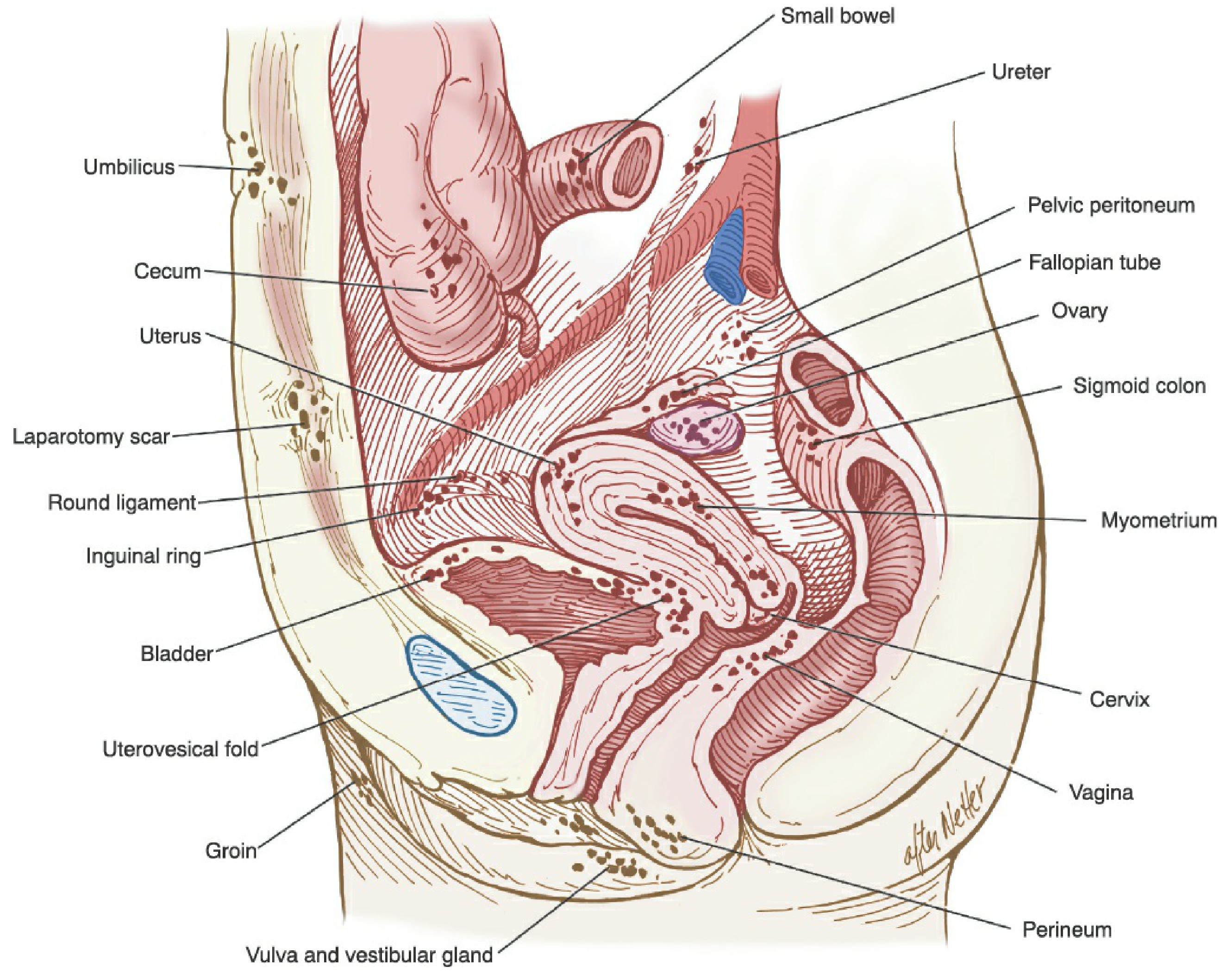
- Peritoneal lesions: "Powder-burn" (black/brown), red flame-like, clear vesicular, or white scarring lesions
- Endometriomas: Ovarian cysts containing thick "chocolate fluid" (hemosiderin)
- Deep infiltrating endometriosis (DIE): Lesions penetrating >5 mm below peritoneum, especially in the posterior cul-de-sac and uterosacral ligaments
- Invasive with ~0.6-1.8/1,000 risk of major complications (bowel perforation, vascular injury)
- Surgeon experience-dependent: subtle lesions (vesicular, atypical) are easily missed
- 24% of typical-appearing lesions are histologically negative - biopsy is essential
- Does not justify routine use as a first diagnostic step
- Berek & Novak's Gynecology
2. Clinical Diagnosis (Non-Surgical)
| Feature | Significance |
|---|---|
| Dysmenorrhea (especially severe, progressive) | Most common presenting symptom |
| Deep dyspareunia | Suggests posterior cul-de-sac or uterosacral involvement |
| Chronic pelvic pain | Present in >70% of cases |
| Infertility | Present in ~30-50% of cases |
| Dyschezia / dysuria (cyclical) | Suggests bowel or bladder DIE |
| Uterosacral nodularity on pelvic exam | Highly specific for DIE |
| Fixed retroverted uterus ("frozen pelvis") | Advanced adhesive disease |
| Cervical displacement, cul-de-sac obliteration | Advanced disease |
3. Imaging: Current Status and Recent Developments
A. Transvaginal Ultrasound (TVUS) - First-Line
| Phenotype | Sensitivity | Specificity |
|---|---|---|
| Endometrioma | 93% | 96% |
| Deep infiltrating (rectovaginal/bladder nodules) | 79% | 94% |
| Peritoneal endometriosis | 65% | 95% |
B. MRI - Growing Role
- Surgical mapping of complex DIE
- Detection of extrapelvic/distant endometriosis (diaphragm, bowel, ureter)
- Patients unable to tolerate TVUS
- Identifying hyperintense iron deposits in endometriotic lesions
- Less operator-dependent than TVUS due to standardized sequences
- Deep and ovarian endometriosis: sensitivity 91-93.5%, specificity 86-87.5%
- A negative eMRI does not exclude peritoneal disease (lesions <5 mm are missed)
- MRI sensitivity: 0.74 vs. TVUS sensitivity: 0.51 (p=0.04) - MRI significantly more sensitive
- Specificity comparable: MRI 0.93 vs. TVUS 0.97 (p=0.22)
C. Bowel and Urological Assessment
- Ureteral involvement is asymptomatic in up to 50% of cases
- Renal/transrectal/transvaginal US, CT urogram, or MRI are recommended to assess ureteral, bladder, and bowel involvement
- No single technique is proven superior; use what the radiologist is most experienced with
4. Biomarkers: The Frontier of Non-Invasive Diagnosis
A. CA-125 - Still Inadequate Alone
- Elevated in moderate-severe endometriosis; normal in mild disease
- Levels in moderate-severe: 13-95 U/mL (non-menstrual); wide overlap with normal (8-22 U/mL)
- Compared to laparoscopy, serum CA125 has no diagnostic value as a standalone test
- CA125 is frequently elevated with endometriomas, complicating ovarian cancer differentiation
- Berek & Novak's Gynecology
B. Neutrophil-to-Lymphocyte Ratio (NLR)
- Pooled sensitivity: 0.66, specificity: 0.63
- Positive LR: 1.79; Negative LR: 0.53
- Conclusion: NLR alone is insufficient but combinations with CA125 show more promising results
C. Proteomics-Based Biomarkers
| Sample Source | Proteins of Interest | Sensitivity / Specificity |
|---|---|---|
| Serum/peripheral blood | Alpha-1-antitrypsin, Albumin, Vitamin D-binding protein, Haptoglobin | 38-100% / 59-99% |
| Urine | Cathepsin G, Vitamin D-binding protein, Albumin | 58-91% / 76-93% |
| Menstrual blood / cervical mucus | Complement C3, S100-A8 | Under evaluation |
D. MicroRNAs (miRNAs) - Most Promising Blood-Based Biomarker
- Pooled sensitivity: 0.82 (95% CI: 0.79-0.84)
- Pooled specificity: 0.79 (95% CI: 0.76-0.82)
- AUC: 0.87 - moderate-to-good diagnostic accuracy
- Multi-miRNA panels outperform single miRNAs
- miR-8: sensitivity 94.8%, specificity 91.9% - highest accuracy but high heterogeneity (I² >90%)
- miR-122: More consistent performance with narrower confidence intervals - better candidate for validation
- Current evidence limited by low GRADE certainty; independent validation cohorts needed before clinical use
E. Non-Coding RNAs (ncRNAs) Broadly
F. Endometrial Biopsy for Nerve Fibers
5. Artificial Intelligence in Endometriosis Diagnosis
- 59 studies reviewed; 6 specifically on endometriosis US
- AI models focus on classification (presence vs. absence of endometriosis) and automated segmentation
- Most models are internally validated only - external validation is lacking
- Combining eMRI and TVUS-derived AI markers may produce powerful diagnostic capability
- AI diagnostic tools combining independent eMRI and eTVUS markers are in early development
- Potential to standardize diagnosis and reduce operator dependency
6. Staging and Classification Systems
7. Special Populations: Adolescents
- Among adolescents clinically suspected of endometriosis undergoing US: 32.8% had at least one sign
- Of ultrasound-confirmed cases: 48.5% had DIE, 45.5% had endometrioma
- MRI showed signs in 49.8% of adolescent cases
- Early recognition is critical: untreated adolescent endometriosis can lead to progressive pelvic disease and fertility compromise
- The threshold for imaging should be low in young women with severe dysmenorrhea
8. Summary: Diagnostic Pathway (2024-2026 Evidence Base)
Symptoms (dysmenorrhea, CPP, dyspareunia, infertility)
         ↓
   Clinical Examination
(uterosacral nodularity, fixed uterus, cul-de-sac obliteration)
         ↓
   TVUS (first-line imaging)
   - Endometrioma: Sens 93%, Spec 96%
   - DIE nodules: Sens 79%, Spec 94%
   - Cannot exclude peritoneal disease
         ↓
   If TVUS negative but strong suspicion:
   eMRI (especially for RVS-DIE, extrapelvic, surgical planning)
   - RVS-DIE sensitivity 74% vs TVUS 51%
         ↓
   Trial of empiric hormonal therapy
   (COCs ± NSAIDs; recommended before laparoscopy if no adnexal mass)
         ↓
   Diagnostic ± Operative Laparoscopy
   (gold standard; biopsy all suspicious lesions)
- miRNA panels (AUC ~0.87) + CA125 + NLR combined scores
- Endometrial nerve fiber biopsy
- Proteomics from urine/serum
- AI-assisted eMRI/TVUS
Key Takeaways
- Gold standard remains laparoscopy + histology - but this is being challenged by better imaging and biomarker research
- TVUS is excellent for endometriomas (93% sensitivity) but misses peritoneal disease; eMRI is superior for rectovaginal DIE
- No single blood test is diagnostically adequate - CA125 and NLR alone are insufficient
- miRNA panels show the most promise among blood biomarkers (AUC 0.87); multi-miRNA combinations are superior to single markers
- AI integration with eMRI and ultrasound is an active frontier but not yet clinically validated
- Diagnostic delay of 6-8 years persists - empiric hormonal therapy before laparoscopy in non-adnexal-mass cases is endorsed to reduce unnecessary surgery