
Type of hydrocele and in detail
types of hydrocele diagram scrotum anatomy
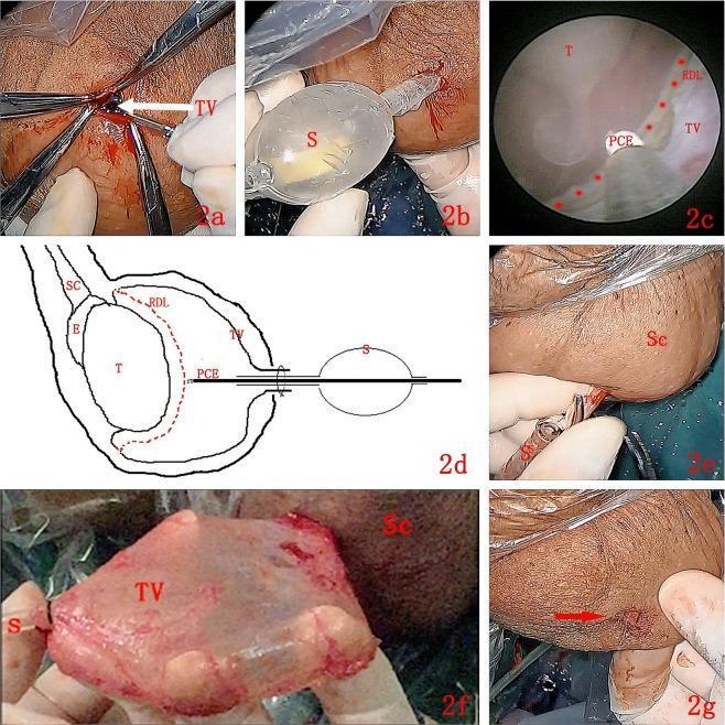
A multi-panel figure illustrating the 'Su-Wang technique' for treating hydrocele via endoscopic resection of the tunica vaginalis (TV). Panel (a) shows a 1 cm transverse incision on the anterior-superior scrotum exposing the parietal TV. (b) Demonstrates the insertion of a specialized working sheath (S) into the vaginal cavity. (c) Provides an endoscopic view where a plasma cylindrical electrode (PCE) is used to create a circular marker (red dotted line, RDL) on the parietal TV, approximately 1–2 cm away from the testis (T). (d) Is a schematic diagram showing the spatial relationship between the sheath, electrode, testis, epididymis (E), and spermatic cord (SC). Panels (e) and (f) depict the blunt separation and complete stripping of the parietal TV from the scrotal wall (Sc) using the sheath for traction. (g) Shows the final 1 cm surgical site immediately after the procedure. This clinical photograph and diagram set demonstrates a minimally invasive urological technique for hydrocele sac resection.
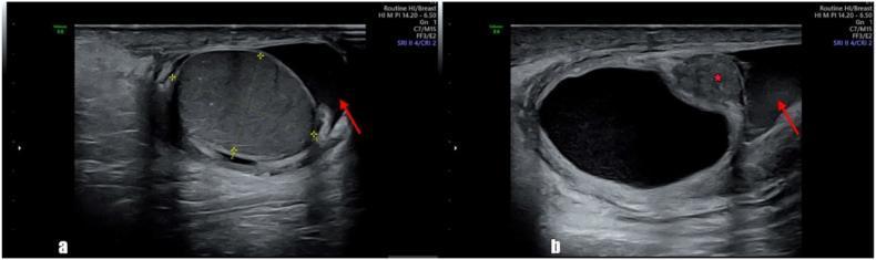
Two-panel diagnostic ultrasonography of the right scrotum demonstrating scrotal anatomy and pathology. Figure (a) displays a grayscale sagittal view of the right testis, showing normal, homogeneous echogenicity and regular borders. A red arrow indicates a small, anechoic fluid collection surrounding the testis, characteristic of a mild right-sided hydrocele. Figure (b) reveals a large, well-circumscribed cystic lesion adjacent to the epididymal head. This cyst is marked with a red asterisk (*) and exhibits thick walls with internal heterogeneity. A second red arrow in panel (b) highlights the concomitant hydrocele fluid. These findings are consistent with an epididymal cyst (spermatocele) which, in this clinical context, demonstrated torsion. This diagnostic imaging serves as an educational example of scrotal ultrasound interpretation for differentiating normal testicular parenchyma from epididymal pathologies and secondary fluid accumulations.
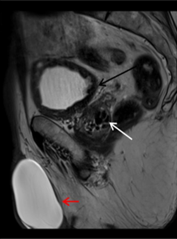
A sagittal T2-weighted MRI of the male pelvis and scrotum demonstrating several urogenital pathologies. In the right scrotum, a large, well-circumscribed, oval lesion (marked with a red arrow) shows homogeneous high T2 signal intensity, characteristic of a hydrocele. The lesion is located within the tunica vaginalis, causing significant scrotal distension. The urinary bladder exhibits a thickened wall with low T2 signal intensity (marked with a black arrow) and an irregular, trabeculated appearance, suggesting chronic bladder outlet obstruction or chronic cystitis; internal layering debris is also noted within the bladder lumen. Within the prostate gland, multiple small, well-defined, low T2 signal intensity foci are visible (marked with a white arrow), representing prostatic calcifications. This image is used for educational purposes to distinguish normal pelvic anatomy from common urological findings such as hydrocele, bladder wall trabeculation, and prostatic lithiasis.
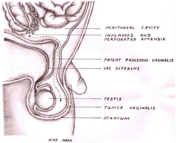
This medical anatomical diagram illustrates the pathophysiology of a scrotal abscess occurring as a rare complication of acute appendicitis. The sagittal view depicts the anatomical relationship between the peritoneal cavity and the scrotum. Key labeled structures include an 'inflamed and perforated appendix' situated superiorly, which is shown shedding purulent material. A dashed arrow traces the pathological pathway of the infection inferiorly through a 'patent processus vaginalis,' an embryonic developmental remnant. The diagram shows this channel passing adjacent to the 'vas deferens' and terminating in the 'scrotum,' where the 'testis' and 'tunica vaginalis' are located. This illustration serves to explain how intra-abdominal infection can track extra-abdominally into the scrotal sac when the processus vaginalis fails to obliterate. The diagram is intended for intermediate to advanced medical education, focusing on surgical anatomy, embryology, and atypical presentations of appendicitis.
Hydrocele - Types and Detailed Overview
Embryological Basis
Classification Diagram
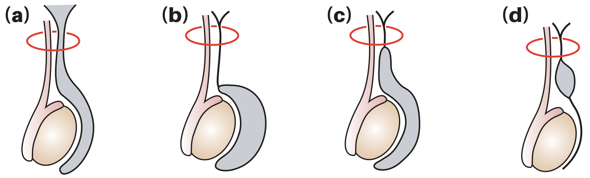
Types of Hydrocele
1. Vaginal (Non-Communicating / Primary) Hydrocele
- Most common type globally
- The processus vaginalis is fully closed with no communication to the peritoneal cavity
- Fluid accumulates around the testis within the tunica vaginalis due to excessive production or defective absorption by the tunica (often from disrupted lymphatic drainage)
- When no cause is found, it is called a primary or idiopathic hydrocele
- Occurs most commonly in middle and later life
- The swelling is typically painless and may grow large before the patient seeks treatment
- Can also occur in older children
2. Communicating (Congenital) Hydrocele
- A patent processus vaginalis persists, allowing free communication between the peritoneal cavity and the tunica vaginalis
- Peritoneal fluid flows down into the scrotum
- Associated with indirect inguinal hernia (if the processus is wide enough, bowel can herniate)
- Common in neonates and infants - majority resolve spontaneously by 18 months
- Fluid may drain back into the peritoneal cavity when the child lies down, making the swelling intermittent
- Pressure on the hydrocele does not always empty it completely
- Bilateral swellings should prompt checking for ascites
3. Infantile Hydrocele
- The distal end of the processus vaginalis closes correctly, but the mid-portion remains patent
- The proximal end is open and communicates with the tunica vaginalis
- Fluid fills from the mid-cord level down to the scrotum
- Essentially an intermediate variant between communicating and non-communicating types
4. Hydrocele of the Cord (Encysted Hydrocele of Cord)
- The distal end closes correctly, the mid-portion remains patent, but the proximal end is also closed
- Results in an isolated, encysted fluid collection along the spermatic cord, separate from both the peritoneal cavity and the tunica vaginalis
- Presents as a smooth oval swelling above the testis, near the spermatic cord
- Liable to be mistaken for an inguinal hernia
- Distinguishing sign: the swelling moves downward and becomes less mobile when the testis is gently pulled downward
5. Abdominoscrotal Hydrocele
- An uncommon variant, accounting for approximately 1.25% of all hydroceles
- A large non-communicating scrotal hydrocele that extends upward into the abdomen through the inguinal canal
- Bilateral in about 30% of cases
- The abdominal component may be palpable as an abdominal mass
- Usually manifests in infancy
- Ultrasound is useful to define the proximal abdominal extent
- Managed surgically via inguinal approach to excise both scrotal and abdominal components
6. Secondary Hydrocele
- A hydrocele that is secondary to an underlying disease or injury
- Causes include:
- Epididymo-orchitis (most common cause of acute secondary hydrocele)
- Testicular torsion
- Testicular tumour (important - must be excluded in young men with acute hydrocele)
- Trauma
- Filariasis (a major cause in endemic tropical regions - Wuchereria bancrofti)
- Radiotherapy
- Post-varicocelectomy (from lymphatic disruption - incidence 3-33%, average ~7%)
- Usually occurs in men over 40 years
Warning: If a tumour is suspected, the hydrocele must NOT be punctured due to risk of malignant needle-track implantation.
Summary Table
| Type | Processus Vaginalis Status | Communication | Age Group | Key Feature |
|---|---|---|---|---|
| Vaginal (Primary) | Fully closed | None | Adults (middle-aged+) | Most common; idiopathic |
| Communicating (Congenital) | Fully patent | Peritoneum ↔ tunica | Neonates/infants | Intermittent; resolves spontaneously |
| Infantile | Mid-portion patent, distal closed, proximal open | Peritoneum → tunica | Infants | Intermediate type |
| Hydrocele of cord | Mid-portion patent, both ends closed | None (encysted) | Any | Swelling above testis, moves with traction |
| Abdominoscrotal | Non-communicating but large | None | Infants | Extends into abdomen |
| Secondary | Variable | Variable | Adults >40 | Underlying disease/injury |
Clinical Features
- Can get above the swelling and palpate a normal spermatic cord (distinguishes from inguinal hernia)
- The testis and epididymis are enclosed within the swelling and may be impalpable
- Transilluminates (the classic sign - light passes through the fluid-filled sac)
- Ultrasound is the key adjunct to assess the testis itself and exclude an underlying tumour
Treatment
| Approach | Indication |
|---|---|
| Watchful waiting | Congenital hydrocele in infants (most resolve by 18 months); small asymptomatic hydroceles |
| Herniotomy (ligation of processus vaginalis) | Congenital/communicating hydrocele persisting beyond 18-24 months |
| Lord's operation (plication) | Thin-walled acquired hydrocele; minimal dissection |
| Jaboulay's operation (eversion of sac) | Common technique for acquired hydrocele with moderate-thickness sac |
| Excision of sac | Thick-walled hydrocele |
| Aspiration +/- sclerotherapy | High-risk surgical patients; palliative; high recurrence rate |
Pericarditis in surgery in detail
pericarditis ECG pericardial effusion constrictive pericarditis
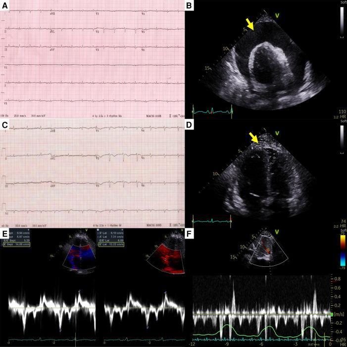
This composite figure illustrates the clinical and diagnostic progression of a patient with effusive-constrictive pericarditis. Panels A and C show 12-lead electrocardiography (ECG). Panel A displays low-voltage QRS complexes, typical of a large pericardial effusion. Panel C, post-pericardiocentesis, shows increased QRS voltage and newly developed T-wave inversions in the anterior leads. Panels B and D provide transthoracic echocardiography (TTE) views. Image B reveals a massive, circumferential anechoic space representing a large pericardial effusion (arrow) causing cardiac tamponade. Image D, following pericardiocentesis, demonstrates significant reduction in effusion but reveals diffuse hyperechoic pericardial thickening and adhesions (arrow). Panels E and F showcase hemodynamic assessment of constrictive physiology. Panel E shows tissue Doppler imaging indicating 'annulus reversus,' where medial early diastolic tissue velocity (e') exceeds lateral velocity. Panel F displays pulse-wave Doppler of hepatic vein flow, highlighting prominent expiratory diastolic flow reversal. Together, these findings characterize the transition from cardiac tamponade to constrictive pericarditis post-drainage.
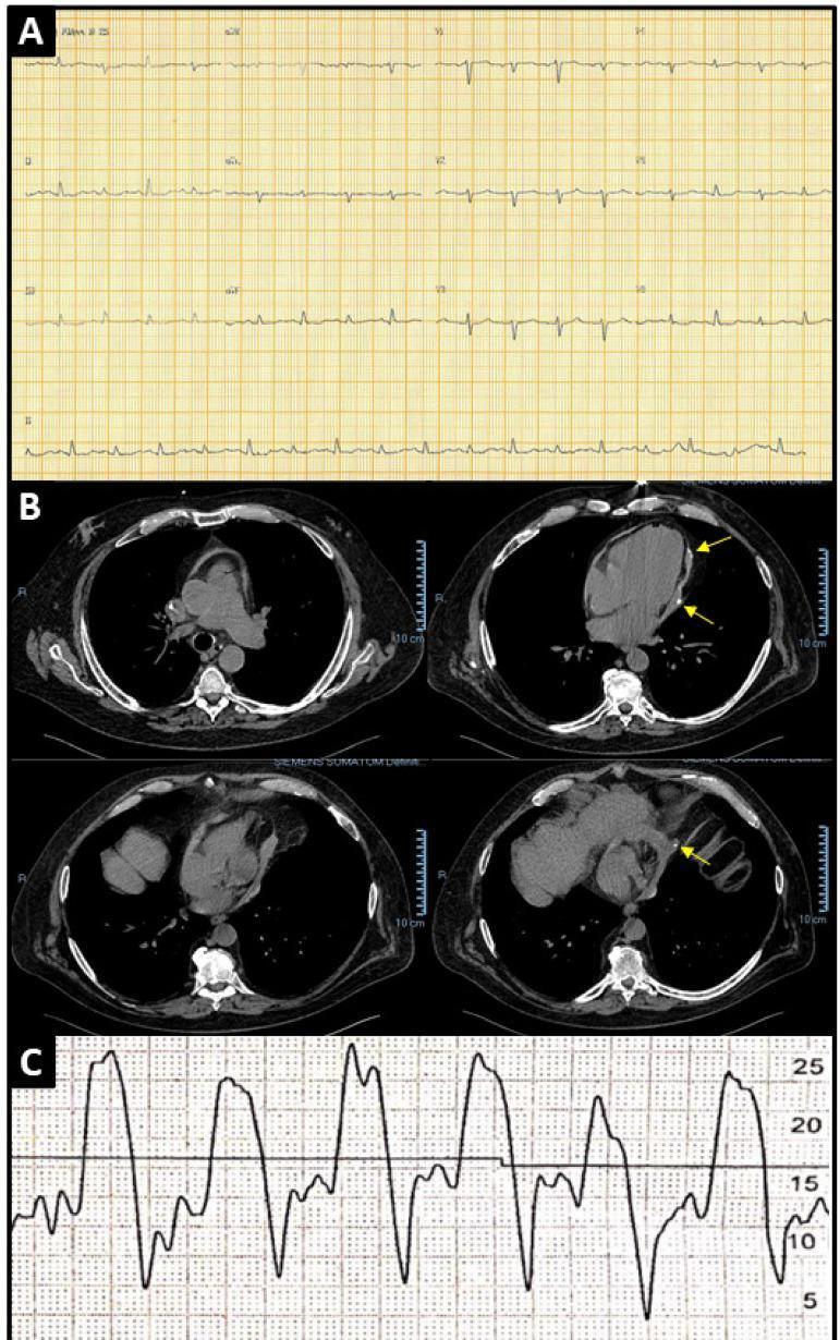
This composite educational image illustrates diagnostic findings for constrictive pericarditis through three distinct modalities. Panel A displays a 12-lead electrocardiogram (ECG) demonstrating a sinus rhythm with diffuse low voltage of the QRS complexes, a classic sign of impaired cardiac conduction due to pericardial thickening or effusion. Panel B presents four axial views from a non-contrast chest CT scan, showing a notably thickened pericardium with high-attenuation hyperdense foci (yellow arrows) consistent with pericardial calcification. Panel C provides a pressure tracing from cardiac catheterization, showing the characteristic 'square root sign' (dip-and-plateau configuration) and equalization of atrial and ventricular diastolic pressures. Collectively, these images teach the hallmark cardiovascular manifestations of constrictive pericarditis, including structural calcification, electrical voltage reduction, and hemodynamic alterations in ventricular filling. The content is suitable for medical education regarding restrictive versus constrictive cardiomyopathy diagnostic protocols.
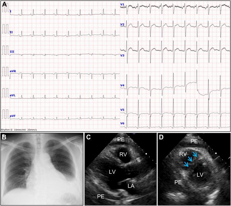
This composite figure illustrates the multimodal diagnostic findings of acute pericarditis with pericardial effusion. Panel A is a 12-lead electrocardiogram (ECG) demonstrating sinus tachycardia, generalized T-wave flattening, and PR-segment depression, with characteristic reciprocal PR-segment elevation in lead aVR. Panel B is a posterior-anterior chest radiograph showing a classic 'flask-shaped' or 'water-bottle' enlarged cardiac silhouette, indicative of large pericardial effusion, alongside a left-sided pleural effusion. Panels C and D are transthoracic echocardiogram frames. Image C (parasternal long-axis view) displays a significant circumferential pericardial effusion (PE) surrounding the right ventricle (RV), left ventricle (LV), and left atrium (LA). Image D (parasternal short-axis view at the mid-ventricular level) highlights the hemodynamic impact through abnormal diastolic septal motion (blue arrows), where the interventricular septum shifts toward the LV. These findings are consistent with constrictive physiology and transient constrictive pericarditis.
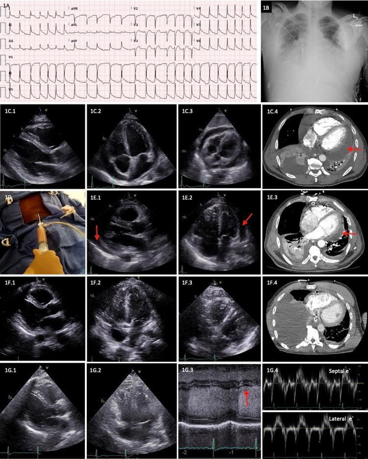
A multi-panel clinical composite illustrating the diagnosis and longitudinal management of purulent pericarditis. Panel A presents a 12-lead ECG showing atrial fibrillation and diffuse ST-segment elevation. Panel B is a frontal chest X-ray displaying cardiomegaly and bilateral interstitial opacities with costophrenic angle blunting. Panels C.1-C.4 (Day 1) show a large circumferential pericardial effusion via transthoracic echocardiography (TTE) in parasternal long-axis, apical 4-chamber, and subcostal views, and an axial CT scan (red arrow). Panel D depicts clinical pericardiocentesis with aspiration of purulent fluid. Panels E.1-E.3 (Day 4) show residual loculated effusion following surgical drainage. Panels F.1-F.4 (Day 8) demonstrate near-complete resolution of the effusion after intrapericardial r-tPA administration. Panels G.1-G.4 represent a 2-year follow-up, using TTE and M-mode (G.3) and Doppler (G.4) imaging to confirm the absence of pericardial effusion or constrictive physiology, such as the absence of early-mid diastolic notching and annulus reversus.
Pericarditis - Detailed Surgical Overview
Definition and Anatomy
Etiology
| Category | Causes |
|---|---|
| Infectious | Viral (most common in West), bacterial, fungal, parasitic, TB (most common in developing world) |
| Post-cardiac injury | Post-MI (Dressler syndrome), post-cardiac surgery, penetrating/blunt trauma, radiation |
| Systemic diseases | SLE, RA, scleroderma, sarcoidosis, amyloidosis, uremia (most common systemic cause) |
| Malignancy | Metastatic (lung, breast, leukemia, lymphoma), primary (mesothelioma) |
| Drugs | Hydralazine, procainamide, isoniazid |
| Idiopathic | 80-90% in Western countries (viral presumed) |
- S. aureus is the most common cause of purulent pericarditis after cardiac surgery in adults
- Tuberculosis is the most common cause of chronic constrictive pericarditis globally
- Uremia is the most common systemic disorder associated with pericarditis
Classification / Types
1. Acute Fibrinous Pericarditis
- Granulocytic and lymphocytic infiltration of the pericardium
- Exudate is fibrinous ("bread and butter" or "shaggy" appearance on the pericardial surface)
- In viral or uremic pericarditis: fibrinous exudate
- In bacterial: fibrinopurulent (suppurative) with pus
- In malignancy: exuberant shaggy exudate + bloody effusion
- Chest pain - sharp, pleuritic, retrosternal; radiates to trapezius ridge or back
- Relieved by sitting forward and leaning ahead
- Worsened by lying supine, deep inspiration, swallowing
- Fever and myalgias are common
- Pericardial friction rub - the hallmark sign
- A "scratchy," leathery sound
- Best heard at the lower left sternal border with the patient leaning forward in full expiration
- Has up to 3 components: atrial systole, ventricular systole, ventricular diastole
- Intermittent and migratory
2. Pericardial Effusion
- Any cause of pericarditis can also produce an effusion
- Other causes: aortic dissection (retrograde bleeding), CHF (transudative), blunt/penetrating trauma
- Hemodynamic significance depends on volume AND rate of accumulation
- Bacterial, fungal, HIV, and malignant effusions have a higher risk of progressing to tamponade
- 20% of large symptomatic effusions of unknown cause are due to undiagnosed cancer
3. Cardiac Tamponade
- Tamponade occurs when pericardial pressure exceeds cardiac filling pressure (usually at 15-20 mmHg)
- Right heart filling is affected first, then left
- The y-descent of the JVP is lost (because no blood can be ejected from the heart at this point)
- Hypotension (low cardiac output)
- Jugular venous distension (elevated venous pressure)
- Muffled heart sounds
- Pulsus paradoxus - fall in systolic BP >10 mmHg with inspiration (ABSENT in LV dysfunction, ASD, positive-pressure ventilation, aortic insufficiency)
- Tachycardia, dyspnoea, diaphoresis
4. Constrictive Pericarditis
- Most common: Cardiac surgery and radiation therapy (especially breast cancer, Hodgkin lymphoma)
- Tuberculosis (most common cause in developing world)
- Subclinical viral pericarditis, connective tissue disease, uremia, neoplasm
- Can be idiopathic
- Inextensible pericardium causes equalization of telediastolic pressure in all cardiac chambers
- Interventricular septum convexity is inverted
- Normal total intracardiac volume is fixed - respiratory variation causes paradoxical septal motion (enhanced RV filling on inspiration, LV on expiration)
- Elevated systemic venous pressures (JVD, Kussmaul's sign)
- Right heart failure: dyspnea, orthopnea, fatigue
- Hepatomegaly, ascites, peripheral edema
- Low cardiac output
- Pericardial knock - early diastolic sound
| Tool | Finding |
|---|---|
| CXR | Pericardial calcification (40-50%), dilated SVC/azygos, straightened cardiac borders |
| Echocardiography | Pericardial thickening, abnormal septal motion, diastolic LV posterior wall flattening, dilated IVC with reduced inspiratory collapse, increased respiratory variation of mitral/tricuspid flow |
| CT | Pericardial thickening >4 mm (suggestive) / >5-6 mm (highly specific); calcification |
| MRI | Direct visualization of morphological abnormalities; delayed hyperenhancement suggests inflammation |
| Cardiac catheterization | "Square root sign" (dip-and-plateau), equalization of diastolic pressures in all chambers |
- This distinction is critical because constrictive pericarditis is treated surgically while restrictive cardiomyopathy is managed medically
- MRI, cardiac catheterization, and tissue Doppler (annulus reversus) are key discriminating tools
- Pericardial resection (pericardiectomy) is the only definitive treatment
- Should be as complete as possible
- Coronary angiography required preoperatively in patients >50 years to exclude co-existing coronary artery disease
- Benefits are progressive over months post-operatively
- Operative mortality: 5-10% even in experienced centers
- Worst outcomes in radiation-induced disease (deep myocardial penetration by fibrosis/calcification)
- Surgery should be performed as early as possible before severe myocardial atrophy sets in
5. Effusive-Constrictive Pericarditis
- Combination of tense pericardial effusion PLUS constrictive thickening
- After pericardiocentesis, physiology changes from tamponade to constriction
- Intrapericardial pressure and CVP decline but not to normal
- Causes: TB, recurrent acute idiopathic pericarditis, radiation, trauma, renal failure, scleroderma, neoplasm
- Treatment: Wide excision of both visceral and parietal pericardium
6. Purulent (Bacterial) Pericarditis
- Most commonly caused by S. aureus (especially post-cardiac surgery)
- Routes: Contamination at surgery, bacteremic seeding, extension from pneumonia/empyema/lung abscess, complication of endocarditis
- Presentation: Fever + severe chest pain + tachycardia + hemodynamic instability
- Can progress rapidly to septic shock or cardiac tamponade
- Treatment: Surgical drainage (pericardiotomy) + IV antibiotics + pericardiocentesis
7. Tuberculous Pericarditis
- Common cause of chronic pericardial effusion and constrictive pericarditis in developing world, especially with HIV
- Presentation: Chronic systemic illness + weight loss + fever + enlarging cardiac silhouette
- Diagnosis: Pericardial fluid culture, pericardial biopsy (ideally via limited thoracotomy); granulomas with caseation
- Treatment: Anti-tuberculous chemotherapy (4-drug regimen)
- If biopsy shows thickened pericardium after 2-4 weeks of ATT → pericardiectomy to prevent constriction
- Tubercular constriction = surgery performed while patient is on ATT
8. Post-Cardiac Injury Syndromes (Surgical Context)
- Occurs weeks after myocardial infarction
- Due to antibodies against injured myocardium (autoimmune)
- Also occurs after cardiac surgery, pericardiotomy, or trauma
- Specifically post-cardiac surgery
- Fever + pericardial friction rub + chest pain appearing days to weeks after surgery
- Treated with NSAIDs and colchicine
ECG Stages in Acute Pericarditis
| Stage | Timing | ECG Findings |
|---|---|---|
| Stage 1 | Hours to days | Diffuse saddle-shaped (concave) ST elevation; PR segment depression (most leads); reciprocal ST depression + PR elevation in aVR and V1 |
| Stage 2 | Days | ST and PR segments normalize; T waves flatten |
| Stage 3 | Weeks | Deep, symmetrical T wave inversions |
| Stage 4 | Weeks-months | ECG reverts to normal (T wave inversions may be permanent) |
- ST elevation in pericarditis is concave (saddle-shaped) vs convex in STEMI
- Simultaneous T wave inversions NOT seen in Stage 1 pericarditis
- Not confined to a single coronary distribution
- No Q wave development
- Ventricular dysrhythmias are rare - their presence should raise suspicion for concomitant myocarditis
Diagnostic Criteria (for Acute Pericarditis)
- Characteristic chest pain (pleuritic, positional)
- Pericardial friction rub on auscultation
- New widespread ST elevation / PR depression on ECG
- New or worsening pericardial effusion on imaging
Treatment Summary
| Type | Treatment |
|---|---|
| Acute/Idiopathic | NSAIDs (ibuprofen 600 mg TID) + Colchicine (0.5 mg BD x 3 months); restrict physical activity |
| Recurrent | Colchicine is cornerstone; consider IL-1 blockers (anakinra) for recalcitrant cases |
| Bacterial/Purulent | Surgical drainage + IV antibiotics |
| Tuberculous | Anti-TB chemotherapy; pericardiectomy if constriction develops |
| Uremic | Dialysis; NSAIDs; pericardiectomy for refractory cases |
| Cardiac tamponade | Pericardiocentesis (emergency); surgical drainage if required |
| Constrictive | Pericardiectomy (definitive - operative mortality 5-10%) |
| Malignant effusion | Pericardiocentesis for symptom relief; pericardial window creation |