
PPD infectious
tuberculin skin test PPD interpretation
tuberculin skin test PPD Mantoux induration reading
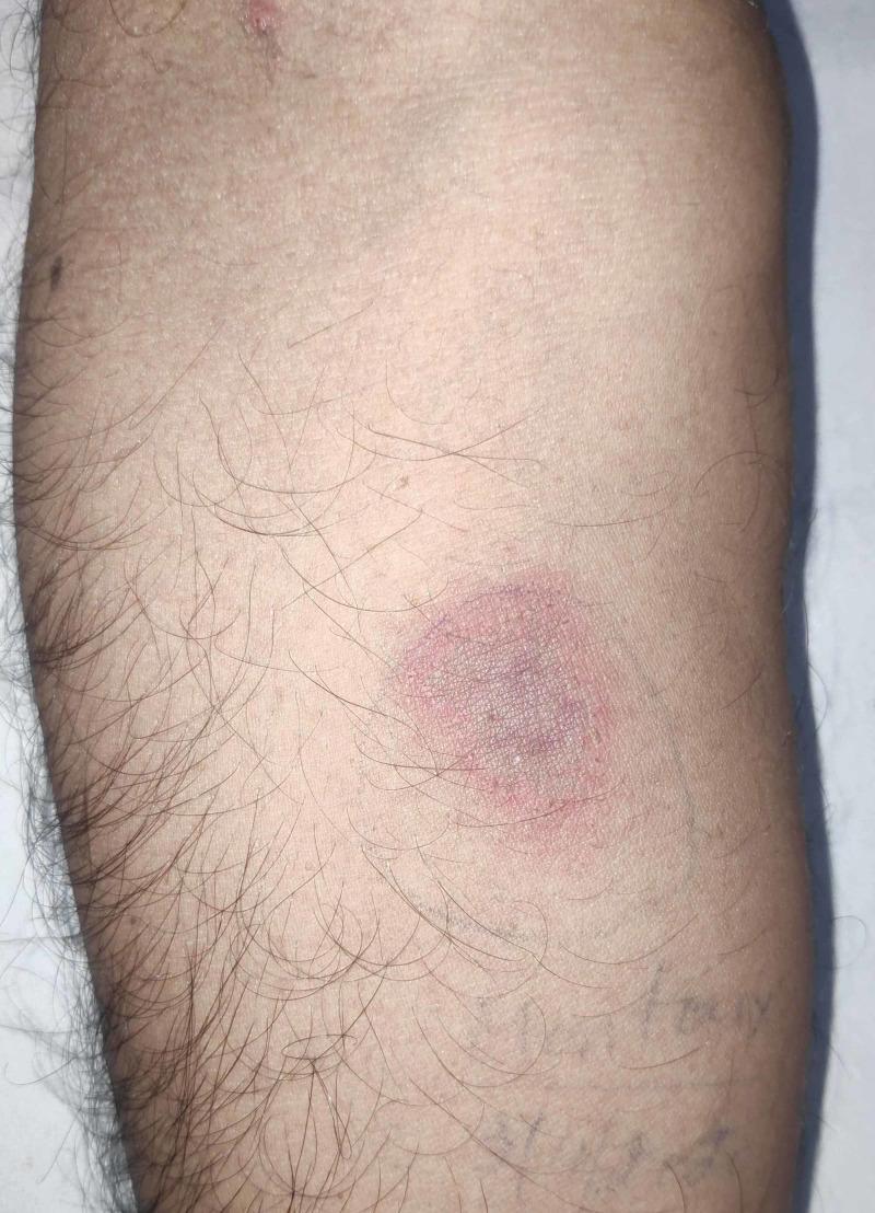
This clinical photograph displays a Mantoux tuberculin skin test site on the volar aspect of a human forearm, 72 hours post-intradermal injection of purified protein derivative (PPD). The visual findings demonstrate a positive reaction characterized by a central, well-defined zone of induration exhibiting a reddish-purple, dusky hue. Surrounding the induration is a peripheral ring of lighter pink erythema. The lesion is roughly circular in morphology, measuring approximately 24 mm in diameter. The surrounding skin appears otherwise normal with standard follicular distribution. This image is a classic educational representation used in infectious disease and pulmonology to illustrate delayed-type hypersensitivity (Type IV) reactions and screening for Mycobacterium tuberculosis infection. Key diagnostic features highlighted include the palpable firmness (induration) and the significant size threshold indicating a positive result in clinical diagnostic protocols.
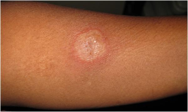
This clinical photograph displays a positive Mantoux tuberculin skin test on the volar aspect of a patient's forearm. The visual hallmark is a central, well-demarcated area of induration, appearing as a raised, firm, and palpable plaque. The indurated center exhibits a slightly lighter, yellowish-tan coloration with a textured, peau d'orange appearance compared to the surrounding skin. Encircling this central induration is a broad, poorly defined halo of erythema (redness). The reaction site is roughly circular, and a small central puncture mark from the intradermal injection is visible. In the clinical context provided, this measure of 28 mm represents a strongly positive reaction, indicating a delayed-type hypersensitivity response to Purified Protein Derivative (PPD). Such a robust reaction is highly suggestive of infection with Mycobacterium tuberculosis and, in this specific case, correlates with systemic findings of ocular and pulmonary tuberculosis.
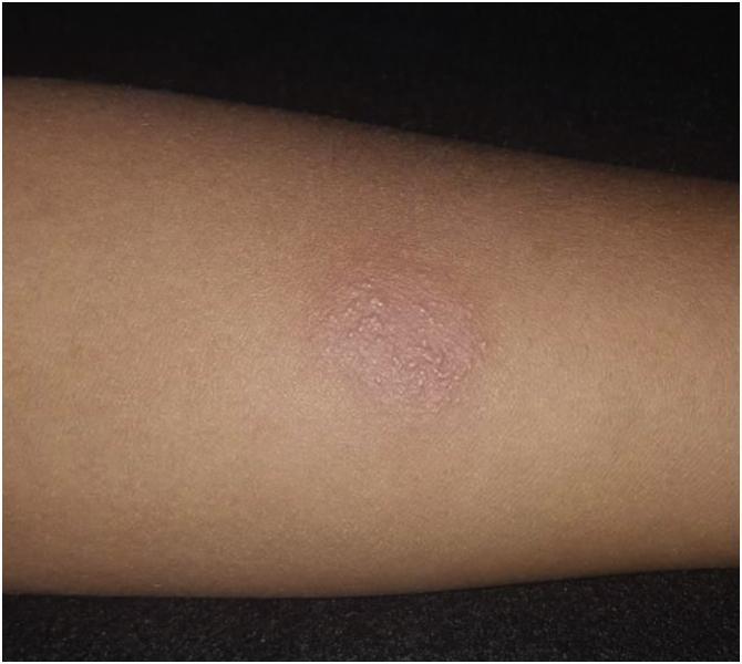
This clinical photograph displays a positive Tuberculin Skin Test (Mantoux test) on the forearm of a patient. The image shows a well-defined, raised erythematous plaque characterizing a strongly reactive 19 mm PPD (purified protein derivative) result. The lesion is primarily a reddish-pink circular area of induration, exhibiting a slightly bumpy, textured surface with fine papular elevations. The surrounding skin shows mild peripheral erythema that fades into the normal skin tone. This visual represents a delayed-type hypersensitivity reaction (Type IV), indicative of current or past infection with Mycobacterium tuberculosis. The educational focus is on the clinical assessment of induration rather than just erythema to determine a positive diagnostic result in tuberculosis screening.
PPD (Tuberculin Skin Test / Mantoux Test) - Infectious Disease
What Is PPD?
- Robbins & Kumar Basic Pathology and Robbins, Cotran & Kumar Pathologic Basis of Disease both confirm: A positive tuberculin test "signifies T cell-mediated immunity to mycobacterial antigens but does not differentiate between infection and active disease."
How It Works - Mechanism
- Intracutaneous injection of 0.1 mL of PPD (5 tuberculin units) into the volar forearm
- A Type IV (delayed-type) hypersensitivity reaction is triggered by sensitized T lymphocytes
- Read at 48-72 hours - measure the induration (not erythema/redness)
- Use a ruler or ballpoint pen technique to measure the raised, palpable area in millimeters
Interpretation - Positivity Thresholds
| Induration | Group |
|---|---|
| ≥ 5 mm | HIV-positive individuals; recent close contacts of active TB; fibrotic changes on CXR consistent with old TB; organ transplant recipients; immunosuppressed patients (e.g., on ≥15 mg/day prednisone) |
| ≥ 10 mm | Recent immigrants from high-prevalence countries (<5 years); IV drug users; residents/employees of high-risk congregate settings (prisons, nursing homes, hospitals); mycobacteriology lab personnel; children <4 years; those with medical risk factors (diabetes, silicosis, CKD, malignancy, low body weight) |
| ≥ 15 mm | Any person with no known risk factors (general population screening) |
False Negatives (Anergy)
- HIV infection / AIDS (especially low CD4 count)
- Active overwhelming TB disease itself (paradoxically)
- Malnutrition
- Sarcoidosis
- Hodgkin lymphoma
- Recent viral infections (measles, varicella, influenza)
- Corticosteroid or immunosuppressive therapy
- Very young age (<6 months)
False Positives
- BCG vaccination (attenuated M. bovis) - common in many countries; can cause false positives, though reactions >15 mm are unlikely to be due to BCG alone
- Nontuberculous mycobacteria (NTM) infections - atypical mycobacteria (e.g., M. avium, M. kansasii) can cross-react
- Incorrect technique (measuring erythema instead of induration)
PPD vs. IGRA (Interferon-Gamma Release Assay)
| Feature | PPD (TST) | IGRA (QuantiFERON, T-SPOT) |
|---|---|---|
| Method | Intradermal injection, read at 48-72h | In vitro blood test |
| BCG interference | Yes - can cause false positive | Minimal/no |
| NTM interference | Yes | Less |
| Requires 2 visits | Yes | No |
| Anergy affected | Yes | Yes |
| Differentiates latent vs. active | No | No |
| Cost | Lower | Higher |
Clinical Significance
- A positive PPD in a previously negative individual ("tuberculin conversion") indicates new infection - 3-4% will develop active TB in the first year after conversion
- All newly positive individuals should receive clinical evaluation and chest X-ray
- Latent TB infection (LTBI) treatment is indicated for positive tests in appropriate risk groups (typically 9 months isoniazid, or shorter rifamycin-based regimens)
- In pre-biologic therapy screening (e.g., anti-TNF agents like infliximab), PPD or IGRA is mandated before initiation - as noted in Firestein & Kelley's Textbook of Rheumatology
Epidemiology
- ~80% of the population in certain Asian and African countries is tuberculin positive
- In 2019, approximately 5% of the U.S. population was positive
- An estimated 13 million cases of latent tuberculosis exist in the United States as of 2019
- TB remains the most common cause of death from a single endemic infectious agent worldwide (WHO) - 1.3 million deaths and 5.8 million new cases in 2021
- Robbins & Kumar Basic Pathology (9780323790185)
- Robbins, Cotran & Kumar Pathologic Basis of Disease (9780443264528)
- Medical Microbiology 9e (9780323673228)

Tell me about these tests for my infectious ward
Gram stain bacteria purple red microscopy
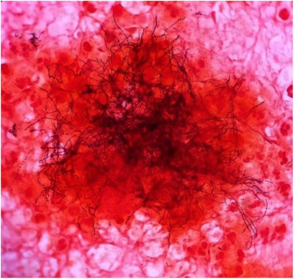
This light microscopy image demonstrates a Gram stain of an excised clinical sample, typically used in microbiology for diagnostic identification. The central focus shows a dense cluster of Gram-variable filamentous rods, characteristic of Actinomyces species. These organisms appear as dark, elongated, branching, thread-like structures. Some filaments exhibit a beaded appearance, staining more intensely (Gram-positive/purple) or less intensely (Gram-negative/pink) along their length. The background consists of host cellular debris and inflammatory cells, which are counterstained pink/red with safranin. These background elements include various eukaryotic cells and likely polymorphonuclear leukocytes. This visual is highly representative of Actinomycosis, demonstrating the classic 'sulfur granule' or 'tangled mass' morphology often seen in abscess aspirates or tissue samples. It serves as a critical educational tool for distinguishing filamentous bacteria from standard cocci or bacilli and highlights the importance of Gram stain variability in clinical diagnostics.
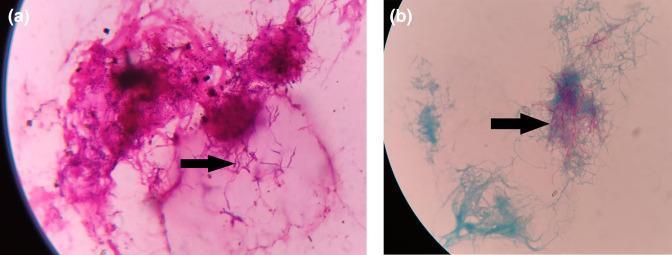
This composite diagnostic image presents two light microscopy views of pleural fluid highlighting the microbiological identification of Nocardia species. Panel (a) shows a Gram stain at high magnification, demonstrating Gram-positive (purple-stained), thin, beaded, and branching filamentous bacteria. Panel (b) illustrates a modified Ziehl-Neelsen (ZN) stain of the same clinical specimen. The bacteria exhibit weak acid-fast properties, characterized by pink-to-red filamentous structures that retain the primary stain against a light blue counterstained background. Arrows in both panels indicate the characteristic filamentous and branching morphology. This combination of Gram-positive branching filaments and partial acid-fastness is a key diagnostic feature used to distinguish Nocardia from other filamentous bacteria like Actinomyces. These findings are clinically significant in diagnosing thoracic nocardiosis in immunocompromised patients or those with pre-existing pulmonary disease.
- پروسیجرهای ضروری = Essential Procedures
- نحوه ی آموزش (کیس/فیلم...) = Method of Training (Case/Film/...) - for competency sign-off
Essential Procedures - Infectious Disease Ward (پروسیجرهای ضروری)
1. PPD (پیپیدی)
2. تهیه لام خون محیطی - Peripheral Blood Smear Preparation
- Place a small drop of fresh blood (EDTA or fingerstick) near one end of a clean glass slide
- Place a spreader slide at a 30-45° angle, back it into the drop, and push forward in one smooth motion
- The smear should be thin enough that RBCs are in a single layer at the feathered edge
- Air-dry immediately - do NOT blow or heat-fix
- Stain with Wright-Giemsa or Field stain (for malaria, use thick and thin smears)
- Read under 100x oil immersion
| Finding | Significance |
|---|---|
| Ring forms in RBCs | Malaria (P. falciparum, P. vivax, etc.) |
| Toxic granulation, band forms | Bacterial sepsis |
| Atypical lymphocytes | EBV (mononucleosis), CMV |
| Schistocytes | Thrombotic microangiopathy (HUS/TTP) |
| Intraerythrocytic rings/Maltese cross | Babesiosis |
3. رنگ آمیزی گرم - Gram Staining
- Crystal violet - flood the smear, wait 1 minute → all bacteria turn purple
- Gram's iodine - apply as mordant, 1 minute → forms crystal violet-iodine complex
- Decolorizer (95% ethanol or acetone-alcohol) - 5-10 seconds → Gram-negative cells lose purple; Gram-positive cells retain it
- Safranin (counterstain) - 1 minute → Gram-negative cells turn red/pink
| Result | Color | Examples |
|---|---|---|
| Gram-positive cocci in clusters | Purple | S. aureus |
| Gram-positive cocci in chains | Purple | Streptococcus spp. |
| Gram-negative diplococci | Pink | N. meningitidis, N. gonorrhoeae |
| Gram-negative rods | Pink | E. coli, Klebsiella, Pseudomonas |
| Gram-positive rods | Purple | Listeria, Clostridium |
4. احتیاطات استاندارد و ایزولاسیون - Standard Precautions and Isolation
- Hand hygiene - soap and water (for C. difficile and norovirus) or alcohol-based hand rub for most pathogens
- PPE - gloves for contact with blood/body fluids; mask, gown, eye protection when splash risk exists
- Safe needle handling - never recap; use safety devices; sharps containers replaced at 2/3 full
- Respiratory hygiene / cough etiquette
- Aseptic technique for procedures
| Type | Route | PPE Required | Examples |
|---|---|---|---|
| Contact | Touch/fomites | Gloves + gown | MRSA, VRE, C. diff, scabies, impetigo |
| Droplet | Large droplets (>5 µm) | Surgical mask + gloves | Influenza, meningococcal meningitis, mumps, pertussis |
| Airborne | Small droplets (<5 µm) | N95 respirator + negative-pressure room | TB, measles, varicella, SARS-CoV-2 (aerosol-generating procedures) |
| Protective / Reverse | Protect immunocompromised patient | Positive-pressure room, HEPA filtration | Neutropenic patients, BMT recipients |
5. گرفتن نمونه کشت خون - Obtaining Blood Culture Sample
- Gather 2 sets of bottles (each set = 1 aerobic + 1 anaerobic) from 2 separate venipuncture sites (reduces contamination interpretation)
- Skin antisepsis: Clean with 2% chlorhexidine-alcohol (allow to dry 30 seconds) or 2% tincture of iodine
- Draw 8-10 mL per bottle in adults (higher volume improves yield - each 1 mL increase raises yield ~3%)
- Inoculate the anaerobic bottle first when using a butterfly needle (removes air from line), aerobic first with direct syringe
- Time cultures before antibiotics if possible - sensitivity drops significantly after even one antibiotic dose
- Label with time, site, and bottle number
- Transport to lab within 2 hours
- 2 sets detect ~80-90% of bacteremia episodes
- 3 sets detect ~96%
- Common contaminants: CoNS (coagulase-negative staphylococci), Bacillus, diphtheroids - single positive set usually indicates contamination
6. گرفتن نمونه کشت حلق - Obtaining Throat Culture Sample
- Have the patient open mouth wide, use a tongue depressor to depress the tongue
- Use a sterile Dacron (not cotton) swab
- Swab both tonsillar pillars and the posterior pharynx - specifically any areas of exudate, erythema, or membrane
- Avoid touching the tongue, teeth, or lips - these contaminate the specimen
- Place swab into appropriate transport medium immediately
- For GAS, rapid antigen detection tests (RADT) give results in minutes; culture is the gold standard but takes 24-48h
- For diphtheria, use specific media (Loeffler's serum, tellurite agar) and alert lab
7. نمونه از زخم پوستی - Skin Wound Sampling
| Wound Type | Preferred Method |
|---|---|
| Abscess / pustule | Aspirate pus with needle/syringe before opening; send for C&S + Gram stain |
| Open wound / ulcer | Cleanse wound first with saline; use Levine technique - swab the wound base with firm rotation over 1 cm² area for 5 seconds |
| Tissue biopsy | Best for deep infections, leishmaniasis, necrotizing fasciitis - punch biopsy or surgical sample |
| Blister / vesicle | Unroof and swab base (Tzanck smear + viral culture for HSV/VZV) |
| Sinus tract | Probe and culture the deepest accessible area, not the opening |
- Gram stain + aerobic/anaerobic culture
- If actinomycosis or mycobacterium suspected: acid-fast stain and fungal culture
- If leishmaniasis suspected: smear + culture on NNN media
8. لومبار پونکسیون - Lumbar Puncture (Spinal Tap)
- Position: Lateral decubitus (fetal) position with maximal spine flexion, OR seated leaning forward - lateral is preferred for pressure measurement
- Landmark: L4-L5 interspace (line between the posterior iliac crests = level of L4 spinous process) - this is BELOW the spinal cord terminus (conus at L1-L2)
- Sterile technique: full aseptic prep (chlorhexidine + drapes), sterile gloves
- Local anesthesia: 1-2% lidocaine subcutaneously
- Insert spinal needle (20-22G) with stylet in the midline, angled slightly toward the umbilicus
- Feel a "pop" as the needle enters the subarachnoid space
- Measure opening pressure with a manometer (normal: <20 cm H₂O)
- Collect CSF into 4 numbered tubes sequentially (tubes 1&4 for cell count, tube 2 for chemistry, tube 3 for microbiology)
| Finding | Bacterial | Viral | TB/Fungal |
|---|---|---|---|
| Appearance | Turbid/cloudy | Clear | Clear/slightly turbid |
| WBC | >1000/µL, neutrophils | 10-1000/µL, lymphocytes | 100-500/µL, lymphocytes |
| Protein | Very high (>100 mg/dL) | Normal/mildly raised | High (100-500 mg/dL) |
| Glucose | Very low (<45 mg/dL) | Normal | Low |
| Gram stain | Often positive | Negative | Negative (AFB in TB) |
9. تفسیر آزمایشات - Interpretation of Lab Tests
| Panel | Key Interpretation Points |
|---|---|
| CBC | WBC with differential - neutrophilia (bacterial), lymphocytosis (viral), eosinophilia (parasites/fungi), neutropenia (risk for opportunistic infections) |
| CRP / ESR / Procalcitonin | PCT >0.5 ng/mL favors bacterial infection; PCT-guided therapy can reduce antibiotic duration |
| Blood cultures | Identify organism + susceptibilities; 2 positive sets = true bacteremia; single set CoNS = likely contaminant |
| LFTs / Renal function | Drug dosing adjustments; aminoglycosides, vancomycin, acyclovir, fluconazole all require renal dose adjustment |
| Gram stain & culture | Correlate morphology (Gram stain) with colony type and sensitivities |
| Serologies | Acute vs. convalescent titers; IgM = acute infection; IgG = past exposure/immunity |
| CSF analysis | See LP table above |
| HIV serology / viral loads | 4th generation Ag/Ab test detects both p24 antigen and antibody; viral load guides treatment and infectivity |
- Jawetz, Melnick & Adelberg's Medical Microbiology 28e - Gram staining
- Neuroanatomy through Clinical Cases 3e - Lumbar puncture
- Red Book 2021 (AAP) - Standard precautions, blood culture aseptic technique
- Robbins & Kumar Basic Pathology - PPD/tuberculin skin test
Make sure I have every detail that I need
lumbar puncture spinal needle CSF collection technique positioning
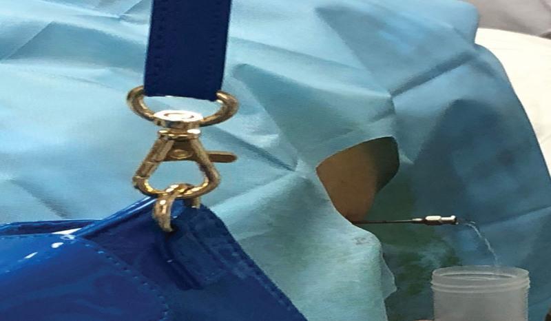
This clinical procedural photograph illustrates a subarachnoid puncture, commonly referred to as a lumbar puncture, being performed for cerebrospinal fluid (CSF) collection or drainage. The image shows a spinal needle inserted into the lumbar region of a patient, specifically at the L2-L3 interspace. The procedural site is prepared with sterile blue surgical drapes. A key clinical finding is the visualization of clear, colorless fluid (CSF) dripping from the hub of the spinal needle into a sterile collection container, confirming successful entry into the subarachnoid space. This visual evidence of 'free-flow' is critical for diagnostic sampling and therapeutic pressure management. The photograph demonstrates proper aseptic technique and the lateral approach for accessing the lumbar cistern. It serves as an educational tool for medical students and clinicians to recognize the appearance of CSF during neurosurgical or neurological procedures, highlighting the correct anatomical positioning and procedural outcomes in a clinical setting.
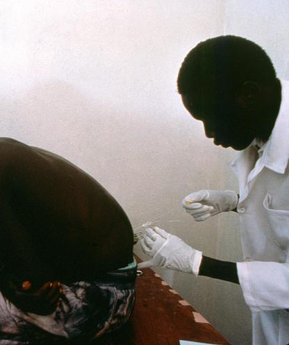
A clinical photograph depicting a healthcare professional performing a lumbar puncture (spinal tap) on a patient in a clinical setting. The patient is positioned in a lateral decubitus posture, lying on their side with the spine flexed and knees drawn toward the chest to increase the interspinous space. The healthcare worker, wearing a white lab coat and sterile gloves, is shown inserting a spinal needle into the lumbar region, specifically targeting the subarachnoid space to collect cerebrospinal fluid (CSF). The visual captures the precise manual technique required for this invasive procedure. Contextually, this diagnostic maneuver is illustrated here as a critical step in staging African Sleeping Sickness (Human African Trypanosomiasis) to detect central nervous system involvement. This image serves as a teaching tool for neuro-diagnostic procedures and clinical manifestations of tropical diseases in resource-limited environments.
blood culture bottles aerobic anaerobic collection venipuncture
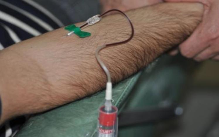
This clinical photograph captures a venipuncture procedure for blood collection, specifically from the brachial vein in the antecubital fossa of a patient's arm. The setup features a winged infusion set (butterfly needle) with a green safety shield, which is inserted into the vein. A flexible plastic extension tube connects the needle to a vacuum collection tube. The collection tube has a red cap, indicating it is likely a plain serum tube without anticoagulants, and is seen partially filled with dark venous blood. The patient's arm is positioned on a green medical cushion, with a practitioner's hand visible providing stabilization. This procedural image demonstrates the initial step in obtaining blood for Platelet-Rich Fibrin (PRF) preparation, highlighting the medical specialty of oral and maxillofacial surgery or regenerative medicine. The educational focus is on the aseptic technique and the specific equipment required for autologous blood harvesting.
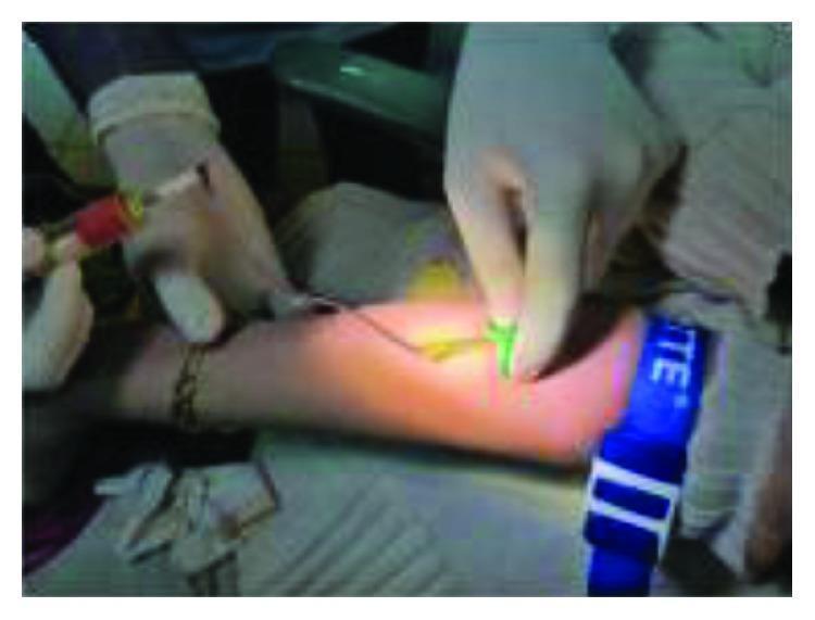
This clinical photograph illustrates a venipuncture procedure for blood collection from the antecubital fossa of a patient's arm. A blue reusable tourniquet is applied to the mid-humerus region to facilitate venous distension. A healthcare professional, wearing white sterile gloves, is performing the procedure using a winged infusion set (butterfly needle), indicated by the green plastic 'wings' held at the puncture site. The needle is connected via flexible tubing to a vacuum-sealed blood collection tube (vacutainer) with a red-stoppered cap. The setup demonstrates standard aseptic technique and equipment used for obtaining venous blood samples, which in this clinical context is being utilized for the subsequent preparation of Leukocyte-Platelet-Rich Fibrin (L-PRF) for surgical use.
COMPLETE INFECTIOUS WARD PROCEDURES - MASTER REFERENCE
1. PPD - Purified Protein Derivative (Mantoux Tuberculin Skin Test)
Mechanism
Indications
- TB screening in close contacts of active TB cases
- Healthcare workers (baseline and periodic)
- HIV-positive patients and other immunocompromised patients
- Pre-biologic therapy (anti-TNF agents: infliximab, adalimumab, etanercept)
- Immigrants from high-prevalence countries
- Residents/staff of congregate settings (prisons, shelters, nursing homes)
- Suspected TB in any patient with compatible symptoms
Contraindications
- Known active TB (no need to test)
- Documented previous strongly positive reaction (severe ulceration/necrosis risk)
- Prior severe reaction to tuberculin
- Extensive burns or eczema at injection site
Technique - Step by Step
- Draw 0.1 mL (5 tuberculin units) of PPD solution into a 1 mL tuberculin syringe with a 26-27G, 3/8-inch bevel-up needle
- Select the volar (inner) forearm, 4 fingerbreadths below the antecubital fossa - avoid veins, scars, and hairy areas
- Cleanse with alcohol and allow to dry
- Stretch the skin taut and insert needle bevel-up at 5-15° angle, just under the skin (intradermally, NOT subcutaneously)
- Inject slowly - a pale, raised bleb 6-10 mm should form. If no bleb forms, the injection was too deep (subcutaneous) - document and repeat at a different site
- Do NOT massage the site after injection
- Record the forearm used, lot number, and time
Reading (48-72 hours)
- Mark the margins of induration (the palpable, raised, firm area) with a ballpoint pen using the "ballpoint pen roll" technique
- Measure the transverse diameter (perpendicular to the long axis of the forearm) in millimeters
- Record erythema separately but base interpretation on induration ONLY
Interpretation Thresholds
| Induration | Population |
|---|---|
| ≥ 5 mm | HIV-positive; recent close contact of active TB; fibrotic CXR changes consistent with old TB; organ transplant; patients on ≥15 mg/day prednisone; TNF inhibitor therapy |
| ≥ 10 mm | Recent immigrants (<5 yrs) from high-prevalence countries; IVDU; residents/employees of congregate settings; mycobacteriology lab staff; children <4 years; medical risk factors (DM, silicosis, CKD, haematologic malignancies, weight loss >10%) |
| ≥ 15 mm | All other persons with no risk factors |
False Negatives (Anergy) - Remember "VMSAAHI"
- Viral infections (measles, EBV, influenza, HIV)
- Malnutrition
- Sarcoidosis
- Active overwhelming TB (paradoxical anergy in miliary TB)
- Age extremes (<6 months, elderly)
- Hodgkin lymphoma / haematologic malignancies
- Immunosuppression (steroids, chemotherapy)
False Positives
- BCG vaccination (especially if recent; reactions >15 mm unlikely to be BCG alone)
- Nontuberculous mycobacteria (NTM) cross-reaction
- Technical error (measuring erythema instead of induration)
Two-Step Testing
Tuberculin Conversion
PPD vs. IGRA
| PPD | IGRA (QuantiFERON, T-SPOT) | |
|---|---|---|
| BCG cross-reaction | Yes | No |
| NTM cross-reaction | Yes | Minimal |
| Requires return visit | Yes (48-72h) | No (1 visit, blood test) |
| Reader variability | Yes | No |
| Preferred when | Low-cost setting, <5 yrs old | BCG-vaccinated, unreliable follow-up |
2. تهیه لام خون محیطی - Peripheral Blood Smear Preparation
Indications in the Infectious Ward
- Suspected malaria (most important - this is a medical emergency)
- Sepsis evaluation (toxic granulation, left shift)
- Infectious mononucleosis (atypical lymphocytes)
- Haemolytic anaemia secondary to infection
- Babesiosis, trypanosomiasis, relapsing fever (Borrelia)
- Microfilaria (nocturnal periodicity - collect at midnight)
- Baseline in any critically unwell febrile patient
Equipment
- Clean glass slides (pre-degreased)
- Fresh EDTA blood or fingerstick
- Spreader slide or coverslip
- Wright-Giemsa stain (preferred) or Field stain (for malaria)
- Immersion oil, light microscope
Technique - Thin Smear (for identification)
- Place a small drop (2-3 µL) of blood 1-2 cm from the frosted end of the slide
- Place spreader slide at 30-45° angle, back it into the drop until blood spreads along the full width of the spreader
- Push forward in one smooth, even, rapid stroke - do NOT pause or lift
- The smear should show a feathered edge (cells in monolayer at the thin end)
- Air-dry immediately - do NOT heat-fix or blow; this distorts morphology
- Fix with methanol for 1-2 minutes, then stain with Giemsa (1:20 dilution for 45 min, or rapid Field stain)
- Read under 10x (scan feathered edge), then 40x, then 100x oil immersion
Technique - Thick Smear (for sensitivity in malaria)
- Place a large drop of blood in the centre of a slide and spread in a circular motion to a 1-2 cm circle
- Air-dry for 30+ minutes (do NOT fix - the RBCs must lyse to expose parasites)
- Stain with Giemsa (without methanol fixation)
- Thick smears are ~20x more sensitive than thin smears for detecting parasites but cannot identify Plasmodium species
Standard Practice for Malaria
- Make BOTH thick and thin smears simultaneously
- If first smear is negative, repeat every 12-24 hours for 3 sets before ruling out malaria
- Never rule out malaria on a single negative smear in a febrile returning traveller
Key Findings on Peripheral Smear in Infectious Disease
| Finding | Significance |
|---|---|
| Ring forms within RBCs (multiple rings per cell, banana-shaped gametocytes) | P. falciparum - malaria, SEVERE |
| Enlarged RBCs with ring + Schüffner's dots | P. vivax or P. ovale |
| RBCs normal size, ring + "Maltese cross" (tetrad form) | Babesiosis |
| Trypomastigotes (C-shaped organisms in plasma) | African trypanosomiasis |
| Spiral organisms between RBCs | Relapsing fever (Borrelia) |
| Toxic granulation (dark granules in neutrophils) | Bacterial sepsis |
| Döhle bodies (blue cytoplasmic inclusions in neutrophils) | Severe bacterial infection, sepsis |
| Left shift (bands > 10%, metamyelocytes, myelocytes) | Bacterial infection, sepsis |
| Atypical lymphocytes (large, lobulated, monospot) | EBV mononucleosis, CMV, acute HIV |
| Neutropenia | Typhoid, viral infections, bone marrow suppression |
| Reactive thrombocytosis | Post-infection, asplenic patients |
| Thrombocytopenia | Malaria, dengue, HUS, sepsis, DIC |
| Schistocytes + thrombocytopenia | HUS, TTP (associated with STEC infection, HIV) |
3. رنگ آمیزی گرم - Gram Staining
Purpose
Complete Step-by-Step Protocol
- Prepare smear: Spread specimen thinly on a clean glass slide; air-dry
- Heat-fix: Pass the slide quickly through a flame 2-3 times, OR methanol-fix (more gentle, less distortion)
- Crystal violet (primary stain): Flood slide, wait 60 seconds, rinse with tap water
- Gram's iodine (mordant): Flood slide, wait 60 seconds - forms crystal violet-iodine complex inside all bacteria; rinse
- Decolorizer (95% ethanol or acetone-alcohol): Apply for 5-10 seconds only - the most critical and error-prone step. Too long = over-decolorize Gram-positive cells (they appear Gram-negative); too short = Gram-negatives appear Gram-positive. Rinse immediately with water
- Safranin (counterstain): Flood slide, wait 60 seconds; rinse; blot dry (do NOT rub)
- Examine under 100x oil immersion
Results
| Colour | Interpretation |
|---|---|
| Purple/violet | Gram-positive (thick peptidoglycan retains crystal violet-iodine complex) |
| Pink/red | Gram-negative (thin peptidoglycan, outer membrane loses complex on decolorization) |
Key Organisms by Morphology
| Morphology | Common Organisms | Clinical Context |
|---|---|---|
| Gram+ cocci in clusters | Staphylococcus aureus, CoNS | Abscess, bacteremia, endocarditis |
| Gram+ cocci in chains | Streptococcus spp., Enterococcus | Pharyngitis, pneumonia, UTI |
| Gram+ diplococci (lancet-shaped) | S. pneumoniae | Pneumonia, meningitis |
| Gram- diplococci (coffee bean, intracellular) | Neisseria meningitidis, N. gonorrhoeae | Meningitis, urethritis, joint infection |
| Gram- rods (straight) | E. coli, Klebsiella, Pseudomonas, Proteus | UTI, pneumonia, wound infection |
| Gram+ rods (large, box-car shaped) | Clostridium, Bacillus | Wound infection, gas gangrene |
| Gram+ rods (small, tumbling motility) | Listeria monocytogenes | Meningitis in neonates, elderly, immunocompromised |
| Branching Gram+ filaments | Actinomyces, Nocardia | Abscess, pulmonary infection |
| No organisms visible | Consider: anaerobes, mycobacteria, fungi, viruses - use special stains |
Special Stains (when Gram stain is insufficient)
| Stain | Used For | Result |
|---|---|---|
| Ziehl-Neelsen (AFB stain) | Mycobacteria, Nocardia | AFB = red/pink; background = blue |
| India ink | Cryptococcus neoformans in CSF | Clear halo (capsule) around organism |
| KOH preparation | Fungi | Clear fungal elements on dissolved background |
| Giemsa | Malaria, Chlamydia, Rickettsia | Varies |
| PAS | Fungi, Pneumocystis | Magenta cell walls |
Common Gram Stain Errors
| Error | Cause | Effect |
|---|---|---|
| Over-decolorized | Too long with acetone-alcohol | Gram+ appear pink (false Gram-) |
| Under-decolorized | Too little acetone-alcohol | Gram- appear purple (false Gram+) |
| Too thick smear | Overlapping cells | Poor resolution, missed organisms |
| Old culture | Aged bacteria lose Gram+ wall integrity | Gram-variable result |
4. احتیاطات استاندارد و ایزولاسیون - Standard Precautions and Isolation
Standard Precautions (apply to EVERY patient, ALWAYS)
-
Hand hygiene - the single most effective infection control measure
- Alcohol-based hand rub (ABHR): before/after patient contact, after touching patient surroundings, before clean/aseptic procedures, after body fluid exposure
- Soap and water: when hands are visibly soiled, after C. difficile exposure (alcohol does NOT kill spores), after norovirus exposure
- Minimum 20 seconds for soap, 15-25 seconds for ABHR (rub until dry)
-
Gloves: for contact with blood, body fluids, mucous membranes, non-intact skin
-
Mask + eye protection/face shield: when splash/spray of blood or body fluids is possible
-
Gown/apron: when clothing/skin may become contaminated
-
Safe sharps handling: never recap by two-handed technique; use safety devices; sharps container at point of use, replaced at 2/3 full
-
Respiratory hygiene/cough etiquette: mask on coughing patients, tissues available, hand hygiene
-
Safe injection practices: one needle, one syringe, one patient, one time
-
Aseptic technique: sterile field, sterile equipment for invasive procedures
-
Environmental cleaning: routine cleaning of surfaces; terminal clean on discharge
Skin Antisepsis for Procedures
- Immunisation and routine venipuncture: 70% isopropyl alcohol (allow to air-dry)
- Blood cultures, incision, IV catheter insertion: 2% chlorhexidine gluconate in 70% isopropyl alcohol (allow to dry 30 seconds) - Red Book (AAP)
- Neonates <2 months and mucous membranes: use povidone-iodine (chlorhexidine is NOT approved for <2 months)
Transmission-Based Precautions
| Type | Transmission Route | PPE Required | Room | Duration | Pathogens |
|---|---|---|---|---|---|
| Contact | Direct skin-to-skin or indirect via fomites | Gloves + gown (on room entry) | Single room or cohort | Until resolved | MRSA, VRE, C. difficile, scabies, impetigo, Shigella, norovirus, RSV |
| Droplet | Large droplets (>5 µm) - travel <1 metre | Surgical mask (on room entry), gloves | Single room; door may stay open | Until resolved (or for influenza: 5 days after symptom onset, 10 days if immunocompromised) | Influenza, meningococcal disease, pertussis, mumps, rubella, Haemophilus influenzae meningitis, SARS-CoV-2 (routine) |
| Airborne | Small droplet nuclei (<5 µm) - remain suspended in air, travel >1 metre | N95 respirator (fit-tested, seal-checked); gown + gloves | Negative-pressure room (≥12 air changes/hour), door kept CLOSED | TB: until 3 negative AFB smears on 3 consecutive days; measles/varicella: until lesions crusted | TB, measles, varicella/disseminated zoster, SARS-CoV-2 (aerosol-generating procedures) |
| Protective (Reverse) | Protect immunocompromised patient FROM environment | Mask on visitors + staff, gown + gloves | Positive-pressure room, HEPA filtration, avoid fresh flowers/plants | Duration of immunosuppression | Neutropenic patients (ANC <500), BMT recipients, severe combined immunodeficiency |
Donning (Putting On) Order
Doffing (Removing) Order - MOST CRITICAL (self-contamination risk)
Clostridium difficile - Special Precautions
- Contact precautions + soap and water hand hygiene (ABHR does NOT kill spores)
- Dedicated equipment (stethoscope, BP cuff, thermometer)
- Environment: clean with 10% bleach or sporicidal agent
- Maintain precautions for at least 48 hours after diarrhoea resolves
5. گرفتن نمونه کشت خون - Blood Culture Collection
When to Order
- Fever ≥38.3°C or hypothermia <36°C in a hospitalised patient
- Suspected bacteremia or fungemia
- Before starting empiric antibiotics whenever possible
- Suspected endocarditis, septic arthritis, meningitis, osteomyelitis
- New onset of rigors/chills
- Unexplained hypotension or organ dysfunction
Equipment
- 2 sets of bottles (each set = 1 aerobic + 1 anaerobic)
- Sterile gloves, tourniquet
- 2% chlorhexidine-alcohol swabs (or povidone-iodine for neonates)
- 20-22G needle or butterfly (winged infusion set)
- 20-50 mL syringe or vacutainer system
Technique - Step by Step
- Two separate sites: Draw each set from a different venipuncture site (different arm, or arm + other site) - this is mandatory for interpretation of contaminants
- Antiseptic prep of bottles: Wipe the bottle rubber tops with 70% isopropyl alcohol; allow to dry
- Skin antisepsis: Apply 2% chlorhexidine-alcohol in a concentric circle from centre outward; wait 30 seconds to dry completely; do NOT touch the site again
- Draw blood:
- Adults: 8-10 mL per bottle (10 mL optimum)
- Paediatrics: 1-3 mL per bottle (or paediatric bottles with 1-5 mL)
- Total adult draw: typically 20 mL (10 mL × 2 bottles per set)
- Inoculation order:
- With a syringe: aerobic bottle first (to prevent introducing air into anaerobic)
- With a butterfly/winged set: anaerobic bottle first (the tubing contains air - fill aerobic last after air is flushed)
- Gently invert bottles 8-10 times to mix blood with broth (prevents clotting)
- Label with: patient ID, date, time, collection site (e.g., "right antecubital fossa"), bottle number
- Transport to microbiology lab within 2 hours at room temperature (NOT refrigerated)
Volume Effect on Yield
- 5 mL/bottle → ~50-60% sensitivity for bacteremia
- 10 mL/bottle → ~80-90% sensitivity
- 2 sets (20-30 mL total) → 80-90% detection
- 3 sets → ~96% detection
Timing
- Draw before antibiotics whenever possible - even one dose reduces yield by 30-40%
- In suspected endocarditis: draw 3 sets over 30-60 minutes before starting antibiotics
- In septic shock: draw 2 sets simultaneously, do NOT delay antibiotics beyond 1 hour for culture collection
- In suspected fungemia: hold bottles for up to 5 weeks (fungi grow slowly; standard incubation is 5 days)
Interpreting Results
| Organism | 1/2 bottles positive | 2/2 sets positive |
|---|---|---|
| S. aureus | Likely true (always treat) | Definite bacteremia |
| E. coli, Klebsiella, Pseudomonas | Likely true | Definite bacteremia |
| CoNS (S. epidermidis) | Likely contaminant | Consider true if indwelling catheter |
| Bacillus, Corynebacterium, Propionibacterium | Almost always contaminant | Rarely true pathogen |
| Candida spp. | Always significant - fungemia | Mandates antifungal therapy |
| S. pneumoniae, Listeria | Always significant | Always treat |
Common Contaminants ("SPACE")
- Staphylococcus epidermidis (CoNS)
- Propionibacterium acnes
- Aerococcus / Alpha-haemolytic streptococci (viridans group)
- Corynebacterium (diphtheroids)
- Enterococcus (when single set only)
6. گرفتن نمونه کشت حلق - Throat Culture
Indications
- Suspected Group A Streptococcus (GAS) pharyngitis (most common)
- Suspected diphtheria (grey membrane, bull neck, toxicity)
- Suspected gonococcal pharyngitis (N. gonorrhoeae)
- Peritonsillar abscess (pus swab)
- Suspected Candida pharyngitis
- Epidemiological screening for carriers (MRSA nares + throat)
- Contact investigation for diphtheria or meningococcal carriage
Equipment
- Sterile Dacron or rayon swab (NOT cotton - inhibits some organisms)
- Tongue depressor
- Light source
- Transport medium (Amies or Stuart's, or specific media for gonococci: Thayer-Martin)
- Rapid antigen detection test (RADT) kit if available
Technique - Step by Step
- Explain the procedure, reassure the patient
- Position patient with mouth wide open and head tilted back; use good lighting
- Use tongue depressor to depress the tongue firmly - expose the posterior pharynx and tonsils
- Ask patient to say "Aaah" - this elevates the uvula and exposes the posterior wall
- Swab in this order: tonsillar crypts (right and left) → any visible exudate or membrane → posterior pharyngeal wall
- Apply firm, rotating pressure - vigorous sampling improves yield
- AVOID: tongue, teeth, lips, buccal mucosa, uvula, saliva - all are contaminants
- Place swab in transport medium immediately
- If diphtheria is suspected: send a second swab and specifically alert the laboratory ("Please culture for Corynebacterium diphtheriae on Loeffler's/tellurite agar")
Rapid Antigen Detection Test (RADT) for GAS
- Sensitivity: 70-90%; Specificity: 95-99%
- Positive RADT: treat without culture
- Negative RADT in adults: may accept (low GAS prevalence in adults means negative predictive value is high)
- Negative RADT in children: confirm with culture (guidelines recommend backup culture in children due to higher GAS rates and rheumatic fever risk)
- Read result at exactly the time specified (usually 5 minutes)
Media for Throat Culture
| Organism | Media |
|---|---|
| GAS (S. pyogenes) | Blood agar (5% sheep blood) - beta haemolysis at 24-48h |
| Diphtheria | Loeffler's serum slope + Tellurite (CTBA) agar |
| N. gonorrhoeae | Modified Thayer-Martin (chocolate agar with antibiotics) - transport in CO₂ |
| Candida | Sabouraud dextrose agar |
Interpretation
- GAS on culture: treat with amoxicillin (10 days) or penicillin V. Failure to treat → risk of acute rheumatic fever and post-streptococcal glomerulonephritis
- Do NOT swab in suspected epiglottitis (hot potato voice, drooling, stridor, tripod position) - risk of laryngospasm. Go directly to airway management and ENT/anaesthesia
Centor/McIsaac Criteria (for GAS probability)
- Score ≤1: No testing, no antibiotics
- Score 2-3: RADT; treat only if positive
- Score ≥4: Treat empirically (or test and treat)
7. نمونه از زخم پوستی - Skin and Wound Sampling
Indications
- Cellulitis with purulent component (abscess, furuncle, carbuncle)
- Infected surgical or traumatic wound
- Diabetic foot ulcer
- Pressure ulcer with signs of infection (purulence, odour, surrounding cellulitis)
- Suspected necrotizing fasciitis
- Cutaneous TB, leishmaniasis, atypical mycobacteria
- Infected bite wounds (human, animal)
- Ecthyma, impetigo not responding to empiric therapy
Technique by Wound Type
- Clean skin with chlorhexidine-alcohol
- Insert a 18-20G needle and aspirate pus
- Send aspirate for Gram stain, aerobic + anaerobic culture
- Send additional samples for AFB if mycobacterium suspected
- Aspiration before incision gives the cleanest, most diagnostic specimen
- Irrigate wound with sterile saline first to remove surface bacteria (surface swabs are unreliable and show colonisers, not pathogens)
- Press a Dacron swab firmly over a 1 cm² area of viable wound bed tissue
- Rotate swab for 5 seconds with enough pressure to express fluid from the wound tissue
- Avoid necrotic slough, eschar, or pus pockets (these give misleading results)
- Obtain under sterile conditions (punch biopsy 4-6 mm, or surgical excision)
- Place in sterile container with a small amount of sterile saline (NOT formalin for culture)
- Send half to microbiology (C&S, AFB, fungal) and half to histopathology
- Best for: necrotizing fasciitis, osteomyelitis, leishmaniasis, cutaneous TB, actinomycosis
- Unroof the blister with a needle
- Swab the base (not the blister fluid - virus is in the cells at the base)
- Send for: Tzanck smear (HSV shows multinucleated giant cells), viral PCR or DFA for HSV/VZV
- For intact vesicles: aspirate fluid with a fine needle
- Do NOT swab the opening - this reflects surface colonisers only
- Probe the tract and sample the deepest accessible tissue
- Best approach: aspirate from the deepest part or biopsy
Special Stains / Tests for Skin Specimens
| Suspected Organism | Test |
|---|---|
| Mycobacterium (TB, NTM) | AFB smear + culture on Lowenstein-Jensen media (4-8 weeks); PCR |
| Leishmania | Giemsa smear (look for amastigotes in macrophages); NNN media culture; PCR |
| Fungi (dermatophytes) | KOH prep; Sabouraud agar |
| HSV/VZV | Tzanck smear; PCR (most sensitive) |
| Actinomyces | Gram stain (Gram+ branching filaments); anaerobic culture |
| Nocardia | Modified AFB stain (weakly acid-fast); aerobic culture |
Key Pitfalls
- Always sample BEFORE cleaning the wound with antibacterials
- Superficial swabs of chronic wounds represent colonisation only - they should NOT drive antibiotic prescribing without clinical correlation
- Necrotizing fasciitis: send peroperative tissue from the advancing edge - do not wait for microbiological confirmation before surgery
8. لومبار پونکسیون - Lumbar Puncture
Indications
- Suspected meningitis or encephalitis (most urgent)
- Suspected subarachnoid haemorrhage with negative CT
- Suspected CNS TB, cryptococcal meningitis, carcinomatous meningitis
- Neurosyphilis, Lyme neuroborreliosis
- Measuring intracranial pressure (e.g., idiopathic intracranial hypertension)
- Intrathecal drug delivery (antibiotics, chemotherapy)
- Myelography/cisternography
When to Do CT FIRST (before LP)
- Papilledema (note: absent in up to 15% adults and 50% children with early raised ICP)
- Altered level of consciousness
- Focal neurological signs (focal deficit, gaze palsy, arm/leg drift, facial palsy)
- Seizure within 1 week
- Age ≥60 years with new neurological symptoms
- Immunocompromised state
- History of CNS disease (mass lesion, stroke, focal infection)
- Midline shift
- Loss of suprachiasmatic or basilar cisterns
- Posterior fossa mass or cistern obliteration
Absolute Contraindications
- Local skin/soft tissue infection at puncture site
- Uncorrected coagulopathy (INR >1.4) or severe thrombocytopenia (platelets <20,000; relative if dropping rapidly)
Relative Contraindications
- Unstable patient (hypotension, shock, status asthmaticus) - stabilise first
- Anticoagulation (warfarin, heparin, NOACs) - consider risk/benefit; use smallest gauge needle if proceeding
- Spinal stenosis, prior lumbar surgery (may need fluoroscopic guidance)
Equipment
- Sterile drape and gloves, chlorhexidine prep
- 20-22G spinal needle with stylet (22G preferred - lower post-LP headache rate)
- Manometer and 3-way stopcock
- 4 sterile collection tubes (numbered 1-4)
- Local anaesthetic (1% or 2% lidocaine, 25G needle)
Technique - Step by Step
- Lateral decubitus (lying on side): preferred for pressure measurement. Patient lies in the fetal position - hips, knees, and neck maximally flexed to open posterior intervertebral spaces. An assistant can help the patient maintain flexion.
- Seated position: sit patient upright, leaning forward with arms crossed on a pillow. Good for obese patients. Pressure measurement less reliable (hydrostatic column of CSF adds to pressure).
- Draw an imaginary line connecting the tops of the posterior superior iliac crests - this crosses the spine at the L4 spinous process (or L4-L5 interspace)
- Insert at L3-L4 or L4-L5 (both are safe - the conus medullaris ends at L1-L2)
- Palpate the spinous processes and identify the midline and interspinous space
- Prep skin with chlorhexidine in concentric circles; allow to dry; apply sterile drape
- Infiltrate local anaesthetic into skin and subcutaneous tissues at the chosen space
- Insert spinal needle bevel-UP (bevel parallel to dural fibres - reduces post-LP headache) in the midline, angled slightly cranially (toward umbilicus)
- Advance slowly - feel resistance as you pass through skin, subcutaneous fat, supraspinous ligament, interspinous ligament, ligamentum flavum, then a distinct "pop" as you enter the epidural and then subarachnoid space
- Remove the stylet and check for CSF flow
- Measure opening pressure with manometer in cm H₂O (patient must be relaxed, legs extended slightly for accuracy)
- Normal: <20 cm H₂O (or <25 cm H₂O in some references)
- Elevated in: bacterial meningitis, cryptococcal meningitis, SAH, IIH, cerebral venous sinus thrombosis
- Collect CSF into 4 numbered tubes, 1-2 mL each, in sequential order:
- Tube 1: Cell count and differential (may have RBCs from traumatic tap at start)
- Tube 2: Biochemistry (protein, glucose - always send paired serum glucose simultaneously)
- Tube 3: Microbiology (Gram stain, C&S, AFB, India ink, cryptococcal antigen, viral PCR)
- Tube 4: Cell count again (compare to Tube 1 to distinguish traumatic tap from true haemorrhage)
- Replace stylet before withdrawing needle (reduces post-LP headache)
- Apply pressure dressing; patient may lie flat for 1 hour
CSF Interpretation Table
| Parameter | Normal | Bacterial Meningitis | Viral Meningitis | TB/Fungal Meningitis |
|---|---|---|---|---|
| Appearance | Clear, colourless | Turbid/cloudy, may be purulent | Clear | Clear to slightly turbid/xanthochromic |
| Opening pressure | <20 cm H₂O | Markedly elevated (>30 cm H₂O) | Normal or mildly elevated | Elevated |
| WBC (cells/µL) | 0-5 (lymphocytes) | >1000, often >10,000 | 10-1000 | 100-500 |
| Cell type | Lymphocytes | Neutrophils (>80%) | Lymphocytes | Lymphocytes |
| Protein (mg/dL) | 15-45 | Very high: >100-500 | Normal or slightly raised (45-100) | High: 100-500 |
| Glucose (mg/dL) | 50-80 (or >60% of serum) | Very low: <40 (often <20) | Normal (>60% serum) | Low: <45 |
| CSF:serum glucose ratio | >0.6 | <0.4 | >0.6 | <0.5 |
| Gram stain | Negative | Positive in 60-80% | Negative | Negative (AFB positive in <40%) |
| Organisms | None | Bacteria | None | India ink: Crypto; AFB: TB |
| Other tests | Bacterial antigen panel; PCR | Viral PCR (HSV, enterovirus) | Cryptococcal Ag; AFB culture; ADA level |
Traumatic Tap vs. True Subarachnoid Haemorrhage
| Feature | Traumatic Tap | SAH |
|---|---|---|
| RBCs in CSF | Present | Present |
| RBC count across tubes 1→4 | Decreasing | Unchanged |
| Xanthochromia (after centrifugation) | Absent (if processed immediately) | Present (develops 2-4 hours after bleed) |
| Clotting in tubes | May occur | Rarely clots |
Complications
- Post-LP headache (1-70%): positional, bilateral, frontal-occipital, worsens upright, relieved supine. Usually resolves in days. Treatment: bed rest, oral hydration, caffeine, NSAIDs. Refractory: epidural blood patch (10-20 mL autologous blood) - most effective treatment, >90% success.
- Herniation (rare if contraindications are respected)
- Local back pain (common, transient)
- Bleeding (epidural haematoma - rare, most relevant in coagulopathic patients)
- Infection (bacterial meningitis, epidural abscess - rare with strict asepsis)
- Nerve root irritation / radicular pain (needle touches a nerve root - withdraw and redirect)
9. تفسیر آزمایشات - Interpretation of Lab Tests
CBC with Differential - Infectious Ward Interpretation
| WBC Pattern | Significance |
|---|---|
| Neutrophilia (>7,500/µL) with left shift | Bacterial infection, stress, steroid use, early sepsis |
| Band forms > 10% | Bacterial infection, early sepsis ("left shift") |
| Neutropenia (<1,500/µL) | Viral infections (typhoid, dengue, influenza), drugs, sepsis-induced bone marrow suppression |
| Lymphocytosis | Viral infections (EBV, CMV, acute HIV, pertussis), TB |
| Atypical lymphocytes > 10% | EBV, CMV, acute HIV, toxoplasmosis, hepatitis |
| Monocytosis | TB, brucellosis, typhoid, fungal infections, EBV |
| Eosinophilia | Parasitic infections (helminths esp.), fungal infections, drug reaction |
| Basophilia | Rare in infection; consider haematologic malignancy |
| Thrombocytopenia | Dengue (hallmark), malaria, sepsis-induced DIC, HUS, typhoid, HIV |
| Thrombocytosis (reactive) | Post-infectious, post-splenectomy |
Key Infection Biomarkers
| Biomarker | Reference Range | Interpretation |
|---|---|---|
| CRP | <5 mg/L | Rises within 6 hours, peaks at 48h. Rises with any inflammation (bacterial, viral, tissue damage). Not specific. Serial values more useful than single value. |
| ESR | <20 mm/hr (men), <30 mm/hr (women) | Slow to rise (24-48h), slow to normalise. Useful for chronic infections (TB, osteomyelitis, infective endocarditis) and monitoring treatment response. |
| Procalcitonin (PCT) | <0.1 ng/mL | PCT >0.5 ng/mL = suggestive of bacterial infection. PCT <0.1 ng/mL = bacterial infection less likely. PCT-guided protocols can safely reduce antibiotic duration in respiratory infections and sepsis. Caution: low PCT does NOT exclude severe bacterial infection - Washington Manual. Elevated in: bacterial sepsis, invasive fungal infection, severe burns/trauma. NOT elevated in: viral infections, localised infections (abscess without bacteremia), autoimmune disease. |
| Lactate | <2 mmol/L | >2 mmol/L = tissue hypoperfusion. Lactate ≥4 = septic shock. Serial lactate guides resuscitation (normalisation target per Surviving Sepsis Guidelines). |
| Ferritin | Variable | Markedly elevated (>10,000 µg/L) in: haemophagocytic lymphohistiocytosis (HLH), adult-onset Still's disease, severe sepsis, cytokine storm. |
Serology Interpretation
| Pattern | Meaning |
|---|---|
| IgM positive, IgG negative | Acute/primary infection (within 1-2 weeks) |
| IgM positive, IgG positive | Recent infection (1-4 weeks, still in acute phase) |
| IgM negative, IgG positive | Past infection or vaccination (immunity) |
| Both negative | Not infected, no immunity |
| 4-fold rise in IgG titre (paired acute + convalescent, 2-4 weeks apart) | Confirms recent infection even if IgM is equivocal |
HIV Testing
- 4th generation Ag/Ab combo test: detects both HIV p24 antigen AND antibodies → shortens window period to ~18 days from exposure. Reactive result requires confirmatory testing (HIV-1/HIV-2 differentiation assay + HIV RNA viral load).
- Window period: Ag/Ab test = 18-45 days. If exposure is <18 days ago, test is unreliable - retest at 45 days and 90 days.
- Viral load: undetectable (<50 copies/mL on ART) = treatment success + prevents transmission. Detectable on ART = assess adherence, resistance testing.
- CD4 count: <200/µL = AIDS-defining; <50/µL = risk for CMV retinitis, MAC, Cryptococcus, PCP.
Dengue Serology (NS1 + IgM/IgG)
| Day of Illness | NS1 Ag | IgM | IgG | Interpretation |
|---|---|---|---|---|
| Days 1-5 | + | - | - | Early primary dengue |
| Days 5-9 | ± | + | - | Primary dengue |
| Days 5-9 | ± | + | + | Secondary dengue (worse prognosis, risk of severe dengue) |
| Day >10 | - | + | + | Late primary or secondary dengue |
Urinalysis in Infection
- Pyuria (>5 WBC/hpf) + bacteriuria + symptoms = UTI
- Sterile pyuria (WBC without bacteria): TB, partly-treated UTI, renal TB, Chlamydia urethritis
- Nitrites: positive = Gram-negative bacteria (Enterobacteriaceae); absence does not rule out UTI (Enterococcus, S. saprophyticus, Pseudomonas are nitrite-negative)
- Leucocyte esterase: proxy for WBCs; positive + nitrites = ~85% sensitivity for UTI
Renal Function - Antibiotic Dose Adjustment
| Drug | Adjustment Required in Renal Impairment |
|---|---|
| Aminoglycosides (gentamicin, amikacin) | Reduce dose OR extend interval; monitor trough levels; target trough <1 mg/L (gentamicin); nephrotoxic + ototoxic |
| Vancomycin | Reduce frequency (AUC/MIC guided dosing); target AUC 400-600 mg·h/L; monitor troughs or AUC |
| Acyclovir / Valacyclovir | Reduce dose; risk of crystalline nephropathy and encephalopathy |
| Fluconazole | Reduce dose by 50% if eGFR <50; IV formulation contains cyclodextrin - avoid if eGFR <50 |
| Piperacillin-tazobactam | Reduce dose/extend interval if eGFR <40 |
| Carbapenems | Dose reduction needed (all) |
| Colistin | Careful dose adjustment; highly nephrotoxic |
- Robbins & Kumar Basic Pathology - PPD/tuberculin test
- Robbins, Cotran & Kumar Pathologic Basis of Disease - TB, PPD interpretation
- Jawetz, Melnick & Adelberg's Medical Microbiology 28e - Gram staining
- Neuroanatomy through Clinical Cases 3e - Lumbar puncture technique
- Pfenninger & Fowler's Procedures for Primary Care - LP contraindications
- Red Book 2021 (AAP Committee on Infectious Diseases) - Standard precautions, blood culture antisepsis
- The Washington Manual of Medical Therapeutics - Procalcitonin, sepsis biomarkers
- Fishman's Pulmonary Diseases - Procalcitonin in respiratory infection
Give me a pdf of this
I didn't get the induration threshold in ppd