
Giddinesses evaluation
dizziness vertigo evaluation diagnosis
Dix-Hallpike maneuver BPPV vertigo test
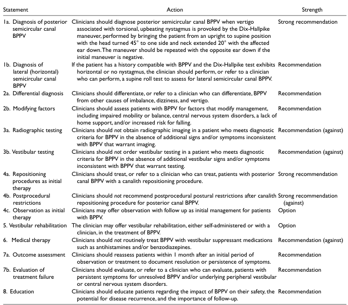
<table> <tr> <th>Statement</th> <th>Action</th> <th>Strength</th> </tr> <tr> <td>1a. Diagnosis of posterior semicircular canal BPPV</td> <td>Clinicians should diagnose posterior semicircular canal BPPV when vertigo associated with torsional, upbeating nystagmus is provoked by the Dix-Hallpike maneuver, performed by bringing the patient from an upright to supine position with the head turned 45° to one side and neck extended 20° with the affected ear down. The maneuver should be repeated with the opposite ear down if the initial maneuver is negative.</td> <td>Strong recommendation</td> </tr> <tr> <td>1b. Diagnosis of lateral (horizontal) semicircular canal BPPV</td> <td>If the patient has a history compatible with BPPV and the Dix-Hallpike test exhibits horizontal or no nystagmus, the clinician should perform, or refer to a clinician who can perform, a supine roll test to assess for lateral semicircular canal BPPV.</td> <td>Recommendation</td> </tr> <tr> <td>2a. Differential diagnosis</td> <td>Clinicians should differentiate, or refer to a clinician who can differentiate, BPPV from other causes of imbalance, dizziness, and vertigo.</td> <td>Recommendation</td> </tr> <tr> <td>2b. Modifying factors</td> <td>Clinicians should assess patients with BPPV for factors that modify management, including impaired mobility or balance, central nervous system disorders, a lack of home support, and/or increased risk for falling.</td> <td>Recommendation</td> </tr> <tr> <td>3a. Radiographic testing</td> <td>Clinicians should not obtain radiographic imaging in a patient who meets diagnostic criteria for BPPV in the absence of additional signs and/or symptoms inconsistent with BPPV that warrant imaging.</td> <td>Recommendation (against)</td> </tr> <tr> <td>3b. Vestibular testing</td> <td>Clinicians should not order vestibular testing in a patient who meets diagnostic criteria for BPPV in the absence of additional vestibular signs and/or symptoms inconsistent with BPPV that warrant testing.</td> <td>Recommendation (against)</td> </tr> <tr> <td>4a. Repositioning procedures as initial therapy</td> <td>Clinicians should treat, or refer to a clinician who can treat, patients with posterior canal BPPV with a canalith repositioning procedure.</td> <td>Strong recommendation</td> </tr> <tr> <td>4b. Postprocedural restrictions</td> <td>Clinicians should not recommend postprocedural postural restrictions after canalith repositioning procedure for posterior canal BPPV.</td> <td>Strong recommendation (against)</td> </tr> <tr> <td>4c. Observation as initial therapy</td> <td>Clinicians may offer observation with follow up as initial management for patients with BPPV.</td> <td>Option</td> </tr> <tr> <td>5. Vestibular rehabilitation</td> <td>The clinician may offer vestibular rehabilitation, either self-administered or with a clinician, in the treatment of BPPV.</td> <td>Option</td> </tr> <tr> <td>6. Medical therapy</td> <td>Clinicians should not routinely treat BPPV with vestibular suppressant medications such as antihistamines and/or benzodiazepines.</td> <td>Recommendation (against)</td> </tr> <tr> <td>7a. Outcome assessment</td> <td>Clinicians should reassess patients within 1 month after an initial period of observation or treatment to document resolution or persistence of symptoms.</td> <td>Recommendation</td> </tr> <tr> <td>7b. Evaluation of treatment failure</td> <td>Clinicians should evaluate, or refer to a clinician who can evaluate, patients with persistent symptoms for unresolved BPPV and/or underlying peripheral vestibular or central nervous system disorders.</td> <td>Recommendation</td> </tr> <tr> <td>8. Education</td> <td>Clinicians should educate patients regarding the impact of BPPV on their safety, the potential for disease recurrence, and the importance of follow-up.</td> <td>Recommendation</td> </tr> </table>
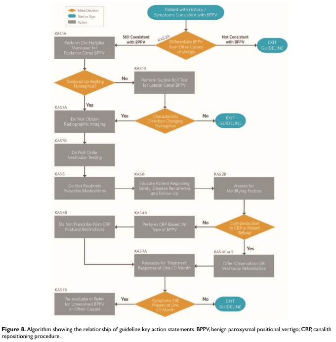
Summary : This flowchart presents a clinical decision-making algorithm for diagnosing and managing benign paroxysmal positional vertigo (BPPV) based on guideline key action statements. It guides clinicians through history-taking, diagnostic maneuvers, differentiation from other causes, and subsequent management steps. flowchart: # Nodes : • Start (rectangle): "Patient with History / Symptoms Consistent with BPPV" • Decision (diamond): "Differentiate BPPV from Other Causes of Vertigo" • Action (rectangle): "Perform Dix-Hallpike Maneuver for Posterior Canal BPPV" • Decision (diamond): "Torsional Up-Beating Nystagmus?" • Action (rectangle): "Perform Supine Roll Test for Lateral Canal BPPV" • Decision (diamond): "Characteristic Direction Changing Nystagmus?" • Action (rectangle): "Do Not Obtain Radiographic Imaging" • Action (rectangle): "Do Not Order Vestibular Testing" • Action (rectangle): "Do Not Routinely Prescribe Medications" • Action (rectangle): "Educate Patient Regarding Safety, Disease Recurrence and Follow-Up" • Action (rectangle): "Do Not Prescribe Post-CRP Postural Restrictions" • Action (rectangle): "Perform CRP Based On Type of BPPV" • Decision (diamond): "Contraindication to CRP or Patient Refusal" • Action (rectangle): "Assess for Modifying Factors" • Action (rectangle): "Offer Observation OR Vestibular Rehabilitation" • Decision (diamond): "Reassess for Treatment Response at One (1) Month" • Decision (diamond): "Symptoms Still Present at One (1) Month" • Action (rectangle): "Re-evaluate or Refer for Unresolved BPPV or Other Causes" • End (rectangle): "EXIT GUIDELINE" # Connectors : • Arrows indicate progression based on clinical findings and decisions. • Orange diamonds represent decision points (e.g., symptom consistency, nystagmus presence, contraindications). • Grey rectangles represent actions (tests, education, treatment). • Blue rectangles indicate start/stop points (entry/exit from guideline). • Branches split based on Yes/No answers at decision nodes, leading to different subsequent actions or exit points. # Layout : • Top-down, multi-column structure. • Initial entry at the top, with parallel branches for posterior and lateral canal BPPV. • Decision nodes create branches, with some leading to exit points if criteria are not met. • Actions and assessments are sequenced, with periodic reassessment and possible referral for unresolved cases. • Colour coding: Orange (decision), Grey (action), Blue (start/stop). # Analysis : • The algorithm provides a systematic approach for evaluating patients with suspected BPPV, emphasizing evidence-based steps and minimizing unnecessary imaging or medication. • Decision points ensure only patients meeting specific criteria proceed through the guideline, with clear exit points for those not consistent with BPPV. • The flowchart highlights the importance of reassessment and referral for unresolved symptoms, supporting safe and effective patient management. • The structure ensures that contraindications and patient preferences are considered before proceeding with canalith repositioning procedures.
peripheral vs central vertigo nystagmus features comparison
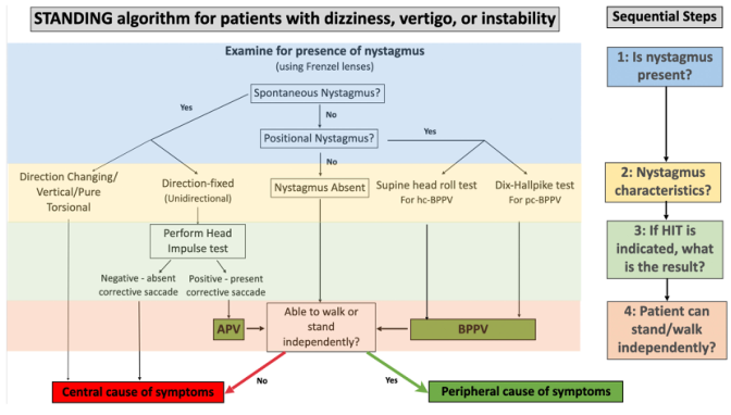
Summary : This figure presents the STANDING algorithm for evaluating patients with dizziness, vertigo, or instability, focusing on the presence and characteristics of nystagmus to differentiate between central and peripheral causes of symptoms. flowchart: # Main Algorithm : • Title: "STANDING algorithm for patients with dizziness, vertigo, or instability" • Initial step: "Examine for presence of nystagmus (using Frenzel lenses)" • First decision: "Spontaneous Nystagmus?" (Yes/No) • If Yes: – "Direction Changing/Vertical/Pure Torsional" → "Central cause of symptoms" (red box) – "Direction-fixed (Unidirectional)" → "Perform Head Impulse test" • "Negative - absent corrective saccade" → "Central cause of symptoms" (red box) • "Positive - present corrective saccade" → "APV" → "Able to walk or stand independently?" – No → "Central cause of symptoms" (red box) – Yes → "Peripheral cause of symptoms" (green box) • If No: – "Positional Nystagmus?" (Yes/No) • If Yes: – "Supine head roll test for hc-BPPV" → "BPPV" (green box) – "Dix-Hallpike test for pc-BPPV" → "BPPV" (green box) • If No: – "Nystagmus Absent" → "Able to walk or stand independently?" – No → "Central cause of symptoms" (red box) – Yes → "Peripheral cause of symptoms" (green box) # Sequential Steps (Right Panel) : • Step 1: "Is nystagmus present?" (blue box) • Step 2: "Nystagmus characteristics?" (yellow box) • Step 3: "If HIT is indicated, what is the result?" (green box) • Step 4: "Patient can stand/walk independently?" (orange box) # Node List : • "Examine for presence of nystagmus" (rectangle) • "Spontaneous Nystagmus?" (diamond) • "Direction Changing/Vertical/Pure Torsional" (rectangle) • "Direction-fixed (Unidirectional)" (rectangle) • "Perform Head Impulse test" (rectangle) • "Negative - absent corrective saccade" (rectangle) • "Positive - present corrective saccade" (rectangle) • "APV" (rectangle) • "Able to walk or stand independently?" (diamond) • "Central cause of symptoms" (red rectangle) • "Peripheral cause of symptoms" (green rectangle) • "Positional Nystagmus?" (diamond) • "Nystagmus Absent" (rectangle) • "Supine head roll test for hc-BPPV" (rectangle) • "Dix-Hallpike test for pc-BPPV" (rectangle) • "BPPV" (green rectangle) # Connectors : • Arrows indicate flow from one decision or test to the next. • Branches split at decision diamonds (Yes/No). • Red arrows lead to "Central cause of symptoms". • Green arrows lead to "Peripheral cause of symptoms". # Layout : • The main algorithm is arranged horizontally, with decision points branching left and right. • The sequential steps are listed vertically on the right side of the figure. # Colour Coding : • Blue background for initial examination. • Yellow for nystagmus characteristics. • Green for peripheral cause outcomes. • Red for central cause outcomes. • Sequential steps use blue, yellow, green, and orange boxes. # Analysis : • The algorithm provides a structured approach to differentiate central from peripheral causes of dizziness based on nystagmus presence, characteristics, head impulse test results, and the patient's ability to stand or walk independently. • Central causes are flagged by direction-changing/vertical/torsional nystagmus, absent corrective saccades, or inability to stand/walk. • Peripheral causes are indicated by unidirectional nystagmus with positive head impulse test, positional nystagmus, or ability to stand/walk. • The flowchart ensures systematic assessment and triage of patients with vertigo or instability.
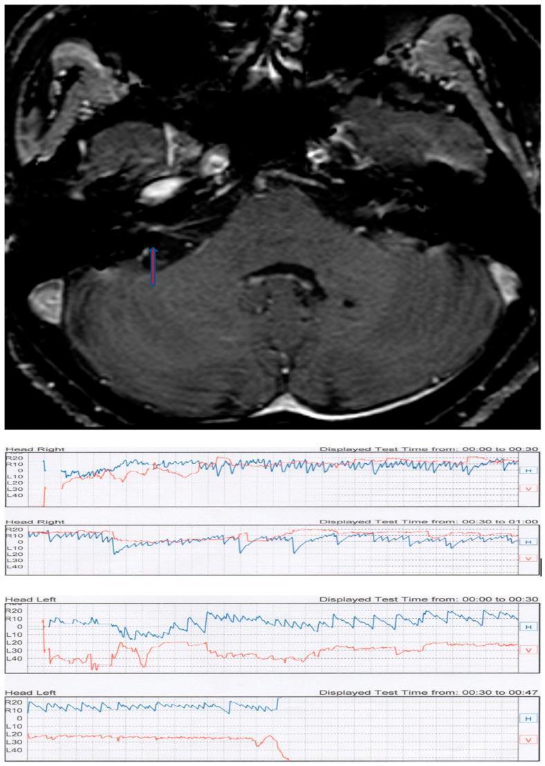
This composite educational figure illustrates the diagnostic findings for a patient with positional vertigo. The top panel features an axial T1-weighted contrast-enhanced MRI scan of the posterior fossa. A blue arrow highlights a small, enhancing lesion within the right internal auditory canal, diagnostic of an intrameatal acoustic neuroma (vestibular schwannoma). The bottom panel contains multiple videonystagmography (VNG) tracings from a Pagnini-McClure positional test. The recordings show horizontal (H, blue line) and vertical (V, red line) eye movements during 'Head Right' and 'Head Left' maneuvers. The tracings demonstrate apogeotropic direction-changing positional nystagmus characterized by rapid, paroxysmal horizontal oscillations. The VNG evidence of central-type positional nystagmus combined with MRI findings demonstrates how a peripheral nerve tumor can manifest with atypical positional vertigo, mimicking or complicating Benign Paroxysmal Positional Vertigo (BPPV) clinical presentations.
Evaluation of Giddiness (Dizziness)
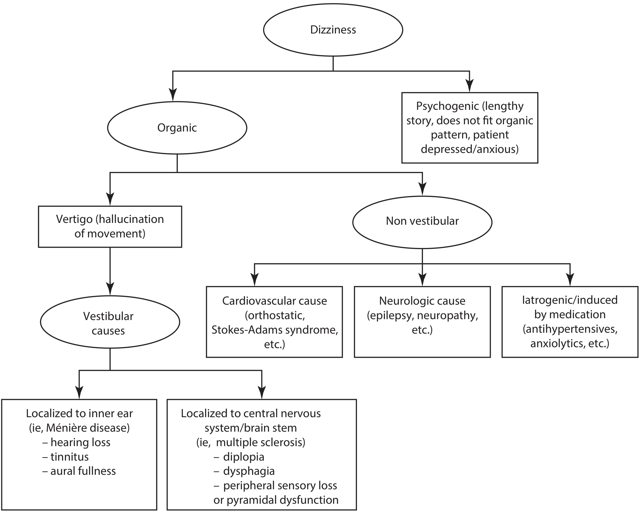
Step 1: Classify the Type of Dizziness
| Category | Description | Common Causes |
|---|---|---|
| Vertigo | Illusion of rotational/linear/tilting movement of self or environment | Inner ear, vestibular nerve, brainstem, cerebellum, panic disorder |
| Presyncope / Lightheadedness | Sensation of impending faint | Orthostatic hypotension, arrhythmia, vasovagal, cardiac insufficiency |
| Disequilibrium | Unsteadiness and imbalance, worse when standing | Multisensory deficits, cerebellar, proprioceptive, extrapyramidal disorders |
| Other / Vague floating | Cannot be neatly categorized | Psychiatric disorders, anxiety, depression, functional disorders (PPPD) |
Step 2: History Taking
- Duration of individual attack: seconds/minutes/hours/days (see temporal pattern table below)
- Frequency: daily, weekly, monthly
- Effect of head movement: better, worse, no effect
- Inducing position or posture (e.g., rolling onto one side in bed)
- Associated aural symptoms: hearing loss, tinnitus, aural fullness/pressure
- Concomitant or prior ear disease or surgery
- Family history: neurofibromatosis, diabetes
- Medications, head trauma, comorbidities
Temporal Pattern and Diagnosis
| Duration Pattern | Likely Cause |
|---|---|
| Seconds to 1 minute, triggered by position change | Benign Paroxysmal Positional Vertigo (BPPV) |
| Hours to days, with hearing loss/tinnitus | Meniere's disease |
| Continuous onset | Stroke (central cause) |
| Recurrent hours-long episodes with migraine features | Vestibular migraine |
| Months of persistent unsteadiness | PPPD / Persistent Postural-Perceptual Dizziness |
Step 3: Physical Examination
- Orthostatic BP and pulse - drop of ≥20 mmHg systolic or ≥10 mmHg diastolic after standing for 2 minutes = orthostatic hypotension
- Ear, nose and throat - signs of infection, otoscopy
- Cardiovascular - murmurs, arrhythmias, carotid bruits
- Neurological - cranial nerves, hearing and vision screening, gait observation, cerebellar testing (finger-nose, heel-shin), Romberg
- Dix-Hallpike maneuver - if BPPV suspected (positive = torsional upbeating nystagmus)
Peripheral vs. Central Vertigo: Key Distinguishing Features
| Feature | Peripheral (e.g., BPPV, Meniere's, labyrinthitis) | Central (e.g., stroke, MS, tumor) |
|---|---|---|
| Nystagmus direction | Unidirectional, horizontal | Direction-changing, vertical, or pure torsional |
| Head Impulse Test (HIT) | Positive (corrective saccade present) | Negative (no corrective saccade) |
| Ability to stand/walk | Preserved | Often impaired |
| Aural symptoms | Common (tinnitus, hearing loss) | Rare |
| Neurological signs | Absent | Present (diplopia, dysphagia, pyramidal signs) |
| Suppression with fixation | Yes | No |

Step 4: Special Clinical Vestibular Tests
- Head Shake Test: Vigorous horizontal shaking at 2 Hz for 15 seconds; post-head-shake nystagmus (HSN) indicates a right-left asymmetry. Fast phase usually directed away from the involved ear. Vertical or rotatory HSN after horizontal shaking = "cross-coupling" → exclude CNS disorder
- Halmagyi Head Thrust (HIT): High-velocity head turn; corrective saccade present = peripheral vestibular lesion
- Dix-Hallpike Maneuver: Patient moved from sitting to supine with head 45° to one side; torsional upbeating nystagmus = posterior semicircular canal BPPV (strong recommendation per BPPV guidelines)
- Supine Roll Test: For horizontal/lateral canal BPPV when Dix-Hallpike shows horizontal or no nystagmus
Step 5: Investigations
- Basic labs: CBC, electrolytes, BUN/creatinine, glucose, serum calcium, LFTs, TSH
- Audiometry: If Meniere's disease or acoustic neuroma suspected
- Neuroimaging: MRI preferred (superior posterior fossa and brainstem visualization); CT if MRI unavailable and stroke urgent
- Cardiac workup: Holter/event monitor for arrhythmia, echocardiogram, carotid Doppler if TIA/stroke suspected
- Vestibular function tests: Caloric testing, videonystagmography (VNG) - only if not meeting clear diagnostic criteria for BPPV
Note: Routine vestibular testing and radiographic imaging should NOT be ordered when the patient clearly meets clinical criteria for BPPV. (BPPV Clinical Practice Guidelines)
Step 6: Specific Conditions and Management Pearls
| Condition | Key Features | Management |
|---|---|---|
| BPPV | Episodic, seconds, head-movement triggered, no aural sx | Canalith repositioning (Epley maneuver) |
| Meniere's disease | Hours-long, tinnitus, fluctuating hearing loss, aural fullness | Low-sodium diet, diuretics, betahistine |
| Vestibular neuritis | Acute prolonged vertigo, no hearing loss, viral prodrome | Corticosteroids, vestibular rehab |
| Orthostatic hypotension | Dizziness on standing, BP drop | Hydration, medication review, fludrocortisone |
| Acoustic neuroma | Unilateral progressive SNHL, tinnitus, mild instability | MRI, referral |
| PPPD | Persistent >3 months, upright posture, visual triggers | Vestibular habituation, CBT, SSRIs |
| Vestibular migraine | Episodic with migraine features | Migraine prophylaxis (see recent systematic review) |
| Drug-induced | Review medication list (antihypertensives, diuretics, anticonvulsants, etc.) | Medication adjustment |
Drugs Commonly Causing Dizziness
Special Populations
- Elderly: Dizziness is a significant risk factor for falls and functional decline. Multisensory dizziness (cumulative deterioration of vision + proprioception + vestibular function + central integration) is especially common with age, diabetes, or prior stroke.
- Psychiatric comorbidity / PPPD: ~20% of specialist dizziness clinic patients have functional dizziness. PPPD is characterized by dizziness or unsteadiness on most days for ≥3 months, exacerbated by upright posture and moving visual stimuli. Psychiatric comorbidity may be absent. Treat with vestibular habituation and psychological therapy. (Bradley and Daroff's Neurology in Clinical Practice)
- K.J. Lee's Essential Otolaryngology, 11e, p. 348
- Textbook of Family Medicine, 9e, pp. 1238-1240
- Goldman-Cecil Medicine, Chapter 366
- Bradley and Daroff's Neurology in Clinical Practice
- Shambaugh Surgery of the Ear
- BPPV Clinical Practice Guidelines (image library)