
Pediatric leukodystrophy
pediatric leukodystrophy classification diagnosis treatment 2024 2025
leukodystrophy pediatric
Pediatric Leukodystrophy
Definition and Core Concept
- Oligodendrocytes (CNS myelin-forming cells)
- Schwann cells (PNS myelin)
- Astrocytes (as in Alexander disease)
- Peroxisomal or lysosomal enzymes critical for myelin lipid metabolism
Classification
| Category | Examples |
|---|---|
| Lysosomal storage disorders | MLD (arylsulfatase A deficiency), Krabbe disease (galactocerebrosidase deficiency) |
| Peroxisomal disorders | X-linked adrenoleukodystrophy (X-ALD), Zellweger spectrum |
| Myelin structural protein defects | Pelizaeus-Merzbacher disease (PLP1 gene) |
| Astrocyte dysfunction | Alexander disease (GFAP mutation) |
| eIF2B / integrated stress response | Vanishing white matter disease |
| Mitochondrial | MERRF, MELAS (overlap) |
| Aminoacidopathy | Canavan disease (aspartoacylase deficiency) |
Key Diseases in Detail
1. Metachromatic Leukodystrophy (MLD)
-
Gene/Enzyme: ARSA gene (chromosome 22q13) → arylsulfatase A (ASA) deficiency; rarely PSAP gene → saposin B deficiency
-
Inheritance: Autosomal recessive
-
Pathophysiology: Sulfatide accumulates in CNS/PNS myelin, causing progressive demyelination; affects both central and peripheral myelin
-
Subtypes by age of onset:
- Late infantile (6 months-2 years) - most common; gait disorder, hypotonia, lower-limb areflexia early; CNS involvement follows
- Juvenile (3-16 years) - intellectual impairment, behavioral difficulties, ataxia, upper motor neuron signs, peripheral neuropathy
- Adult - dementia, psychiatric features (psychosis, hallucinations), dementia; subtle neuropathic signs
-
Diagnosis:
- MRI: large confluent symmetrical T2-hyperintense areas in cerebral white matter, brainstem, cerebellum
- Elevated urinary sulfatide excretion
- Reduced ASA enzyme in leukocytes/fibroblasts (confirmed by molecular testing, since pseudodeficiency exists)
- Elevated CSF protein; reduced nerve conduction velocities
-
Treatment:
- LENMELDY (atidarsagene autotemcel) - FDA-approved in 2024; ex vivo lentiviral HSC gene therapy using the patient's own stem cells transduced with a healthy ARSA gene. Indicated for pre-symptomatic or early-symptomatic late infantile/early juvenile MLD
- Hematopoietic stem cell transplantation (HSCT) can slow progression in pre-symptomatic patients
- Long-term follow-up data (Fumagalli et al., NEJM 2025) support durability of gene therapy
-
Bradley and Daroff's Neurology in Clinical Practice; Goldman-Cecil Medicine
2. Krabbe Disease (Globoid Cell Leukodystrophy)
-
Gene/Enzyme: GALC gene (chromosome 14q31) → galactocerebrosidase (galactocerebroside β-galactosidase) deficiency
-
Inheritance: Autosomal recessive
-
Pathophysiology: Galactocerebroside and psychosine (galactosylsphingosine) accumulate → oligodendrocyte destruction → marked demyelination; characteristic globoid cells (multinucleated macrophages filled with galactocerebroside) in cerebral white matter
-
Clinical features:
- Early infantile (classic): Onset 3-6 months; rapid deterioration, hypertonicity, opisthotonic posturing, optic atrophy, seizures, peripheral neuropathy
- Late-onset: Peripheral neuropathy and spasticity may be the only features
-
Diagnosis: Galactocerebrosidase enzyme assay; NBS programs use lyso-Gb1 as a biomarker; MRI shows symmetric white matter involvement; very slow nerve conduction velocities
-
Treatment: HSCT/cord blood transplantation if performed before symptom onset (newborn screening urgency); no disease-modifying treatment once symptomatic
-
Bradley and Daroff's Neurology in Clinical Practice; Goldman-Cecil Medicine
3. X-Linked Adrenoleukodystrophy (X-ALD)
-
Gene/Enzyme: ABCD1 gene (Xq28) → ALDP (peroxisomal membrane protein) deficiency → VLCFA accumulate (not transported into peroxisomes for beta-oxidation)
-
Inheritance: X-linked recessive
-
Prevalence: ~1 in 20,000 - the most common peroxisomal disorder
-
Phenotypes in males:
- Childhood cerebral ALD (CCALD): Onset 4-8 years; deteriorating school performance, behavioral changes, vision deficits, adrenal insufficiency; symmetrical white matter lesions on MRI; rapidly progressive to total disability and death
- Adrenomyeloneuropathy (AMN): Adult onset; progressive spastic paraparesis, cerebral demyelination, adrenal insufficiency
- Isolated adrenal insufficiency (Addison's disease): ~20% of males
- Carrier females may develop symptoms (spastic paraparesis, sphincter dysfunction) but are typically spared from cerebral ALD and adrenal insufficiency
-
Diagnosis: Elevated plasma very-long-chain fatty acids (VLCFAs), especially C26:0 and C26:0/C22:0 ratio; ABCD1 mutation confirmation; MRI brain
-
Monitoring: Cortisol stimulation testing every 6-9 months for adrenal insufficiency in all males; regular MRI surveillance
-
Treatment:
- Adrenal insufficiency: oral corticosteroid supplementation (life-saving)
- CCALD before MRI lesion progression: HSCT or hematopoietic stem cell gene therapy (Skysona/elivaldogene autotemcel - approved in Europe)
- "Lorenzo's oil" (dietary therapy): has proved disappointing for reversing neurological disease but may slow VLCFA accumulation in asymptomatic boys
-
Tietz Textbook of Laboratory Medicine; Emery's Elements of Medical Genetics and Genomics
4. Pelizaeus-Merzbacher Disease (PMD)
-
Gene: PLP1 gene (X-linked) → proteolipid protein deficiency or dysfunction
-
Inheritance: X-linked recessive
-
Onset: 3 months to 9 years
-
Clinical features: Slowly progressive myelopathy, nystagmus (pendular), cerebellar involvement, spasticity, cognitive impairment; death between 6-25 years in classic form; milder forms (spastic paraplegia type 2) recognized in adults
-
Variants: PMD-like disease type 1 and spastic paraplegia type 44 caused by GJC2 mutations (autosomal recessive)
-
Diagnosis: MRI (hypomyelination pattern - T2 signal does not suppress white matter); genetic testing for PLP1 mutations
-
Treatment: Supportive only; no specific therapy
-
Goldman-Cecil Medicine
5. Canavan Disease
-
Gene/Enzyme: ASPA gene → aspartoacylase deficiency → N-acetylaspartate (NAA) accumulates → brain edema and dysmyelination
-
Inheritance: Autosomal recessive (enriched in Ashkenazi Jewish population)
-
Clinical features: Developmental delay appearing in first months of life, macrocephaly, hypotonia progressing to spasticity, seizures; diffuse symmetrical white matter degeneration involving subcortical areas and globus pallidus on MRI
-
Diagnosis: Elevated NAA on urine organic acids or MR spectroscopy; ASPA gene testing
-
Treatment: No approved treatment; gene therapy trials are ongoing
-
Goldman-Cecil Medicine
6. Alexander Disease
-
Gene: GFAP (glial fibrillary acidic protein) - gain-of-function mutation
-
Inheritance: Usually de novo (sporadic dominant)
-
Pathophysiology: Mutant GFAP accumulates → abundant Rosenthal fibers in periventricular, perivascular, and subpial locations (hallmark)
-
Subtypes:
- Type I (early/infantile): Seizures, motor deficits, macrocephaly, hydrocephalus from CSF pathway obstruction (requires neurosurgical monitoring); frontal-predominant white matter changes on MRI
- Type II (late onset, older child/adult): Bulbar/pseudobulbar features, ataxia, sleep apnea, autonomic dysfunction; spinal cord involvement
-
Diagnosis: MRI (frontal T2 signal with periventricular rim); GFAP mutation; elevated GFAP in CSF/serum (biomarker)
-
Treatment: Supportive; antisense oligonucleotide (ASO) therapy targeting GFAP mRNA is in clinical trials
-
Robbins Pathologic Basis of Disease; PMC5712822
7. Vanishing White Matter Disease (VWM)
-
Gene: EIF2B1-5 genes → eIF2B proteins (involved in integrated cellular stress response)
-
Inheritance: Autosomal recessive
-
Clinical features: Broad spectrum; from rapidly progressive infantile forms to slowly progressive adult disease; characteristic stress-triggered episodes (episodes triggered by fever or minor trauma causing acute/subacute neurological deterioration); vacuolated myelin, cystic appearance on MRI
-
Treatment: Avoidance of triggers (fever, stress, head trauma); no specific therapy; research into the integrated stress response pathway is active
-
Goldman-Cecil Medicine
Clinical Presentation: Red Flags for Leukodystrophy
| Age of Presentation | Common Features |
|---|---|
| Neonatal/early infancy | Hypotonia, seizures, dysmorphic features (Zellweger), failure to achieve milestones |
| Late infancy (6-24 months) | Developmental regression after a period of normal development, gait disturbance, hypotonia |
| Childhood (2-10 years) | Cognitive/behavioral decline, school failure, spasticity, ataxia, visual loss |
| Adolescence/adulthood | Progressive spastic paraparesis, psychiatric features, cognitive decline |
- Developmental regression (not just delay)
- Progressive gait disorder + areflexia in young children (MLD, Krabbe)
- Adrenal insufficiency in boys + neurological decline (X-ALD)
- Macrocephaly + megalencephalic leukoencephalopathy patterns
- White matter changes on MRI that are confluent, symmetric, and periventricular
Diagnosis: Stepwise Approach
- Clinical history and exam: Age of onset, regression vs. delay, family history (consanguinity, ethnicity), adrenal symptoms, peripheral neuropathy
- MRI brain (with spectroscopy): Pattern of white matter involvement guides specific diagnosis; some leukodystrophies have near-pathognomonic MRI patterns
- Metabolic screen:
- Plasma VLCFAs (X-ALD)
- Enzyme assays in leukocytes/fibroblasts (arylsulfatase A, galactocerebrosidase, etc.)
- Urine organic acids (elevated NAA in Canavan; sulfatides in MLD)
- Genetic/genomic testing: Gene panel or whole exome sequencing (WES) - now preferred as first-line for undiagnosed cases
- Newborn screening (NBS): Several states/countries now screen for X-ALD (lysophosphatidylcholine C26:0) and Krabbe (galactocerebrosidase activity); MLD NBS programs are emerging
Treatment Overview
| Disease | Available Treatment |
|---|---|
| MLD | LENMELDY (gene therapy, FDA 2024); HSCT (pre-symptomatic) |
| X-ALD | HSCT or gene therapy (Skysona) for early CCALD; steroids for adrenal insufficiency |
| Krabbe | HSCT (pre-symptomatic only - NBS is critical) |
| PMD | Supportive only |
| Canavan | Gene therapy trials (no approved therapy) |
| Alexander | Supportive; ASO trials |
| VWM | Stress avoidance; supportive |
- Antiepileptic medications for seizures
- Spasticity management (baclofen, botulinum toxin, orthopedic care)
- Nutritional support (gastrostomy if severe dysphagia)
- Physical, occupational, speech therapy
- Palliative/hospice care planning
- Psychological and family support
Recent Evidence (2024-2026)
- LENMELDY (atidarsagene autotemcel) received FDA approval in 2024 for early-onset MLD; long-term NEJM 2025 data confirm durable benefit (Fumagalli et al., NEJM 2025 [PMID 40577679])
- 2024 European consensus recommendations on MLD NBS and clinical management were published
- A 2025 systematic review on POLR3-related leukodystrophies (Michell-Robinson et al., HGG Adv 2025 [PMID 40684265]) provides comprehensive genotype-phenotype data for this subgroup
- Gene therapy platforms (lentiviral HSC, AAV intrathecal, ASOs) represent the most active therapeutic frontier
Key Practical Takeaways (AAP Clinical Report)
- Leukodystrophies are collectively common enough that general pediatricians must be familiar with them
- Some leukodystrophies require urgent treatment - stage of disease determines efficacy (especially Krabbe and MLD)
- Developmental regression - not just delay - is the cardinal warning sign
- NBS for X-ALD and Krabbe is available and critical for treatment timing
- MRI and metabolic/genetic testing should be pursued promptly when a leukodystrophy is suspected
In the context of a radiology pg
pediatric leukodystrophy MRI patterns radiology diagnosis white matter 2024
https://pmc.ncbi.nlm.nih.gov/articles/PMC10885080
Pediatric Leukodystrophy: A Radiology PG Perspective
The Pattern-Based Approach (PG Must-Know Framework)
| Pattern | Key Examples |
|---|---|
| Central/periventricular white matter predominant | MLD, Krabbe, X-ALD (posterior), PMD |
| Subcortical U-fiber predominant | Canavan, MLC, L-2-hydroxyglutaricaciduria |
| Frontal predominant | Alexander disease |
| Posterior predominant | X-ALD (childhood cerebral), Krabbe |
- Enhancement at the leading edge of demyelination → X-ALD (active inflammatory zone)
- Tigroid/leopard skin pattern → MLD
- Macrocephaly on imaging → Alexander, Canavan, MLC
- Basal ganglia involvement → Krabbe (thalami), Canavan (globus pallidus), Alexander (basal ganglia + thalami)
- U-fiber sparing → MLD, Krabbe (early), X-ALD
- Cystic degeneration → VWM, MLC, late Alexander
Disease-by-Disease MRI Features
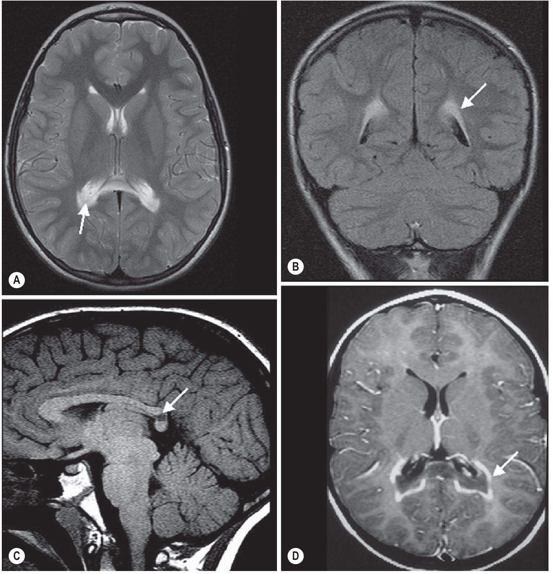
1. X-Linked Adrenoleukodystrophy (X-ALD)
- Location: Posterior > anterior; starts at peritrigonal / splenial region (posterior parietal-occipital white matter and splenium of corpus callosum); progresses anteriorly over time
- Signal: Confluent symmetric T2/FLAIR hyperintensity in posterior periventricular white matter; T1 hypointensity in same regions
- Pathognomonic feature: Three-zone pattern (Loes staging):
- Zone 1 (innermost): central necrosis/gliosis - T1 low, T2 high, no enhancement
- Zone 2 (middle): active demyelination - T2 high, enhances (breakdown of blood-brain barrier, active inflammation)
- Zone 3 (outermost/leading edge): early demyelination - T2 slightly elevated
- Enhancement: Rim/marginal enhancement at the leading edge - characteristic and important for staging
- Corpus callosum: Splenium involved early; involvement bridges across midline
- Loes MRI Severity Score (0-34): Used to stage disease and guide treatment decisions (HSCT indicated when score < 9)
- Adrenal: Look for small adrenal glands on abdominal MRI/CT
2. Metachromatic Leukodystrophy (MLD)
- Location: Bilateral, symmetric periventricular and central white matter; frontal and parietal predominance; corpus callosum involved (especially splenium); internal capsule and corticospinal tracts involved; cerebellar white matter also affected
- U-fibers: Spared initially (subcortical U-fibers preserved until late disease)
- Signal: Confluent T2/FLAIR hyperintensity; confluent sheet-like pattern
- Pathognomonic patterns:
- "Tigroid" pattern: Alternating stripes of demyelinated (T2 bright) and spared (T2 dark) white matter - sparing of perivascular white matter creates a striped appearance; seen in periventricular WM and centrum semiovale
- "Leopard skin" pattern: Punctate foci of T2 sparing within the demyelinated white matter (seen in centrum semiovale)
- Enhancement: Typically no contrast enhancement, even in active disease - important differentiator from X-ALD
- Progression: As disease worsens, inner subcortical WM is eventually involved; corpus callosum, internal capsule, and corticospinal tracts affected
3. Krabbe Disease (Globoid Cell Leukodystrophy)
- Location: Posterior > anterior; starts centrally; specific involvement of:
- Corticospinal tracts as they course through internal capsule and brainstem
- Cerebellar white matter (but dentate nuclei spared)
- Basal ganglia and thalami - characteristically dark thalami on T2 (T2 hypointensity due to iron/mineralization in globoid cells)
- Optic nerves - enlarged/T2 bright in infantile form
- Cervical spinal cord - enlargement in infantile form
- Signal: T2 hyperintensity in white matter; T2 hypointensity in thalami is highly specific
- Enhancement: Can be present in early/active phases
- Infantile form: Very early and severe; optic nerve + cervical cord enlargement is classic
- Adult form: Corticospinal tract hyperintensity prominent
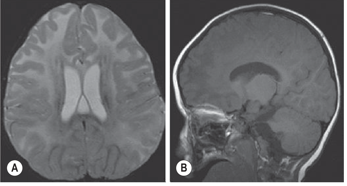
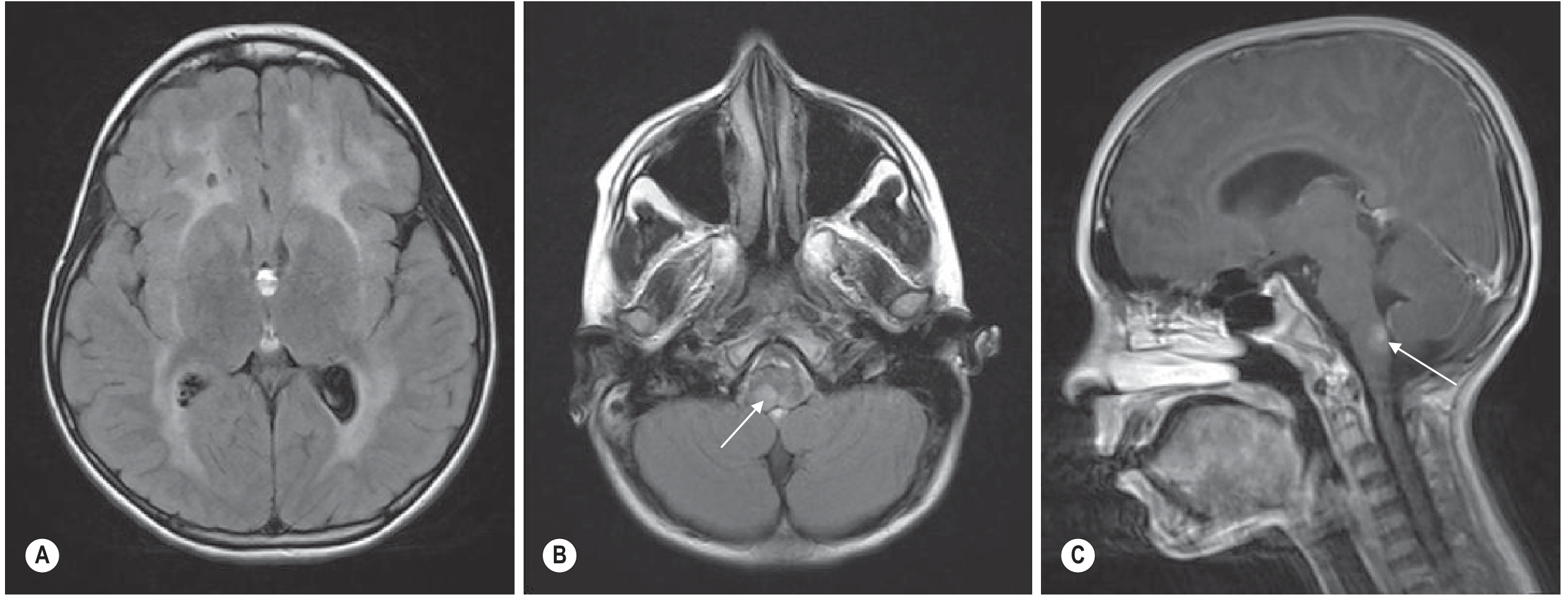
4. Alexander Disease
- Location: Frontal predominant (anterior > posterior) - this is the key differentiator
- Leukoencephalopathy pattern (van der Knaap criteria - 4 of 5 must be present):
- Extensive cerebral white matter change with frontal predominance
- Rim of T1 hyperintensity / T2 hypointensity around the frontal horns (periventricular rim)
- Abnormality of basal ganglia and thalami
- Brainstem abnormalities (midbrain, medullary involvement)
- Contrast enhancement of one or more of: ventricular lining, frontal WM, basal ganglia, brainstem structures
- Macrocephaly: Almost universal in infantile (Type I) - key clinical-imaging correlation
- Infantile (Type I): Frontal white matter swelling + T2 hyperintensity; basal ganglia and thalami involved; periventricular rim; enhancement
- Juvenile/Adult (Type II): Dorsal medulla + upper cervical cord involvement with enhancement is highly characteristic; less frontal predominance; may have cervicomedullary atrophy
- Rosenthal fibers accumulate in periventricular, perivascular, and subpial locations - the histologic hallmark reflected in the periventricular rim sign
5. Canavan Disease
- Location: Subcortical U-fibers involved early (unlike most leukodystrophies); globus pallidus and thalami involved; cerebellar white matter affected
- Signal: Diffuse T2 hyperintensity throughout white matter including subcortical U-fibers; T2 increase in globus pallidus
- Macrocephaly: Present (due to NAA accumulation causing osmotic brain swelling)
- Key MRS finding: Markedly elevated NAA peak (N-acetylaspartate) at 2.0 ppm - pathognomonic; due to aspartoacylase deficiency
- Diffusion: Restricted diffusion in white matter in early disease (cytotoxic component)
- Late stage: Diffuse cerebral atrophy
6. Pelizaeus-Merzbacher Disease (PMD)
- Pattern: Hypomyelination (not demyelination) - this is a key distinction
- MRI appearance: The white matter never properly myelinates - it has the appearance of a newborn/young infant brain regardless of actual age; T2 signal in white matter does not suppress (remains bright like unmyelinated WM); T1 WM remains hypointense
- Distribution: Diffuse; cerebellar and brainstem WM also affected; no clear focal pattern
- No enhancement (no inflammatory component)
- Thinned corpus callosum common
- Key distinguishing point: In demyelinating leukodystrophies, there is initial normal myelination followed by loss. In PMD, myelination is arrested from the start.
7. Vanishing White Matter Disease (VWM)
- Hallmark: White matter eventually becomes isointense with CSF on all sequences (literally vanishes)
- Signal: T2/FLAIR hyperintensity in white matter; as disease progresses the signal follows CSF (T1 dark, T2 bright, FLAIR suppressed)
- Cystic change: WM undergoes vacuolation and cavitation - rarefied/cystic appearance
- Distribution: Diffuse cerebral white matter; relative sparing of subcortical U-fibers and posterior fossa (early)
- Trigger: Episodes of acute neurological deterioration follow febrile illness or head trauma - radiologically, acute worsening of white matter changes is seen after stress
- MRS: Absent NAA and Cho peaks over white matter (no viable tissue); lactate/lipid peaks in cystic areas
The Radiology Differential: Quick Comparison Table (High-Yield for PG)
| Feature | X-ALD | MLD | Krabbe | Alexander | Canavan | PMD | VWM |
|---|---|---|---|---|---|---|---|
| Distribution | Posterior→anterior | Central periventricular | Posterior + central | Frontal predominant | U-fibers + pallidi | Diffuse hypomyelination | Diffuse→cystic |
| Corpus callosum | Splenium early | Splenium involved | Involved | Involved | Diffuse | Thin | Involved |
| U-fibers | Spared early | Spared early | Spared early | Involved later | Involved early | Diffuse | Spared early |
| Enhancement | Yes - leading edge | No | Occasionally | Yes - rim/frontal | No | No | No |
| Thalami/BG | Spared | Spared | T2 dark thalami | Involved | Globus pallidus | Spared | Spared |
| Special sign | 3-zone, Loes score | Tigroid/leopard skin | Dark thalami | Periventricular rim, frontal | Elevated NAA (MRS) | Arrested myelination | WM→CSF signal |
| Macrocephaly | No | No | No | Yes | Yes | No | No |
| Gene | ABCD1 (X-linked) | ARSA (AR) | GALC (AR) | GFAP (AD) | ASPA (AR) | PLP1 (X-linked) | EIF2B (AR) |
MRI Protocol for Suspected Leukodystrophy
- T1 axial + sagittal - assess myelination pattern; corpus callosum
- T2 axial - white matter signal; distribution of lesions
- FLAIR - periventricular lesion conspicuity; cystic vs. solid
- DWI/ADC - active demyelination often shows restricted diffusion at the leading edge; cystic WM shows facilitated diffusion
- Post-gadolinium T1 - enhancement pattern (critical for X-ALD staging; Alexander disease)
- MR Spectroscopy - NAA (Canavan ↑↑↑), Cho/NAA ratio, lactate (mitochondrial), absent peaks (VWM)
- Spinal cord MRI if clinically indicated (X-ALD/AMN, Krabbe infantile - cord enlargement)
Radiology PG Mnemonics
- "Posterior to Anterior" = X-ALD (Parieto-occipital first, then advances forward)
- "Frontal = Alexander" (F for Frontal = F for GFAP/Alexander)
- "Tigroid = MLD" (the only leukodystrophy with this striped pattern, and no enhancement)
- "Dark thalami = Krabbe" (T2 hypointense thalami due to globoid cell accumulation)
- "Macrocephaly + elevated NAA = Canavan"
- "Arrested myelination (not demyelination) = PMD"
- "WM becomes CSF = VWM"
- "Enhancement at leading edge = always think X-ALD first"
Sources
- Grainger & Allison's Diagnostic Radiology: Chapter 76, Inborn Metabolic Brain Disorders
- Adams and Victor's Principles of Neurology, 12th Edition
- Goldman-Cecil Medicine
- Leukodystrophy Imaging review, PMC 2024 (Thakkar et al., 2024 - 22 citations)
Barium swallow and meal radiology findings and everything and pathologies and their appearance in meal and swallow
barium swallow meal radiology findings pathologies esophagus stomach appearances fluoroscopy
achalasia barium esophagus bird beak tapering fluoroscopy
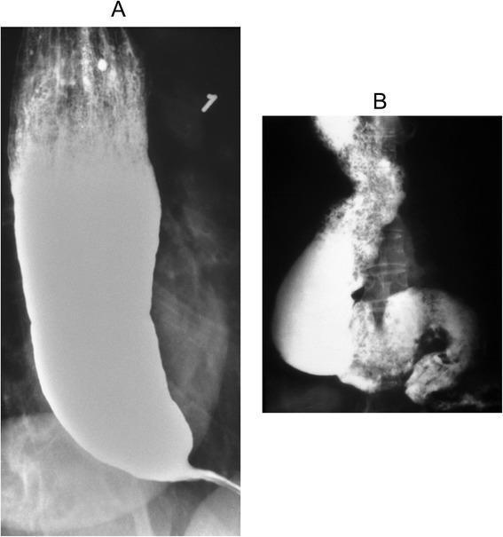
This diagnostic fluoroscopy figure displays two barium swallow (esophagogram) images illustrating the progression of achalasia. Image A shows a classic presentation with significant esophageal dilation and a large column of retained barium. The distal esophagus exhibits a smooth, symmetric tapering toward the gastroesophageal junction, creating the characteristic 'bird's beak' appearance, which indicates failure of the lower esophageal sphincter (LES) to relax. An air-fluid level is visible at the top of the barium column. Image B demonstrates advanced, end-stage achalasia, characterized by a 'sigmoid esophagus.' In this stage, the esophagus is markedly dilated, elongated, and tortuous (deformed). There is a mottled appearance within the barium column caused by a mixture of retained food particles and secretions. These images highlight the loss of primary peristalsis and chronic obstruction resulting in structural remodeling. They serve as key educational examples for gastroenterology and radiology students to differentiate early-stage and decompensated motor disorders of the esophagus.
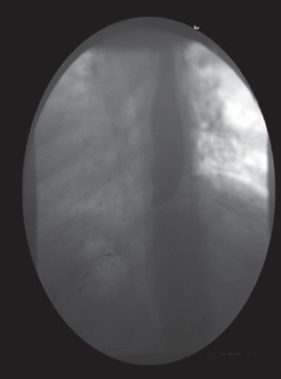
**Imaging Modality:** Barium swallow (esophagram) under fluoroscopy. **Anatomical Region:** Distal esophagus and gastroesophageal junction (GEJ). **Observed Pathology:** The image demonstrates findings highly suggestive of achalasia. There is a distinct "bird’s beak" appearance characterized by smooth, symmetric tapering of the distal esophagus. **Characteristic Visual Features:** - **Proximal Dilation:** The esophageal body shows significant proximal luminal dilation above the level of obstruction. - **Distal Tapering:** Sharp, conical narrowing at the gastroesophageal junction. - **Contrast Flow:** Abrupt cessation of the barium column with only a thin, filiform stream (trickling) of contrast medium passing through the hypertonic lower esophageal sphincter into the gastric cardia. - **Mucosal Integrity:** The margins of the narrowing appear smooth, without evidence of irregular filling defects or mucosal ulceration, typically distinguishing this from malignant pseudoachalasia. **Key Diagnostic Features:** Symmetric tapering ("bird’s beak" sign) and delayed esophageal emptying, indicative of impaired lower esophageal sphincter relaxation and aperistalsis.
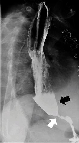
This diagnostic image is a barium swallow fluoroscopy (X-ray radiography) of the upper gastrointestinal tract, specifically the esophagus and stomach. The image demonstrates classic radiological features of achalasia. There is marked dilation of the distal esophagus, which is filled with an opaque accumulation of pooled barium contrast (indicated by the black arrow). At the gastroesophageal junction, there is a smooth, abrupt tapering of the esophageal lumen, creating a 'bird's beak' deformity (indicated by the white arrow) representing the failure of the lower esophageal sphincter (LES) to relax. Minimal contrast is seen trickling into the stomach, indicating delayed bolus transit. The study illustrates the secondary effects of aperistalsis and LES dysfunction on esophageal morphology and emptying. This material is highly relevant for medical education in gastroenterology and radiology for identifying esophageal motility disorders and obstructive pathologies at the gastroesophageal junction.
esophageal carcinoma barium swallow irregular stricture shouldering rat tail
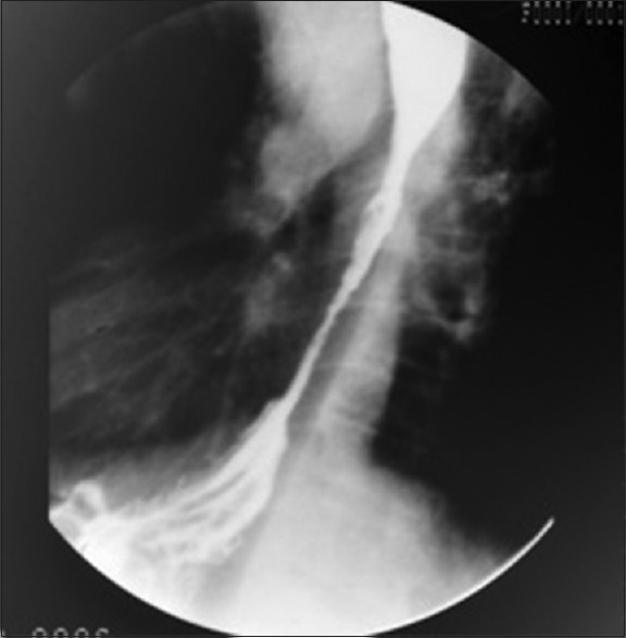
**Imaging Modality:** Fluoroscopic contrast study (Barium Swallow/Esophagram). **Anatomical Region:** Lower esophagus and gastroesophageal junction. **Observed Pathology:** There is a significant, high-grade irregular narrowing involving the distal segment of the esophagus. The lesion demonstrates asymmetric mucosal irregularities and abrupt luminal tapering, characteristic of a malignant stricture. **Characteristic Visual Features:** - **Apple-core configuration:** The circumferential narrowing displays "shouldering" at the proximal margin where the normal esophageal lumen meets the stenotic segment. - **Proximal Dilation:** Mild pre-stenotic dilation is visible superior to the narrowing, indicating chronic partial obstruction. - **Mucosal Pattern:** The contrast column within the narrowed segment appears jagged and thread-like (rat-tail appearance), suggesting infiltrative mucosal destruction rather than a smooth, benign process. **Diagnostic Significance:** These features are highly suggestive of primary esophageal neoplasia (e.g., adenocarcinoma or squamous cell carcinoma) or secondary malignant infiltration. The image provides critical visual evidence for differentiating malignant strictures from benign esophageal webs, rings, or peptic strictures based on the irregularity and shouldered margins.
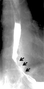
**Imaging Modality:** Barium swallow (fluoroscopic contrast esophagram). **Anatomical Region:** Lower third of the esophagus extending to the gastroesophageal junction (GEJ). **Observed Pathology:** Long-segment, high-grade luminal narrowing (stricture) of the distal esophagus. The stricture appears irregular with significant reduction in the diameter of the barium column. **Characteristic Visual Features:** * **Proximal Dilatation:** There is mild pre-stenotic dilatation of the mid-esophagus above the level of the narrowing. * **Stricture Morphology:** The narrowing demonstrates tapered, "rat-tail" or "bird-beak" like appearance as it approaches the diaphragm, though the mucosal irregularity suggests an organic rather than purely functional etiology. * **Contrast Flow:** Delayed passage of contrast media through the stenotic segment into the gastric cardia. * **Mucosal Pattern:** Loss of normal longitudinal mucosal folds within the strictured zone, replaced by an irregular, thread-like contrast trail. **Diagnostic Context:** Findings are highly suggestive of an organic lower esophageal stricture, frequently associated with advanced gastroesophageal reflux disease (peptic stricture) or malignant infiltration (esophageal carcinoma). The imaging differentiates this from achalasia by the degree of luminal irregularity and the length of the involved segment.
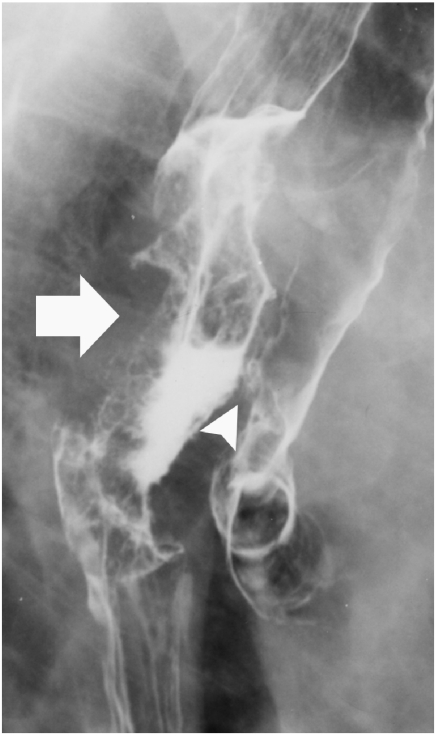
**Imaging Modality:** Fluoroscopic esophagram (barium swallow), spot film. **Anatomical Region:** Mid-to-distal esophagus and adjacent tracheobronchial tree. **Observed Pathology:** The image demonstrates a high-grade malignant esophageal stricture secondary to squamous cell carcinoma. There is an irregular, circumferential mucosal pattern with associated luminal narrowing and "apple-core" apple-core-like shouldering (indicated by the large arrow). **Characteristic Visual Features:** - **Esophageal Findings:** Severe luminal narrowing with irregular, nodular mucosal surfaces and abrupt transition zones. - **Tracheoesophageal Fistula (TEF):** A definitive fistulous tract is visible (indicated by the arrowhead), where contrast medium extravasates anteriorly from the esophageal lumen into the trachea and primary bronchi. - **Contrast Distribution:** Radiopaque contrast outlines the bronchial tree, confirming aspiration or fistulous communication between the digestive and respiratory tracts. **Diagnostic Differentiating Features:** The presence of irregular mucosal destruction and luminal eccentricity is highly suggestive of primary esophageal malignancy. The visualization of barium within the tracheobronchial tree following esophageal opacification is the pathognomonic finding for an acquired tracheoesophageal fistula, a known complication of advanced esophageal carcinoma.
gastric ulcer barium meal niche crater lesser curvature
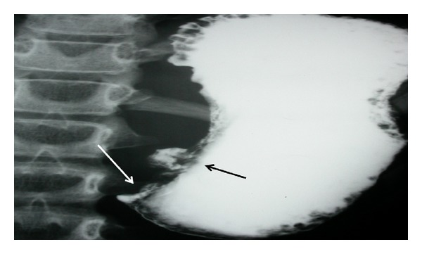
**Imaging Modality:** Barium meal (upper gastrointestinal fluoroscopy with oral contrast). **Anatomical Region:** Distal stomach, specifically the antrum and pyloric region. **Observed Pathology:** The image demonstrates a significant gastric outlet obstruction. A prominent barium-filled collection, characteristic of an ulcer crater, is visible along the lesser curvature of the stomach (indicated by the black arrow). Distal to this, there is a marked narrowing and abrupt termination of the contrast column at the pylorus (indicated by the white arrow), suggesting luminal obstruction. **Characteristic Visual Features:** * **Ulcer Niche:** An out-pouching of contrast material representing a localized mucosal defect or crater. * **Luminal Narrowing:** Severe attenuation of the gastric lumen in the prepyloric region. * **Contrast Retention:** Significant pooling of barium within a dilated gastric body and antrum, with minimal to no contrast passing into the duodenum. **Clinical Significance:** These findings are highly suggestive of chronic peptic ulcer disease leading to cicatricial pyloric stenosis or acute inflammatory edema resulting in gastric outlet obstruction. This description is optimized for retrieval in databases concerning gastrointestinal radiology and obstructive gastric pathologies.
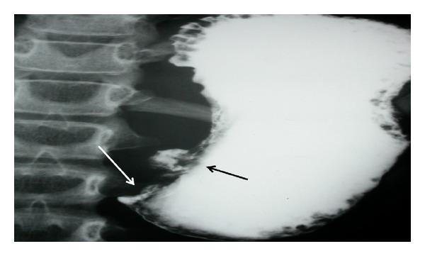
This diagnostic image is an upper gastrointestinal barium meal radiograph (X-ray) focusing on the stomach and pyloric region. The contrast study reveals a significantly distended stomach filled with radiopaque barium. Two key pathological findings are annotated: a black arrow indicates a localized, irregular projection of contrast along the lesser curvature, representing an ulcer crater. A white arrow points to the distal antrum/pylorus, where there is an abrupt, tapered termination of the contrast column with no visible passage into the duodenum, characteristic of a high-grade gastric outlet obstruction. The vertebral column is visible to the left of the gastric shadow. This radiographic appearance is clinically significant for diagnosing mechanical obstruction and mucosal lesions, such as those caused by peptic ulcer disease or gastric malignancy. The image is intended for intermediate to advanced medical learners studying gastrointestinal radiology and surgical pathology.
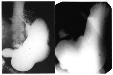
This diagnostic image consists of two side-by-side contrast-enhanced X-ray radiographs (barium swallow/upper GI series) illustrating clinical features of von Presser syndrome. Both cases demonstrate massive gastric dilatation (gastromegaly) involving the fundus and body, with a markedly elongated greater curvature occupying a significant portion of the abdominal cavity. A key diagnostic feature visible in both images is a distinct gastric ulcer located at the lesser curvature, specifically at the incisura angularis. This is characterized by a radiopaque niche or barium-filled outpouching projecting beyond the normal gastric contour, indicative of an active ulcer crater. The pyloric region appears relatively normal in diameter despite the proximal dilatation. These radiographic findings are used in gastroenterology and radiology to identify specific patterns of chronic gastric stasis associated with benign peptic ulcer disease at the incisura.
hiatal hernia barium swallow sliding gastric fundus above diaphragm
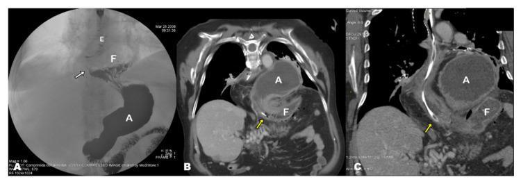
This composite figure demonstrates the progression and diagnosis of gastric pathologies using fluoroscopy and computed tomography (CT). Panel A is an anteroposterior barium swallow image showing a sliding hiatal hernia. The gastroesophageal junction (indicated by a white arrow) and a portion of the gastric fundus (F) are seen herniated above the diaphragm, while the gastric antrum (A) remains in the subdiaphragmatic abdominal cavity. Panels B and C are coronal-oblique contrast-enhanced CT reconstructions during an acute presentation of gastric volvulus. These images reveal a significant anatomical shift: a severely dilated gastric antrum (A) has herniated into the thoracic cavity, while the fundus (F) is located inferiorly below the diaphragm. A nasogastric tube (yellow arrows) is visible traversing the esophageal hiatus, likely for gastric decompression. This comparison highlights the transition from a simple hiatal hernia to an acute, complicated mesentero-axial gastric volvulus characterized by abnormal rotation and displacement of gastric segments relative to the diaphragm.
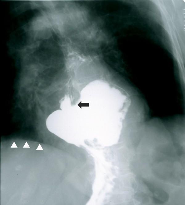
This diagnostic image is an upper gastrointestinal (UGI) barium swallow radiograph. It demonstrates a large sliding hiatal hernia characterized by the intrathoracic migration of a significant portion of the stomach. The barium contrast medium clearly outlines a distended gastric pouch residing in the posterior mediastinum, well above the level of the diaphragm (indicated by white arrowheads). The gastroesophageal junction, identified by a black arrow, is also displaced superiorly into the thoracic cavity, confirming the diagnosis. Key anatomical landmarks include the vertebral column, ribs, and the distinct interface between the contrast-filled herniated stomach and the radiolucent lung fields. This clinical photograph is highly relevant for gastroenterology and radiology education, illustrating the radiographic appearance of a large hiatus hernia and the loss of the normal subdiaphragmatic positioning of the stomach. The image is optimized for teaching diagnostic imaging patterns related to esophageal and gastric motility disorders.
duodenal ulcer barium meal deformity duodenal cap clover leaf
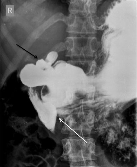
**Imaging Modality:** Fluoroscopic barium meal study, supine view. **Anatomical Region:** Upper gastrointestinal tract, specifically the stomach and duodenum. **Observed Pathology and Visual Features:** - **Duodenal Obstruction:** A distinct, linear vertical filling defect is visible across the proximal third (D3 segment) of the duodenum. This extrinsic compression results in significant pre-stenotic dilatation of the second part (D2 segment) of the duodenum, characteristic of Superior Mesenteric Artery (SMA) syndrome. - **Duodenal Bulb Deformity:** A "trifoliate" or "clover-leaf" deformity of the duodenal bulb is present. This morphology is characterized by central narrowing with multiple surrounding pseudodiverticula or sacculations. **Key Diagnostic Features:** - The vertical indentation on the transverse duodenum suggests extrinsic vascular compression. - The clover-leaf deformity is a pathognomonic radiologic sign of a chronic, healed duodenal ulcer with secondary scarring and retraction of the bulb. **Clinical Significance:** The image demonstrates dual pathology: mechanical duodenal obstruction consistent with SMA syndrome and structural remodeling from prior peptic ulcer disease.
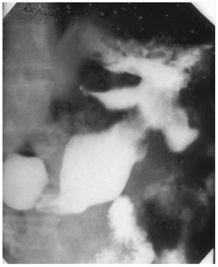
This diagnostic fluoroscopic image is a barium meal study of the upper gastrointestinal tract, specifically focusing on the stomach. The image reveals a significant structural deformity in the median third of the gastric body. A giant ulcer is visible, characterized by a large, deep, and irregular barium-filled cavity that projects beyond the normal gastric contour. This lesion exhibits a classic 'clover-leaf deformity,' where the barium collection is divided into multiple lobes or outpouchings, often resulting from extensive tissue loss and associated scarring or muscular contraction. The surrounding gastric anatomy appears distorted by the size and depth of the crater. This radiographic presentation is typical of a pseudoneoplastic benign giant gastric ulcer, which can mimic malignant processes due to its morphology and extent. Key educational concepts illustrated include contrast radiography for peptic ulcer disease, recognition of the clover-leaf sign, and the differentiation of giant benign ulcers from neoplastic gastric lesions.
carcinoma stomach barium meal linitis plastica leather bottle
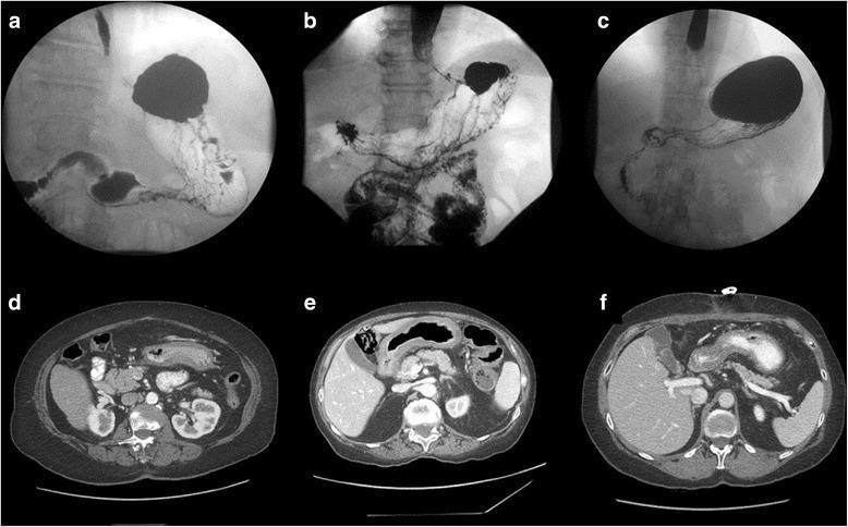
This composite medical image presents diagnostic features of linitis plastica (scirrhous gastric carcinoma) in three different patients using two imaging modalities. The top row (a–c) displays upper gastrointestinal (UGI) contrast studies (barium meals), while the bottom row (d–f) shows corresponding axial computed tomography (CT) scans. In the UGI images, the stomach exhibits characteristic 'leather bottle' morphology, marked by significant loss of distensibility, tubular contraction, and irregular mucosal outlines (especially visible in patient 2). The contrast flow highlights restricted gastric volume and rigid wall structure. The CT scans demonstrate circumferential, diffuse gastric wall thickening across all patients. Patient 1 (d) shows heterogeneous wall thickening, while Patient 2 (e) and Patient 3 (f) exhibit more uniform thickening, with some haziness in the perigastric fat suggesting inflammatory or infiltrative changes. These visual markers illustrate the infiltrative nature of Borrmann type IV tumors, emphasizing the primary pathological features of submucosal and muscularis propria involvement typical of advanced gastric cancer.
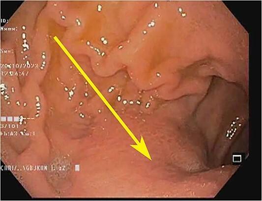
This endoscopic image displays the gastric antrum and distal stomach, highlighting key diagnostic features of linitis plastica. The mucosal surface shows a marked loss of rugosity, where the typical gastric folds are absent or flattened, resulting in a smooth and rigid appearance. There is widespread mucosal erythema, indicating inflammation. A yellow arrow points to an extramucosal bulge, suggesting a submucosal or infiltrating process. These findings are clinically significant as they represent diffuse infiltration of the gastric wall by signet-ring cell carcinoma, characterized by a lack of stomach expansion during insufflation. The image serves as a clinical example of the 'leather bottle' appearance of the stomach in advanced gastric malignancy, often associated with Krukenberg tumors in a systemic context.
Barium Swallow & Barium Meal: Complete Radiology PG Guide
PART 1: OVERVIEW AND TECHNIQUE
Barium Swallow (Esophagram)
- Studies: Pharynx + esophagus (oro- to gastroesophageal junction)
- Technique: Single contrast (thick barium) or double contrast (barium + gas-forming agent)
- Views: Fluoroscopy in real-time; spot films in AP, oblique, lateral positions; patient upright then prone (Trendelenburg to detect reflux)
- Indications: Dysphagia, odynophagia, suspected foreign body, GERD, stricture, suspected perforation (use water-soluble contrast - Gastrografin first)
- Contraindications: Known/suspected perforation (use Gastrografin); aspiration risk; pregnancy
Barium Meal (Upper GI Series)
- Studies: Stomach + duodenum (sometimes includes distal esophagus)
- Technique: Double contrast preferred (high-density barium + gas granules = mucosal coating + distension); single contrast for suspected obstruction
- Positions: Erect, supine, oblique; right lateral decubitus (to fill duodenum)
- Special variants:
- Hypotonic duodenography: IV hyoscine/glucagon → atonic duodenum → delineates pancreatic head pathology
- Barium meal follow-through: Serial images over 2-4 hours → small bowel
PART 2: BARIUM SWALLOW - PATHOLOGIES
1. Achalasia
- Dilated esophagus (massively so in advanced disease) with retained food/fluid level
- "Bird's beak" / "Rat's tail" tapering at the gastroesophageal junction - smooth, symmetric, progressive narrowing
- Air-fluid level above the barium column
- Absence of gastric air bubble (classic)
- "Sigmoid esophagus" in advanced/end-stage disease - markedly dilated, tortuous, deformed esophagus
- Poor/absent primary peristalsis - no stripping wave seen on fluoroscopy
- Contrast trickles through the LES in small amounts
2. Carcinoma of the Esophagus
- Irregular mucosal destruction - rat-tail/thread-like contrast column within the stricture
- "Shouldering" (Apple-core lesion) - abrupt transition between normal and narrowed lumen with overhanging/shelf-like edges at proximal margin
- Pre-stenotic dilatation above the lesion
- Mucosal irregularity - nodular, destroyed folds
- Asymmetric narrowing (vs. smooth in benign strictures)
- Tracheoesophageal fistula - barium enters the tracheobronchial tree (complication of advanced disease)
3. Esophageal Leiomyoma
- Smooth, semilunar/crescent-shaped filling defect - moves with swallowing
- Sharply demarcated from adjacent mucosa
- Makes an acute angle at its margins with the esophageal wall (submucosal origin)
- Mucosa overlying the lesion is intact and smooth
- No shouldering or mucosal destruction
4. Hiatal Hernia
- Type I (Sliding): GEJ + gastric cardia herniate above diaphragm (most common, 95%)
- Type II (True paraesophageal): GEJ stays below diaphragm; fundus herniates through
- Type III (Mixed): Both GEJ and fundus herniate
- Gastric folds seen above the diaphragm
- GEJ above the level of the hiatus (>2 cm above diaphragm)
- "B-ring" (Schatzki ring) - mucosal ring at GEJ
- "A-ring" (muscular ring) may also be visible
- Widened esophageal hiatus (>3.5 cm)
- Gastric fundus herniated into the thoracic cavity
- GEJ at or below the diaphragm (key differentiator from sliding)
- Risk of gastric volvulus (organoaxial rotation)
5. Pharyngeal/Zenker's Diverticulum
- Posterior outpouching at the pharyngoesophageal junction (Killian's dehiscence)
- Barium-filled pouch seen projecting posteriorly on lateral view
- May displace the esophagus anteriorly when large
- Retention of barium within the pouch after swallowing
6. Esophageal Web / Stricture
- Web: Thin, shelf-like defect projecting from the anterior wall, usually upper esophagus (Plummer-Vinson syndrome in women with iron deficiency)
- Peptic stricture: Smooth, tapered narrowing at the distal esophagus (reflux-related); no shouldering; may be short segment
- Post-corrosive stricture: Long, smooth, irregular narrowing; affects mid-esophagus preferentially
7. Esophageal Varices
- Serpentine/worm-like filling defects in the lower esophagus
- Parallel longitudinal columns of varices creating irregular wavy filling defects
- Best seen on prone oblique view with collapsed (non-distended) esophagus
- Mucosal folds appear thickened and tortuous
8. Esophageal Foreign Body
- Opaque FBs visible on plain X-ray; barium used for radiolucent FBs
- Coin: Coronal orientation in esophagus (vs. sagittal in trachea) on AP view
- Filling defect on barium; proximal hold-up of contrast
PART 3: BARIUM MEAL - GASTRIC PATHOLOGIES
1. Gastric (Peptic) Ulcer
- Ulcer niche/crater - projecting bud of barium beyond the normal gastric outline on lesser curvature (en face = ring shadow of barium)
- "Halo sign" - radiolucent zone surrounding the crater (mound of edema around the ulcer)
- Hampton's line - thin radiolucent line across the mouth of the ulcer (overhanging mucosa)
- Mucosal folds converging (radiating) toward the crater
- Persistent spasm on the greater curvature opposite the ulcer ("incisura" on greater curvature = pointing arrow toward the ulcer site)
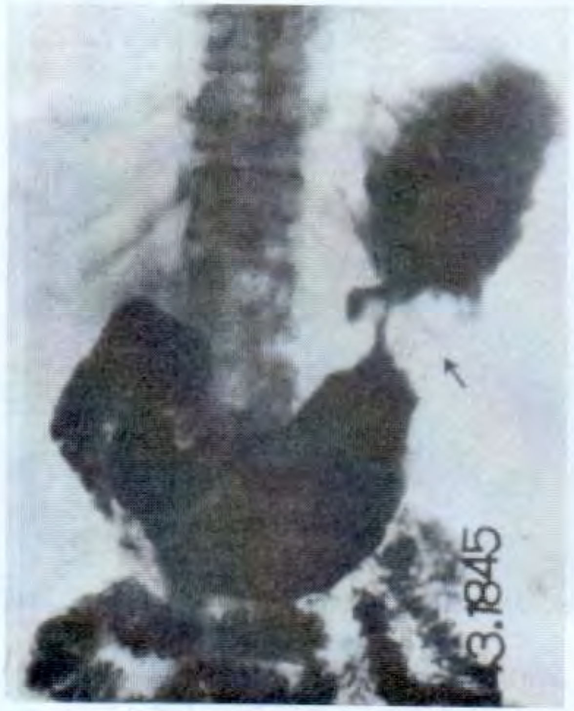
- Hourglass deformity of stomach (with healing/fibrosis of mid-body ulcer)
- Delayed gastric emptying
2. Carcinoma of the Stomach (Gastric Carcinoma)
- Irregular filling defect (constant in all films - key!)
- Lobulated soft-tissue mass
- Mucosal destruction, absent peristalsis in that segment
- Malignant ulcer - inside the lumen (vs. benign ulcer projecting outside)
- "Carman's meniscus sign" - large ulcer with a concave inner margin + radiolucent halo (Kirklin complex) = malignant ulcer
- Codman's triangle - mucosal folds stop abruptly at the edge of the ulcer (no converging folds)
- Mucosal folds are amputated/clubbed, not radiating toward the crater
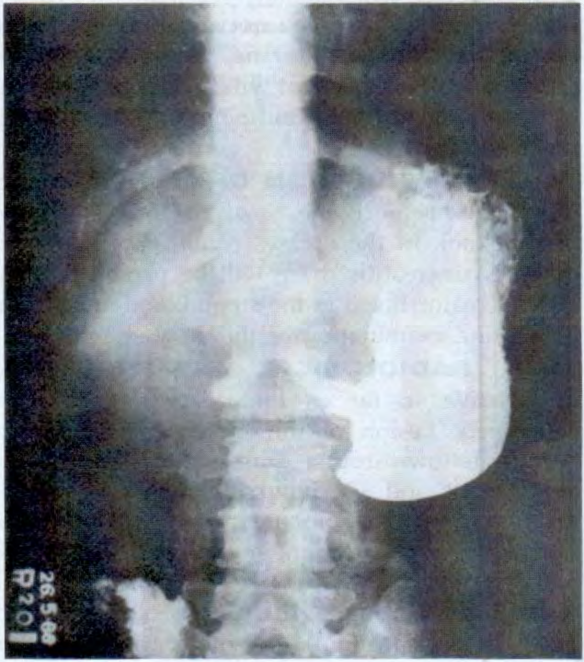
- "Leather bottle stomach" - markedly shrunken, rigid, non-distensible stomach
- Loss of normal rugal folds throughout
- Stomach does not expand with insufflation/peristalsis is absent
- Narrowed lumen; resembles a rigid tube
| Feature | Benign (Peptic) | Malignant |
|---|---|---|
| Location | Outside gastric outline | Inside gastric outline |
| Mucosal folds | Converge toward ulcer | Amputated/stop at edge |
| Surrounding mucosa | Normal | Nodular/destroyed |
| Halo | Hampton's line (thin line) | Wide mound of tissue |
| Wall | Pliable, peristalsis present | Rigid, loss of peristalsis |
| Emptying | Normal | Slow or obstructed |
3. Duodenal Ulcer
- Ulcer crater in the duodenal cap (1st part) - seen as a niche or fleck of barium
- Often on the posterior wall - seen in profile on lateral view
- Persistent deformity of duodenal cap - most reliable sign
- "Cloverleaf deformity" - chronic scarring deforms the duodenal cap into 3 pseudodiverticula (patho-gnomonic of chronic healed DU)
- Rapid emptying of stomach
- Localized tenderness over duodenum on fluoroscopy
- Pyloric spasm
- Residue at 6 hours = pyloric obstruction; residue at 24 hours = organic pyloric stenosis (almost certain)
4. Pyloric Stenosis
- Prolonged gastric emptying
- Dilated stomach with food residue
- Narrowed pyloric channel
- "String sign" - thin thread of barium passing through elongated, narrow pyloric canal
- "Mushroom/umbrella sign" - inverted V indentation on the base of duodenal cap by the hypertrophied pylorus
- "Shoulder sign" - pyloric muscle bulges into antrum making shoulder impression
- NB: Ultrasound is now preferred over barium for IHPS
5. Gastric Volvulus
- Organoaxial volvulus: Stomach rotated along its long axis; greater curvature appears above lesser curvature; barium in a double-bubble configuration within the thorax
- Mesenteroaxial volvulus: Antrum and pylorus flip upward; pylorus seen above the gastric body
PART 4: DUODENAL AND ADJACENT PATHOLOGIES ON BARIUM MEAL
1. Carcinoma of the Head of Pancreas
- "Pad sign" (Frostberg's inverted 3 / epsilon sign): Barium-filled duodenal loop is widened; duodenal sweep is widened medially
- The reversed 3/ε sign on duodenum - indentation on both the inner margin of the 2nd part of duodenum superiorly and inferiorly with central convexity (due to tumor mass)
2. Carcinoma of the Ampulla of Vater
- "Reversed 3" (ε sign) - filling defect at the ampullary region with symmetric indentations above and below
3. Pseudopancreatic Cyst
- Lateral view: Anterior displacement of the stomach by the retrogastric cystic mass
- On AP: Gastric body displaced anteriorly
4. Chronic Pancreatitis (Hypotonic Duodenography)
- Early: Definite flattening of the medial margin of the duodenal loop
- Late: Inverted "3" sign = thickened folds with ulceration at the major papilla (Frostberg's sign)
- Small filling defects or areas of constriction in carcinoma
PART 5: BARIUM MEAL FOLLOW-THROUGH
| Condition | Barium Appearance |
|---|---|
| Crohn's disease | "String sign of Kantor" - long narrow string-like terminal ileum; skip lesions; "cobblestone" mucosa; fistulae |
| Ileocaecal TB | Cecum displaced upward; distal ileum narrowed and near vertical; "Stierlin's sign" = rapid emptying of the cecum; conical/shrunken cecum |
| Appendicitis | Non-filling of appendix; fixity and tenderness on fluoroscopy when filled |
| Carcinoma of cecum | Irregular filling defect in cecum |
| Intussusception | "Coiled spring" appearance - barium passes around the intussusceptum |
PART 6: KEY SIGNS - QUICK REFERENCE TABLE
| Sign | Appearance | Condition |
|---|---|---|
| Bird's beak / Rat tail | Smooth symmetric distal tapering | Achalasia |
| Sigmoid esophagus | Tortuous massively dilated esophagus | Advanced achalasia |
| Shouldering / Apple-core | Abrupt overhanging edges at tumor margins | Esophageal/gastric carcinoma |
| Rat tail (malignant) | Irregular thread-like narrowing | Esophageal carcinoma |
| Niche / Crater | Barium pouch projecting outside gastric outline | Benign gastric ulcer |
| Carman's meniscus sign | Concave inner ulcer margin + halo | Malignant gastric ulcer |
| Hampton's line | Thin lucent line across ulcer mouth | Benign peptic ulcer |
| Mucosal convergence | Rugal folds radiating to crater | Benign gastric ulcer |
| Leather bottle | Rigid, shrunken non-distensible stomach | Linitis plastica (Borrmann IV) |
| Hourglass stomach | Mid-body constriction | Chronic peptic ulcer/fibrosis |
| String sign of Kantor | Thread-like terminal ileum | Crohn's disease |
| Cloverleaf deformity | Trilobed duodenal cap | Chronic healed DU |
| Pad sign / ε sign | Widened C-loop with indentation | Ca head of pancreas |
| Reversed 3 (ε) | Symmetric indentations in 2nd part D | Carcinoma of ampulla |
| String sign (IHPS) | Thin pyloric channel | Infantile hypertrophic pyloric stenosis |
| Mushroom/umbrella sign | Duodenal cap base indentation | IHPS |
| Serpentine filling defects | Worm-like defects in lower esophagus | Esophageal varices |
| Stierlin's sign | Rapid emptying of cecum | Ileocaecal TB |
| Coiled spring | Spiral barium around intussusceptum | Intussusception |
PART 7: BENIGN vs. MALIGNANT FILLING DEFECT - KEY DIFFERENTIATORS
| Feature | Benign | Malignant |
|---|---|---|
| Margins | Smooth, rounded | Irregular, shouldered, nodular |
| Peristalsis | Present | Absent in involved segment |
| Mucosal folds | Normal or converging | Destroyed, amputated |
| Wall | Flexible | Rigid |
| Consistency | Changes with peristalsis | Constant in all films |
| Ulcer position | Projects outside lumen | Within lumen |
- S. Das Manual of Clinical Surgery, 13th Edition
- Schwartz's Principles of Surgery, 11th Edition
- Mulholland & Greenfield's Surgery, 7th Edition
- Radiology Info - Upper GI Examination
Create a study PDF with all pathognomonic barium signs and images
esophageal varices barium swallow worm-like filling defects
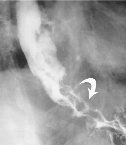
**Imaging Modality:** Fluoroscopic contrast study (Esophagram/Barium Swallow). **Anatomical Region:** Distal esophagus and gastroesophageal junction. **Observed Pathology:** Varicoid carcinoma of the distal esophagus. **Characteristic Visual Features:** The image demonstrates multiple, prominent serpiginous and longitudinal filling defects within the contrast-filled esophageal lumen. These defects create a tortuous, "cobblestone" or "worm-like" appearance, highlighted by a curved white arrow in the distal segment. Unlike traditional esophageal varices, these findings represent a fixed mucosal/submucosal neoplastic infiltration. **Key Diagnostic Features:** - **Pattern:** Serpiginous, longitudinal filling defects mimicking the appearance of esophageal varices (varicoid pattern). - **Distribution:** Concentrated in the distal third of the esophagus. - **Differentiating Factors:** Fixed nature of filling defects on fluoroscopy (typical of varicoid carcinoma) as opposed to the dynamic changes in caliber seen with true venous varices during peristalsis or Valsalva maneuvers. **Clinical Context:** Squamous cell carcinoma or adenocarcinoma presenting with varicoid morphology, necessitating differentiation from portal hypertension-related varices.
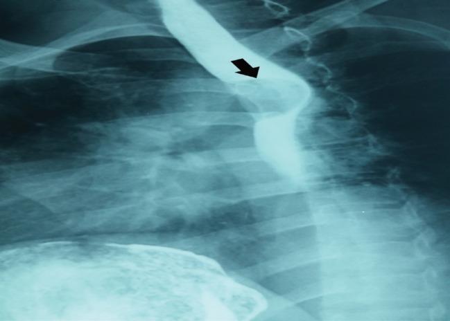
This diagnostic image is a barium swallow radiograph (upper GI series) showing the thoracic esophagus. The image demonstrates a well-defined, semilunar (half-moon shaped) filling defect in the upper thoracic esophagus, indicated by a black arrow. The defect has smooth, regular margins and causes a localized displacement of the contrast column, suggesting an intramural or extramural mass rather than an intraluminal primary mucosal lesion. The surrounding esophageal lumen remains patent but distorted. Relevant anatomical landmarks include the posterior ribcage and the distal trachea. Such findings are classically associated with benign esophageal tumors, such as leiomyomas, which present as smooth filling defects with obtuse angles to the esophageal wall. The image is an essential teaching tool for identifying esophageal filling defects and distinguishing between mucosal and submucosal pathologies in gastroenterology and radiology.
Crohn disease barium string sign Kantor terminal ileum cobblestone
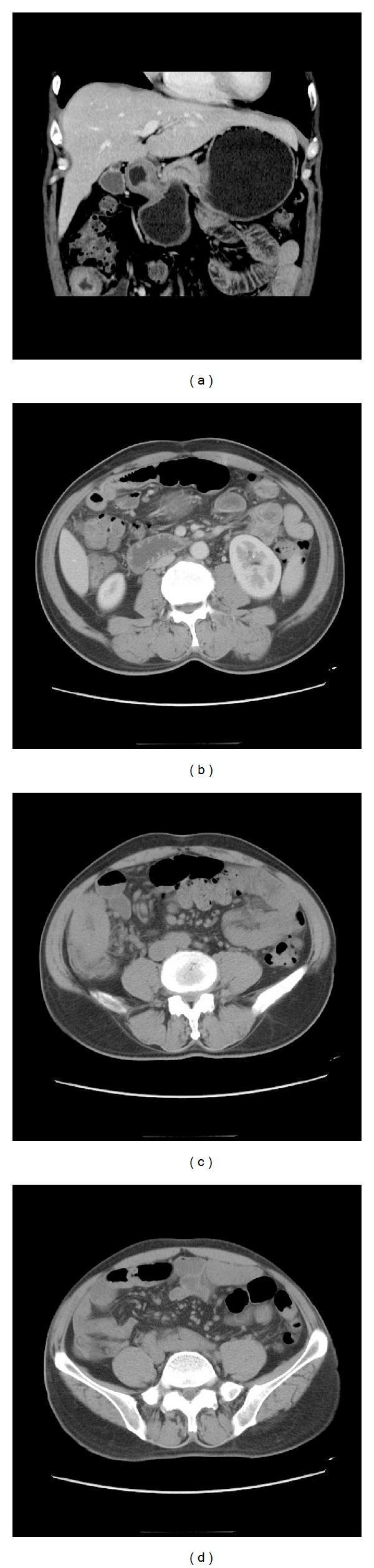
This composite of abdominal Computed Tomography (CT) images, including coronal (a) and axial (b-d) views, demonstrates classic radiographic findings associated with inflammatory bowel disease, specifically Crohn's disease. The primary anatomical focus is the terminal ileum, which exhibits significant small-bowel wall thickening and severe segmental narrowing of the intestinal lumen, often referred to as the 'string sign'. The mucosal surface of the affected ileal segment shows a 'cobblestone appearance,' representing alternating areas of deep ulceration and intramural edema. Associated mesenteric features include mesenteric adenopathy, characterized by enlarged, rounded lymph nodes within the surrounding fat. These diagnostic features illustrate chronic transmural inflammation and are clinically significant for identifying intestinal stenosis and potential fistulization in a gastroenterological context. The images serve as an educational resource for medical students and radiology residents to recognize the complications of chronic granulomatous inflammation in the gastrointestinal tract.
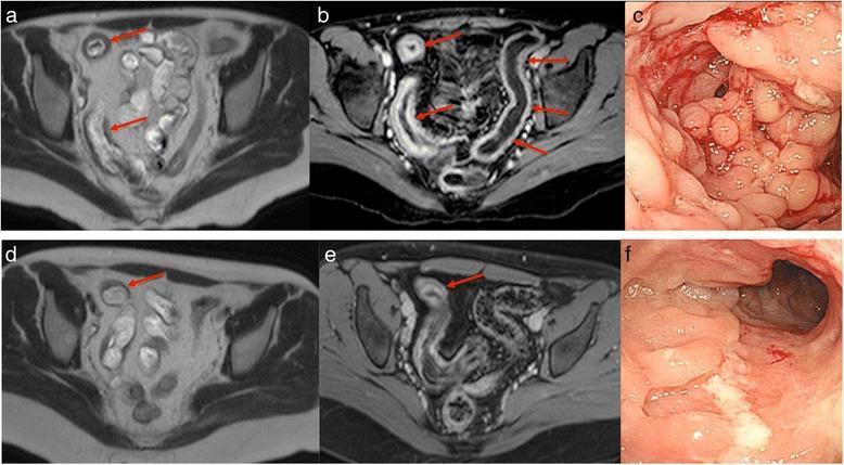
This composite figure illustrates the progression and monitoring of Crohn's disease (CD) in the distal ileum using magnetic resonance imaging (MRI) and endoscopy. Panels (a, b) show axial MRI baseline scans demonstrating severe active inflammation characterized by marked bowel wall thickening, mural edema, and prominent stratified hyperenhancement (target sign) in the terminal ileum. Panel (c) provides the corresponding baseline endoscopic view, revealing classic 'cobblestone' mucosal patterns and luminal narrowing (stricturing). Following 26 weeks of infliximab (IFX) treatment, panels (d, e) show interval improvement on MRI, with reduced wall thickness and milder contrast enhancement, though persistent moderate disease is noted. Panel (f) shows the week 26 endoscopic follow-up, where the cobblestone appearance has transitioned to irregular longitudinal ulcers. This comparison highlights the use of the Magnetic Resonance Index of Activity (MaRIA) score and Simple Endoscopic Score for Crohn’s Disease (SES-CD) to quantify therapeutic response and mucosal healing.
pyloric stenosis infantile barium string sign mushroom sign
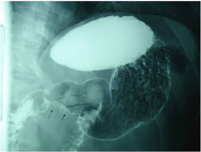
**Imaging Modality:** Fluoroscopic X-ray; Upper Gastrointestinal (UGI) Series with oral barium contrast (Barium Meal). **Anatomical Region:** Upper abdomen, specifically the distal stomach, pylorus, and proximal duodenum. **Observed Pathology:** The image demonstrates significant narrowing of the pyloric canal, appearing as a thin, elongated track of barium contrast (indicated by black arrows). This finding is classically referred to as the "string sign." **Characteristic Visual Features:** - **Stomach:** The gastric body and antrum appear dilated and filled with radiopaque barium contrast. - **Pylorus:** There is a marked luminal narrowing at the gastroduodenal junction. - **Duodenum:** Limited contrast is visible distal to the narrowing, suggesting partial gastric outlet obstruction. - **Mucosal Pattern:** Prominent rugal folds are visible in the gastric body. **Clinical Context & Differential:** The radiographic findings are highly characteristic of Hypertrophic Pyloric Stenosis (HPS), typically seen in pediatric populations, or secondary pyloric cicatrization/stenosis due to chronic peptic ulcer disease or malignancy in adults. The "string sign" serves as a key diagnostic cue for identifying significant pyloric luminal constriction.
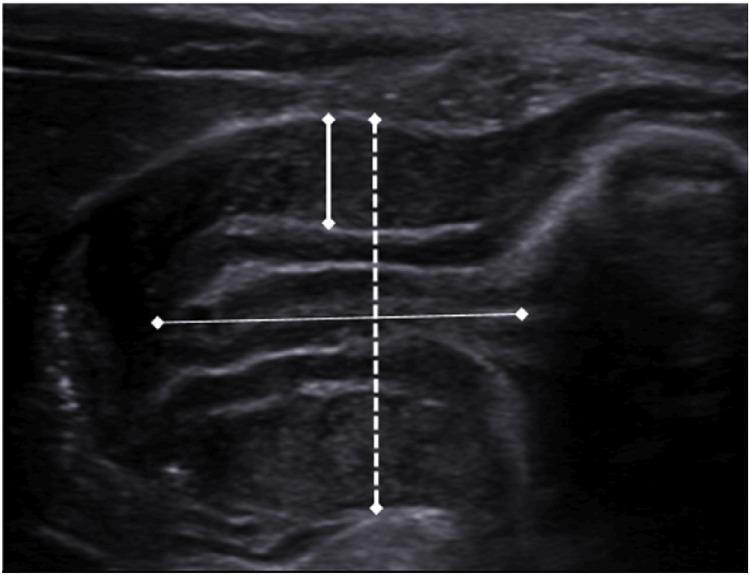
This abdominal ultrasound image captures a longitudinal view of the pylorus in a pediatric patient. The imaging displays classic diagnostic signs of Infantile Hypertrophic Pyloric Stenosis (IHPS). The pyloric muscle is significantly thickened, appearing hypoechoic and edematous, while the central mucosal layer is echogenic and compressed. White calipers demonstrate critical diagnostic measurements: a vertical measurement indicating a pyloric muscle thickness of 4.5 mm and a horizontal measurement indicating a pyloric canal length of 15 mm. The 'cervix sign' is evident due to the elongation and narrowing of the pyloric canal. Clinical significance includes the identification of gastric outlet obstruction, where the thickened muscle prevents the passage of gastric contents into the duodenum. This visual material is used to teach diagnostic criteria for IHPS, focusing on the threshold measurements (muscle thickness >3 mm and canal length >14-15 mm) essential for radiologists and pediatricians.
ls -la /tmp/workspace/ 2>/dev/null || mkdir -p /tmp/workspace && echo "workspace ready"
mkdir -p /tmp/workspace/barium-study && cat > /tmp/workspace/barium-study/build_pdf.py << 'PYEOF' import requests import os from io import BytesIO from reportlab.lib.pagesizes import A4 from reportlab.lib import colors from reportlab.lib.units import cm, mm from reportlab.lib.styles import getSampleStyleSheet, ParagraphStyle from reportlab.lib.enums import TA_LEFT, TA_CENTER, TA_JUSTIFY from reportlab.platypus import ( SimpleDocTemplate, Paragraph, Spacer, Table, TableStyle, Image as RLImage, HRFlowable, PageBreak, KeepTogether ) from reportlab.platypus.flowables import BalancedColumns from reportlab.pdfgen import canvas from PIL import Image as PILImage import urllib.request OUTPUT = "/tmp/workspace/barium-study/Barium_Signs_Study_Guide.pdf" W, H = A4 # ── helper: download image url → reportlab Image ────────────────────────────── def dl_img(url, max_w, max_h): try: req = urllib.request.Request(url, headers={"User-Agent": "Mozilla/5.0"}) data = urllib.request.urlopen(req, timeout=15).read() buf = BytesIO(data) pil = PILImage.open(buf) pil = pil.convert("RGB") out = BytesIO() pil.save(out, "JPEG", quality=85) out.seek(0) iw, ih = pil.size ratio = min(max_w / iw, max_h / ih) return RLImage(out, width=iw*ratio, height=ih*ratio) except Exception as e: print(f" IMG FAIL {url[:60]}: {e}") return None # ── styles ───────────────────────────────────────────────────────────────────── styles = getSampleStyleSheet() COVER_TITLE = ParagraphStyle("CoverTitle", fontName="Helvetica-Bold", fontSize=32, textColor=colors.HexColor("#1a3a5c"), alignment=TA_CENTER, leading=40) COVER_SUB = ParagraphStyle("CoverSub", fontName="Helvetica", fontSize=16, textColor=colors.HexColor("#2e6da4"), alignment=TA_CENTER, leading=22) COVER_INFO = ParagraphStyle("CoverInfo", fontName="Helvetica", fontSize=11, textColor=colors.HexColor("#555555"), alignment=TA_CENTER, leading=16) H1 = ParagraphStyle("H1", fontName="Helvetica-Bold", fontSize=15, textColor=colors.white, alignment=TA_LEFT, leading=20, backColor=colors.HexColor("#1a3a5c"), leftIndent=-8, rightIndent=-8, spaceBefore=14, spaceAfter=6) H2 = ParagraphStyle("H2", fontName="Helvetica-Bold", fontSize=12, textColor=colors.HexColor("#1a3a5c"), alignment=TA_LEFT, leading=16, spaceBefore=10, spaceAfter=4, borderPad=2) BODY = ParagraphStyle("Body", fontName="Helvetica", fontSize=10, textColor=colors.HexColor("#222222"), leading=15, spaceAfter=4, alignment=TA_JUSTIFY) BOLD_BODY = ParagraphStyle("BoldBody", fontName="Helvetica-Bold", fontSize=10, textColor=colors.HexColor("#c0392b"), leading=15, spaceAfter=2) CAP = ParagraphStyle("Cap", fontName="Helvetica-Oblique", fontSize=8.5, textColor=colors.HexColor("#555555"), alignment=TA_CENTER, leading=12, spaceAfter=6) SIGN_NAME = ParagraphStyle("SignName", fontName="Helvetica-Bold", fontSize=10.5, textColor=colors.HexColor("#1a3a5c"), leading=14, spaceBefore=4) SIGN_COND = ParagraphStyle("SignCond", fontName="Helvetica-Bold", fontSize=9.5, textColor=colors.HexColor("#c0392b"), leading=13) SIGN_DESC = ParagraphStyle("SignDesc", fontName="Helvetica", fontSize=9.5, textColor=colors.HexColor("#333333"), leading=14, spaceAfter=3) TABLE_HDR = ParagraphStyle("TblHdr", fontName="Helvetica-Bold", fontSize=9, textColor=colors.white, alignment=TA_CENTER) TABLE_CELL = ParagraphStyle("TblCell", fontName="Helvetica", fontSize=8.5, textColor=colors.HexColor("#222222"), leading=13) TABLE_CELL_B = ParagraphStyle("TblCellB", fontName="Helvetica-Bold", fontSize=8.5, textColor=colors.HexColor("#1a3a5c"), leading=13) NAVY = colors.HexColor("#1a3a5c") BLUE = colors.HexColor("#2e6da4") RED = colors.HexColor("#c0392b") LGRAY = colors.HexColor("#f4f6f9") MGRAY = colors.HexColor("#dce3ec") # ── page numbering canvas ────────────────────────────────────────────────────── def add_page_number(canvas, doc): canvas.saveState() canvas.setFont("Helvetica", 8) canvas.setFillColor(colors.HexColor("#888888")) canvas.drawRightString(W - 1.5*cm, 0.9*cm, f"Page {doc.page}") canvas.drawString(1.5*cm, 0.9*cm, "Barium Signs Study Guide | Radiology PG") canvas.setStrokeColor(MGRAY) canvas.setLineWidth(0.5) canvas.line(1.5*cm, 1.2*cm, W - 1.5*cm, 1.2*cm) canvas.restoreState() # ── sign card helper ─────────────────────────────────────────────────────────── def sign_card(sign, condition, appearance, tip=None, img_url=None, img_w=6*cm, img_h=5*cm): """Returns a list of flowables for one sign card.""" elems = [] # header strip hdr_data = [[Paragraph(f"🔹 {sign}", SIGN_NAME), Paragraph(condition, SIGN_COND)]] hdr_tbl = Table(hdr_data, colWidths=[9.5*cm, 8*cm]) hdr_tbl.setStyle(TableStyle([ ("BACKGROUND", (0,0), (-1,-1), LGRAY), ("LINEBELOW", (0,0), (-1,-1), 1.5, BLUE), ("TOPPADDING", (0,0), (-1,-1), 6), ("BOTTOMPADDING", (0,0), (-1,-1), 5), ("LEFTPADDING", (0,0), (-1,-1), 6), ("RIGHTPADDING", (0,0), (-1,-1), 4), ])) elems.append(hdr_tbl) if img_url: img = dl_img(img_url, img_w, img_h) if img: txt_col = [] txt_col.append(Paragraph("<b>Appearance:</b>", SIGN_DESC)) txt_col.append(Paragraph(appearance, SIGN_DESC)) if tip: txt_col.append(Spacer(1, 3)) txt_col.append(Paragraph(f"<b>⚠ Key tip:</b> {tip}", SIGN_DESC)) body_data = [[img, txt_col]] bw = img_w + 0.3*cm body_tbl = Table(body_data, colWidths=[bw, 17.5*cm - bw]) body_tbl.setStyle(TableStyle([ ("VALIGN", (0,0), (-1,-1), "TOP"), ("TOPPADDING", (0,0), (-1,-1), 5), ("BOTTOMPADDING", (0,0), (-1,-1), 5), ("LEFTPADDING", (0,0), (-1,-1), 5), ("RIGHTPADDING", (0,0), (-1,-1), 5), ("BACKGROUND", (0,0), (-1,-1), colors.white), ])) elems.append(body_tbl) else: elems.append(Spacer(1, 3)) elems.append(Paragraph("<b>Appearance:</b> " + appearance, SIGN_DESC)) if tip: elems.append(Paragraph(f"<b>⚠ Key tip:</b> {tip}", SIGN_DESC)) else: elems.append(Spacer(1, 3)) elems.append(Paragraph("<b>Appearance:</b> " + appearance, SIGN_DESC)) if tip: elems.append(Paragraph(f"<b>⚠ Key tip:</b> {tip}", SIGN_DESC)) elems.append(Spacer(1, 8)) return elems # ── document ─────────────────────────────────────────────────────────────────── doc = SimpleDocTemplate( OUTPUT, pagesize=A4, leftMargin=1.5*cm, rightMargin=1.5*cm, topMargin=1.8*cm, bottomMargin=1.8*cm, title="Barium Signs Study Guide", author="Orris Medical" ) story = [] # ══════════════════════════════════════════════════════════════════════════════ # COVER PAGE # ══════════════════════════════════════════════════════════════════════════════ story.append(Spacer(1, 3.5*cm)) story.append(Paragraph("Pathognomonic Barium Signs", COVER_TITLE)) story.append(Spacer(1, 0.4*cm)) story.append(Paragraph("Barium Swallow & Barium Meal", COVER_SUB)) story.append(Spacer(1, 0.5*cm)) # decorative rule story.append(HRFlowable(width="80%", thickness=3, color=BLUE, spaceAfter=0.4*cm)) story.append(Paragraph("Complete Radiology PG Study Guide", COVER_INFO)) story.append(Spacer(1, 0.3*cm)) story.append(Paragraph("Esophagus · Stomach · Duodenum · Small Bowel", COVER_INFO)) story.append(Spacer(1, 2.5*cm)) # summary box cov_table_data = [ [Paragraph("<b>40+</b>", ParagraphStyle("N", fontName="Helvetica-Bold", fontSize=22, textColor=NAVY, alignment=TA_CENTER)), Paragraph("<b>10</b>", ParagraphStyle("N", fontName="Helvetica-Bold", fontSize=22, textColor=NAVY, alignment=TA_CENTER)), Paragraph("<b>Real</b>", ParagraphStyle("N", fontName="Helvetica-Bold", fontSize=22, textColor=NAVY, alignment=TA_CENTER))], [Paragraph("Classic Signs", ParagraphStyle("L", fontName="Helvetica", fontSize=10, alignment=TA_CENTER, textColor=colors.HexColor("#555"))), Paragraph("Pathologies Covered", ParagraphStyle("L", fontName="Helvetica", fontSize=10, alignment=TA_CENTER, textColor=colors.HexColor("#555"))), Paragraph("Fluoroscopy Images", ParagraphStyle("L", fontName="Helvetica", fontSize=10, alignment=TA_CENTER, textColor=colors.HexColor("#555")))] ] cov_table = Table(cov_table_data, colWidths=[5.5*cm, 5.5*cm, 5.5*cm]) cov_table.setStyle(TableStyle([ ("BACKGROUND", (0,0), (-1,-1), LGRAY), ("BOX", (0,0), (-1,-1), 1, MGRAY), ("INNERGRID", (0,0), (-1,-1), 0.5, MGRAY), ("TOPPADDING", (0,0), (-1,-1), 10), ("BOTTOMPADDING", (0,0), (-1,-1), 8), ("ALIGN", (0,0), (-1,-1), "CENTER"), ("VALIGN", (0,0), (-1,-1), "MIDDLE"), ])) story.append(cov_table) story.append(Spacer(1, 3*cm)) story.append(HRFlowable(width="80%", thickness=1, color=MGRAY)) story.append(Spacer(1, 0.5*cm)) story.append(Paragraph("For Radiology & Surgery Postgraduate Examination Preparation", COVER_INFO)) story.append(Paragraph("Compiled from Grainger & Allison, Schwartz's Surgery, S. Das Surgery, Bradley & Daroff Neurology", COVER_INFO)) story.append(PageBreak()) # ══════════════════════════════════════════════════════════════════════════════ # SECTION 1 : BARIUM SWALLOW # ══════════════════════════════════════════════════════════════════════════════ story.append(Paragraph(" SECTION 1: BARIUM SWALLOW — ESOPHAGEAL SIGNS", H1)) story.append(Spacer(1, 0.3*cm)) story.append(Paragraph( "The barium swallow evaluates the pharynx and esophagus under real-time fluoroscopy. " "Double-contrast technique (barium + gas) gives optimal mucosal detail. " "Use Gastrografin (water-soluble) first if perforation is suspected.", BODY)) story.append(Spacer(1, 0.4*cm)) # ── Sign 1: Bird's Beak ─────────────────────────────────────────────────────── story.append(Paragraph("1. ACHALASIA", H2)) story.extend(sign_card( sign = "Bird's Beak Sign / Rat Tail Sign", condition = "Achalasia (failure of LES relaxation)", appearance = ( "Smooth, symmetric tapering of the distal esophagus at the GEJ creating a narrow " "beak-like point. The esophageal body is massively dilated with retained food " "and fluid, forming an air-fluid level above the barium column. " "Absent primary peristalsis on fluoroscopy. Gastric air bubble absent." ), tip = "Smooth margins = achalasia. Irregular/shouldered margins = pseudoachalasia (carcinoma). Always endoscope to exclude malignancy.", img_url = "https://cdn.orris.care/cdss_images/pmc_clinical_VQA_cd56ea611443e7a0e323f93d0387b93c6b0de1579d3d25688d35a5674830e7b4.jpg", img_w=7*cm, img_h=5.5*cm )) story.extend(sign_card( sign = "Sigmoid Esophagus", condition = "Advanced / End-stage Achalasia", appearance = ( "Massively dilated, tortuous, deformed esophagus resembling a sigmoid colon. " "Mottled appearance within the barium column from mixed food, secretions, and barium. " "Represents chronic progressive obstruction and structural remodeling." ), tip = "End-stage achalasia. Treatment is more complex; surgical myotomy or POEM required.", img_url = "https://cdn.orris.care/cdss_images/roco_radiology_ROCO_72355_1766650038825.png", img_w=5.5*cm, img_h=5*cm )) # ── Sign 2: Esophageal Carcinoma ────────────────────────────────────────────── story.append(Paragraph("2. ESOPHAGEAL CARCINOMA", H2)) story.extend(sign_card( sign = "Apple-Core / Shouldering Sign", condition = "Carcinoma of the Esophagus (SCC or Adenocarcinoma)", appearance = ( "Abrupt, irregular circumferential narrowing with overhanging shelf-like edges (shoulders) " "at the proximal transition zone. The contrast column within the stricture appears jagged, " "thread-like (rat-tail). Pre-stenotic dilation above the lesion. Mucosal folds are " "destroyed and irregular. Asymmetric narrowing." ), tip = "Key differentiator from benign stricture: shouldered/overhanging margins + mucosal destruction + absence of tapering.", img_url = "https://cdn.orris.care/cdss_images/roco_radiology_ROCO_24674_1766647457787.png", img_w=6*cm, img_h=5*cm )) story.extend(sign_card( sign = "Tracheoesophageal Fistula on Barium", condition = "Advanced Esophageal Carcinoma (complication)", appearance = ( "Barium extravasates from the esophagus into the tracheobronchial tree, outlining " "the bronchi with contrast. Irregular mucosal destruction + luminal narrowing present " "at the fistula site. Apple-core stricture may be visible at the primary tumor site." ), tip = "CONTRAINDICATION: Do NOT give barium if TEF is suspected clinically — use Gastrografin first.", img_url = "https://cdn.orris.care/cdss_images/roco_radiology_ROCO_47774_1766648713959.png", img_w=6.5*cm, img_h=5.5*cm )) # ── Sign 3: Leiomyoma ───────────────────────────────────────────────────────── story.append(Paragraph("3. ESOPHAGEAL LEIOMYOMA", H2)) story.extend(sign_card( sign = "Smooth Semilunar / Crescent Filling Defect", condition = "Leiomyoma (most common benign esophageal tumor)", appearance = ( "Smooth, semilunar or crescent-shaped filling defect on barium swallow. " "Moves with swallowing. Sharply demarcated from adjacent mucosa with ACUTE angles " "at the margins (submucosal origin). Overlying mucosa intact and smooth. " "No mucosal destruction or shouldering." ), tip = "Submucosal lesion = ACUTE angle at margins. Intraluminal polyp = OBTUSE angle. Never biopsy leiomyoma transoesophageally (causes adhesions before surgery).", img_url = "https://cdn.orris.care/cdss_images/pmc_clinical_VQA_c2ca065dfa3734b0a870eb7acd80b7b792575678971cd3b3339cfdf3e3687a10.jpg", img_w=6*cm, img_h=5*cm )) # ── Sign 4: Hiatal Hernia ───────────────────────────────────────────────────── story.append(Paragraph("4. HIATAL HERNIA", H2)) story.extend(sign_card( sign = "Intrathoracic Gastric Pouch", condition = "Sliding Hiatal Hernia (Type I) — most common (95%)", appearance = ( "Gastric mucosal folds visible above the diaphragm. GEJ displaced >2 cm above " "the hiatus. Schatzki (B-ring) mucosal ring at GEJ may be seen. " "Widened esophageal hiatus (>3.5 cm). On Trendelenburg view: free gastroesophageal reflux." ), tip = "Sliding hernia: GEJ + cardia above diaphragm. Paraesophageal (Type II): GEJ stays below, fundus herniates — risk of strangulation.", img_url = "https://cdn.orris.care/cdss_images/pmc_clinical_VQA_b6a484b8e9db1f71b4b6973a06e28966fb0e74c7a307f4c80eee8b8ee64cdcd3.jpg", img_w=6*cm, img_h=5*cm )) # ── Sign 5: Varices ─────────────────────────────────────────────────────────── story.append(Paragraph("5. ESOPHAGEAL VARICES", H2)) story.extend(sign_card( sign = "Serpiginous / Worm-like Filling Defects", condition = "Esophageal Varices (portal hypertension)", appearance = ( "Multiple parallel longitudinal, serpentine/worm-like filling defects in the lower " "esophagus. Best seen on prone oblique view with collapsed (non-distended) esophagus. " "Defects change in size with peristalsis and Valsalva (dynamic nature). " "Mucosal folds appear thickened and tortuous." ), tip = "Dynamic = true varices. Fixed on all films = varicoid carcinoma (malignancy mimicking varices). Clinical context (portal HTN history) critical.", img_url = "https://cdn.orris.care/cdss_images/roco_radiology_ROCO_39460_1766648325629.png", img_w=6*cm, img_h=5*cm )) story.append(PageBreak()) # ══════════════════════════════════════════════════════════════════════════════ # SECTION 2 : BARIUM MEAL — GASTRIC SIGNS # ══════════════════════════════════════════════════════════════════════════════ story.append(Paragraph(" SECTION 2: BARIUM MEAL — GASTRIC SIGNS", H1)) story.append(Spacer(1, 0.3*cm)) story.append(Paragraph( "The barium meal evaluates the stomach and duodenum. Double-contrast technique " "(high-density barium + gas-forming agent) gives optimal mucosal coating. " "Single contrast used in suspected obstruction. Patient positioned in multiple obliquities.", BODY)) story.append(Spacer(1, 0.4*cm)) # ── Sign 6: Gastric Ulcer ───────────────────────────────────────────────────── story.append(Paragraph("6. GASTRIC (PEPTIC) ULCER", H2)) story.extend(sign_card( sign = "Ulcer Niche / Crater", condition = "Benign Gastric Ulcer (lesser curvature most common)", appearance = ( "Projecting bud of barium beyond the normal gastric outline — the direct sign of a benign ulcer. " "Hampton's line: thin radiolucent line across the ulcer mouth (overhanging mucosa). " "Halo sign: radiolucent zone of edema surrounding the crater. " "Mucosal folds CONVERGE toward the crater like spokes of a wheel." ), tip = "Ulcer projects OUTSIDE the gastric lumen (en face = fleck of barium in crater + corona of converging folds). Any ulcer on greater curvature = suspect malignancy.", img_url = "https://cdn.orris.care/cdss_images/pmc_clinical_VQA_8c18bed971fb2e6920a01379e27e3b7a8518fd04e9740805d0057ffae0824ddf.jpg", img_w=7*cm, img_h=5.5*cm )) # ── Sign 7: Gastric Carcinoma ───────────────────────────────────────────────── story.append(Paragraph("7. CARCINOMA OF THE STOMACH", H2)) story.extend(sign_card( sign = "Irregular Filling Defect (Constant in All Films)", condition = "Gastric Carcinoma — Polypoid / Fungating (Borrmann I/II)", appearance = ( "Persistent irregular filling defect constant in all films (key!). " "Mucosal pattern destroyed; segmental loss of peristalsis. " "Mucosal folds AMPUTATED/stop abruptly at edge (not converging). " "Carman's meniscus sign (malignant ulcer): large ulcer WITHIN the gastric lumen " "with concave inner margin + wide radiolucent halo (Kirklin complex)." ), tip = "Constant = carcinoma. Must be present on ALL films. Segmental peristalsis loss = strong sign of malignancy. Malignant ulcer lies WITHIN lumen vs. benign outside.", img_url = "https://cdn.orris.care/cdss_images/c73b027678ea06f117dfa3f9a3e1327ed2640eb2ab0af18e5755b027da005f6e.png", img_w=6*cm, img_h=5.5*cm )) story.extend(sign_card( sign = "Leather Bottle Stomach (Linitis Plastica)", condition = "Diffuse Infiltrating Gastric Carcinoma — Borrmann IV (Signet Ring Cell)", appearance = ( "Markedly shrunken, rigid, non-distensible stomach resembling a leather bottle. " "Complete loss of normal rugal folds. Stomach does not expand with barium or gas. " "Absent peristalsis throughout. Narrowed tubular lumen. " "Diffuse circumferential wall thickening on CT correlation." ), tip = "The stomach FAILS TO DISTEND — the key observation on fluoroscopy. Submucosal/intramural infiltration means endoscopy biopsy may be NEGATIVE.", img_url = "https://cdn.orris.care/cdss_images/pmc_clinical_VQA_f318855109f1131211ae48e11a1561688ed5f82976a00a944adaa5ac1bb01f7c.jpg", img_w=7.5*cm, img_h=5.5*cm )) story.extend(sign_card( sign = "Hour-glass Contracture", condition = "Gastric Carcinoma (mid-body) / Healing Peptic Ulcer", appearance = ( "Mid-gastric constriction dividing the stomach into upper and lower pouches " "giving an hour-glass shape. Caused by carcinoma infiltrating the mid-body or " "cicatricial fibrosis from chronic benign ulcer healing." ), tip = "Carcinoma: asymmetric, rigid, mucosal destruction. Benign: symmetric, mucosal folds present, no filling defect.", img_url = "https://cdn.orris.care/cdss_images/b65f59807e1e1ddabab5ba7550149dc96b5f4e384c6000d267fd0807c75e5564.png", img_w=6*cm, img_h=5.5*cm )) story.append(PageBreak()) # ══════════════════════════════════════════════════════════════════════════════ # SECTION 3 : DUODENAL SIGNS # ══════════════════════════════════════════════════════════════════════════════ story.append(Paragraph(" SECTION 3: BARIUM MEAL — DUODENAL SIGNS", H1)) story.append(Spacer(1, 0.3*cm)) story.append(Paragraph("8. DUODENAL ULCER", H2)) story.extend(sign_card( sign = "Cloverleaf / Trifoliate Deformity of Duodenal Cap", condition = "Chronic Healed Duodenal Ulcer", appearance = ( "The duodenal bulb (cap) is deformed into 3 pseudodiverticula or sacculations " "by chronic scarring and retraction — 'cloverleaf' appearance. " "Central narrowing with surrounding outpouchings. " "Direct ulcer crater (fleck of barium in cap) may or may not be visible." ), tip = "Cloverleaf = PATHOGNOMONIC of chronic healed DU. Rapid gastric emptying and cap tenderness on fluoroscopy = supportive signs.", img_url = "https://cdn.orris.care/cdss_images/roco_radiology_ROCO_27829_1766647703507.png", img_w=6.5*cm, img_h=5.5*cm )) story.append(Paragraph("9. CARCINOMA OF HEAD OF PANCREAS", H2)) story.extend(sign_card( sign = "Inverted '3' Sign / Epsilon (ε) Sign [Frostberg's Sign]", condition = "Carcinoma of Head of Pancreas / Ampullary Carcinoma", appearance = ( "On hypotonic duodenography: the widened C-loop of duodenum shows symmetric indentations " "on the inner (medial) wall of the 2nd part, above and below the ampulla, " "with central convexity — resembling a reversed '3' or Greek letter epsilon (ε). " "Caused by the tumor mass pressing into both aspects of the duodenal wall. " "The 'Pad sign' = widened duodenal sweep due to pancreatic head enlargement." ), tip = "Epsilon sign at the ampulla = ampullary carcinoma. Pad sign = any pancreatic head mass. Best seen on hypotonic duodenography (IV hyoscine/glucagon).", img_url = None )) story.append(Paragraph("10. PYLORIC STENOSIS", H2)) story.extend(sign_card( sign = "String Sign of Pylorus", condition = "Infantile Hypertrophic Pyloric Stenosis (IHPS) / Adult Pyloric Stenosis", appearance = ( "A thin, elongated thread-like track of barium passing through the elongated, " "markedly narrowed pyloric canal. The gastric body and antrum are dilated. " "Additional signs in IHPS: Mushroom/umbrella sign (hypertrophied pyloric muscle " "bulging into duodenal cap base), Shoulder sign (muscle indenting the antrum), " "Caterpillar sign (visible peristaltic waves in distended stomach)." ), tip = "Ultrasound is now first-line for IHPS (pyloric muscle thickness >3 mm, canal length >14 mm). Barium used if USS inconclusive.", img_url = "https://cdn.orris.care/cdss_images/roco_radiology_ROCO_44219_1766648543165.png", img_w=6*cm, img_h=5*cm )) story.append(PageBreak()) # ══════════════════════════════════════════════════════════════════════════════ # SECTION 4 : SMALL BOWEL / FOLLOW-THROUGH # ══════════════════════════════════════════════════════════════════════════════ story.append(Paragraph(" SECTION 4: BARIUM MEAL FOLLOW-THROUGH", H1)) story.append(Spacer(1, 0.3*cm)) story.append(Paragraph("11. CROHN'S DISEASE", H2)) story.extend(sign_card( sign = "String Sign of Kantor", condition = "Crohn's Disease (terminal ileum)", appearance = ( "Severe transmural inflammation causes the terminal ileum to appear as a long, " "narrow string-like thread on barium follow-through (markedly narrowed lumen). " "Skip lesions (normal bowel between diseased segments). " "Cobblestone mucosa (longitudinal ulcers + transverse fissures). " "Rose-thorn ulcers. Fistulae. Thickened folds." ), tip = "Skip lesions = Crohn's. Continuous involvement = UC (for colon). Rose-thorn ulcers = deep fissuring ulcers pathognomonic of Crohn's.", img_url = None )) story.append(Paragraph("12. ILEOCAECAL TUBERCULOSIS", H2)) story.extend(sign_card( sign = "Stierlin's Sign / Conical Cecum / Pulled-up Cecum", condition = "Ileocaecal Tuberculosis", appearance = ( "Stierlin's sign: rapid emptying of the cecum — barium fills the cecum but is " "immediately expelled due to irritability. " "Conical/shrunken cecum: cecal fibrosis. " "Cecum pulled upward (displaced superiorly toward the right iliac fossa). " "Distal ileum narrowed and nearly vertical. Thickened ileocaecal valve." ), tip = "Pulled-up cecum + conical cecum + Stierlin's sign = classic TB triad. Crohn's also involves TI but has skip lesions and cobblestone pattern.", img_url = None )) story.append(Paragraph("13. INTUSSUSCEPTION", H2)) story.extend(sign_card( sign = "Coiled Spring Sign", condition = "Intussusception (often ileo-colic in children)", appearance = ( "Barium outlines the lumen around the intussuscepted segment (intussusceptum), " "creating spiral or coiled-spring appearance as barium passes between the walls " "of the intussuscipiens and intussusceptum. " "A soft-tissue mass (leading point) may be seen outlined by barium. " "Hydrostatic barium reduction: gradual retrograde filling of the ileum confirms successful reduction." ), tip = "Air/barium enema can be BOTH diagnostic AND therapeutic in ileocolic intussusception (in children without peritonitis).", img_url = None )) story.append(PageBreak()) # ══════════════════════════════════════════════════════════════════════════════ # MASTER QUICK REFERENCE TABLE # ══════════════════════════════════════════════════════════════════════════════ story.append(Paragraph(" MASTER QUICK REFERENCE: ALL BARIUM SIGNS", H1)) story.append(Spacer(1, 0.3*cm)) hdr = [ Paragraph("Sign", TABLE_HDR), Paragraph("Study", TABLE_HDR), Paragraph("Condition", TABLE_HDR), Paragraph("Key Feature", TABLE_HDR), ] rows = [ # ESOPHAGUS ["Bird's Beak / Rat Tail", "Swallow", "Achalasia", "Smooth symmetric distal tapering; dilated esophagus; absent gastric bubble"], ["Sigmoid Esophagus", "Swallow", "Advanced Achalasia", "Tortuous massively dilated esophagus; mottled barium"], ["Apple-core / Shouldering", "Swallow", "Esophageal Carcinoma", "Irregular circumferential narrowing; overhanging edges; rat-tail lumen"], ["Rat Tail (malignant)", "Swallow", "Esophageal Carcinoma", "Irregular thread-like contrast in stricture; mucosal destruction"], ["TE Fistula", "Swallow", "Advanced Ca Esophagus", "Barium seen in tracheobronchial tree"], ["Smooth Semilunar FD", "Swallow", "Leiomyoma", "Smooth crescent filling defect; acute margins; intact mucosa; moves with swallow"], ["Intrathoracic Gastric Pouch", "Swallow", "Sliding Hiatal Hernia", "Gastric folds above diaphragm; GEJ >2cm above hiatus"], ["Schatzki Ring (B-ring)", "Swallow", "GEJ mucosal ring / Hiatal hernia", "Thin mucosal ring at GEJ; best seen with marshmallow/bread"], ["Serpentine / Worm-like FD", "Swallow", "Esophageal Varices", "Parallel serpentine longitudinal defects; change with Valsalva"], ["Posterior Pharyngeal Pouch", "Swallow", "Zenker's Diverticulum", "Posterior outpouching at Killian's dehiscence; barium retention"], ["Shelf-like web", "Swallow", "Esophageal Web", "Thin anterior wall defect; upper esophagus; Plummer-Vinson"], # STOMACH ["Ulcer Niche / Crater", "Meal", "Benign Gastric Ulcer", "Barium projects OUTSIDE gastric outline; mucosal folds converge"], ["Hampton's Line", "Meal", "Benign Gastric Ulcer", "Thin lucent line across ulcer mouth (overhanging mucosa)"], ["Mucosal Convergence", "Meal", "Benign Gastric Ulcer", "Rugal folds radiating like spokes toward the crater"], ["Carman's Meniscus", "Meal", "Malignant Gastric Ulcer", "Ulcer WITHIN lumen; concave inner margin + wide radiolucent halo"], ["Irregular Filling Defect", "Meal", "Gastric Carcinoma", "Constant on ALL films; loss of peristalsis; amputated folds"], ["Leather Bottle / Linitis", "Meal", "Diffuse Gastric Ca (Borrmann IV)", "Rigid non-distensible stomach; absent rugae; no expansion"], ["Hour-glass Stomach", "Meal", "Ca Stomach / Benign ulcer fibrosis","Mid-body constriction dividing stomach"], ["Anterior Displacement", "Meal", "Pseudopancreatic Cyst", "Lateral view: stomach pushed forward by retrogastric mass"], ["Cascade Stomach", "Meal", "Functional / Positional", "Upper fundus fills last; barium cascades over into dependent portion"], # DUODENUM ["Cloverleaf Deformity", "Meal", "Chronic Healed DU", "3 pseudodiverticula of duodenal cap due to cicatricial scarring"], ["Inverted 3 / Epsilon Sign", "Hypo-D", "Ca Head Pancreas / Ampullary Ca", "Symmetric medial wall indentations above and below papilla"], ["Pad Sign", "Meal", "Ca Head of Pancreas", "Widened C-loop duodenum due to pancreatic head mass"], ["String Sign (Pylorus)", "Meal", "IHPS / Pyloric Stenosis", "Elongated thread-like narrowed pyloric canal"], ["Mushroom / Umbrella Sign", "Meal", "IHPS", "Hypertrophied pylorus indents the duodenal cap base"], ["Shoulder Sign", "Meal", "IHPS", "Pyloric muscle impression on antrum"], # SMALL BOWEL ["String Sign of Kantor", "FT", "Crohn's Disease", "Long thread-like narrowed terminal ileum; skip lesions"], ["Rose-thorn Ulcers", "FT", "Crohn's Disease", "Deep fissuring ulcers extending into bowel wall"], ["Cobblestone Mucosa", "FT", "Crohn's Disease", "Network of transverse + longitudinal ulcers"], ["Stierlin's Sign", "FT", "Ileocaecal TB", "Rapid emptying of cecum; irritable"], ["Conical / Pulled-up Cecum", "FT", "Ileocaecal TB", "Shrunken fibrosed cecum displaced superiorly"], ["Coiled Spring Sign", "Enema/FT","Intussusception", "Spiral barium around intussusceptum"], ["Ribbon Bowel", "FT", "Celiac Disease", "Diluted barium; flocculation; jejunisation of ileum"], ["Lead Pipe Colon", "Enema", "Ulcerative Colitis", "Featureless, rigid, shortened colon; loss of haustra"], ["Apple-core (Colon)", "Enema", "Carcinoma of Colon", "Annular constricting lesion; shouldered edges"], ] table_data = [hdr] for r in rows: row_cells = [ Paragraph(r[0], TABLE_CELL_B), Paragraph(r[1], TABLE_CELL), Paragraph(r[2], TABLE_CELL), Paragraph(r[3], TABLE_CELL), ] table_data.append(row_cells) col_widths = [4.3*cm, 1.8*cm, 4.2*cm, 7.2*cm] ref_table = Table(table_data, colWidths=col_widths, repeatRows=1) ref_table.setStyle(TableStyle([ # header ("BACKGROUND", (0,0), (-1,0), NAVY), ("TEXTCOLOR", (0,0), (-1,0), colors.white), ("FONTNAME", (0,0), (-1,0), "Helvetica-Bold"), ("FONTSIZE", (0,0), (-1,0), 9), # alternating rows *[("BACKGROUND", (0,i), (-1,i), LGRAY) for i in range(1, len(table_data), 2)], *[("BACKGROUND", (0,i), (-1,i), colors.white) for i in range(2, len(table_data), 2)], # grid ("INNERGRID", (0,0), (-1,-1), 0.3, colors.HexColor("#cccccc")), ("BOX", (0,0), (-1,-1), 0.8, NAVY), # padding ("TOPPADDING", (0,0), (-1,-1), 4), ("BOTTOMPADDING", (0,0), (-1,-1), 4), ("LEFTPADDING", (0,0), (-1,-1), 4), ("RIGHTPADDING", (0,0), (-1,-1), 4), ("VALIGN", (0,0), (-1,-1), "TOP"), # section separators (bold rows) ("LINEABOVE", (0,1), (-1,1), 1, BLUE), ("LINEABOVE", (0,12),(- 1,12), 1.2, RED), ("LINEABOVE", (0,21),(-1,21), 1.2, RED), ("LINEABOVE", (0,27),(-1,27), 1.2, RED), ])) story.append(ref_table) story.append(PageBreak()) # ══════════════════════════════════════════════════════════════════════════════ # SECTION 5: BENIGN vs. MALIGNANT DIFFERENTIALS # ══════════════════════════════════════════════════════════════════════════════ story.append(Paragraph(" SECTION 5: BENIGN vs. MALIGNANT — COMPARISON TABLES", H1)) story.append(Spacer(1, 0.4*cm)) story.append(Paragraph("Gastric Ulcer: Benign vs. Malignant", H2)) diff_hdr = [Paragraph(h, TABLE_HDR) for h in ["Feature", "Benign (Peptic) Ulcer", "Malignant Ulcer"]] diff_rows = [ ["Ulcer position", "Projects OUTSIDE gastric lumen", "Lies WITHIN gastric lumen"], ["Margins", "Sharp, regular", "Irregular, raised, nodular"], ["Mucosal folds", "Converge toward crater (spoke-wheel)", "Amputated, stop abruptly at edge"], ["Hampton's line", "Present (thin lucent line)", "Absent"], ["Halo", "Narrow (edema only)", "Wide, irregular (tumor mound)"], ["Wall pliability", "Flexible, peristalsis present", "Rigid, peristalsis absent"], ["Ulcer size", "Usually <2.5 cm", "May be large; any ulcer >1 inch on lesser curve suspect"], ["Gastric emptying", "Normal", "Delayed or obstructed"], ["Greater curvature", "Rare; if present — malignancy UNLESS proven otherwise","Common site for malignant ulcer"], ["Response to PPI", "Heals on treatment", "No healing"], ] diff_data = [diff_hdr] + [[Paragraph(r[0], TABLE_CELL_B), Paragraph(r[1], TABLE_CELL), Paragraph(r[2], TABLE_CELL)] for r in diff_rows] diff_tbl = Table(diff_data, colWidths=[4*cm, 7.5*cm, 7*cm], repeatRows=1) diff_tbl.setStyle(TableStyle([ ("BACKGROUND", (0,0), (-1,0), NAVY), ("BACKGROUND", (0,1), (0,-1), LGRAY), ("TEXTCOLOR", (0,0), (-1,0), colors.white), *[("BACKGROUND", (0,i), (-1,i), colors.HexColor("#e8f4ea")) for i in range(1, len(diff_data), 2)], *[("BACKGROUND", (0,i), (-1,i), colors.white) for i in range(2, len(diff_data), 2)], ("BACKGROUND", (0,1), (0,-1), LGRAY), ("INNERGRID", (0,0), (-1,-1), 0.3, MGRAY), ("BOX", (0,0), (-1,-1), 0.8, NAVY), ("TOPPADDING", (0,0), (-1,-1), 4), ("BOTTOMPADDING",(0,0), (-1,-1), 4), ("LEFTPADDING", (0,0), (-1,-1), 5), ("VALIGN", (0,0), (-1,-1), "TOP"), ])) story.append(diff_tbl) story.append(Spacer(1, 0.7*cm)) story.append(Paragraph("Esophageal Stricture: Benign vs. Malignant", H2)) esd_hdr = [Paragraph(h, TABLE_HDR) for h in ["Feature", "Benign Stricture", "Malignant Stricture"]] esd_rows = [ ["Margins", "Smooth, tapered", "Irregular, shouldered (overhanging)"], ["Transition", "Gradual taper into narrowing", "Abrupt 'step-down' transition"], ["Mucosal folds", "Intact", "Destroyed, absent"], ["Enhancement", "No (benign fibrosis)", "May show enhancement on CT"], ["Wall", "Pliable", "Rigid, fixed"], ["Length", "Usually short segment", "Long segment involvement common"], ["Cause", "GERD, caustic ingestion, post-radiation, web", "SCC (mid), Adenocarcinoma (lower/GEJ)"], ] esd_data = [esd_hdr] + [[Paragraph(r[0], TABLE_CELL_B), Paragraph(r[1], TABLE_CELL), Paragraph(r[2], TABLE_CELL)] for r in esd_rows] esd_tbl = Table(esd_data, colWidths=[3.5*cm, 7.5*cm, 7.5*cm], repeatRows=1) esd_tbl.setStyle(TableStyle([ ("BACKGROUND", (0,0), (-1,0), NAVY), ("TEXTCOLOR", (0,0), (-1,0), colors.white), *[("BACKGROUND", (0,i), (-1,i), colors.HexColor("#fef9e7")) for i in range(1, len(esd_data), 2)], *[("BACKGROUND", (0,i), (-1,i), colors.white) for i in range(2, len(esd_data), 2)], ("INNERGRID", (0,0), (-1,-1), 0.3, MGRAY), ("BOX", (0,0), (-1,-1), 0.8, NAVY), ("TOPPADDING", (0,0), (-1,-1), 4), ("BOTTOMPADDING",(0,0), (-1,-1), 4), ("LEFTPADDING", (0,0), (-1,-1), 5), ("VALIGN", (0,0), (-1,-1), "TOP"), ])) story.append(esd_tbl) story.append(PageBreak()) # ══════════════════════════════════════════════════════════════════════════════ # SECTION 6: HIGH-YIELD MNEMONICS # ══════════════════════════════════════════════════════════════════════════════ story.append(Paragraph(" SECTION 6: HIGH-YIELD MNEMONICS & EXAM TIPS", H1)) story.append(Spacer(1, 0.4*cm)) mnemonics = [ ("Bird's Beak = Achalasia", "Smooth symmetric tapering — the 'beak' is smooth and pointy, not ragged. " "If it's ragged → pseudoachalasia → endoscope to exclude malignancy."), ("Shouldering = Malignancy", "Any stricture with SHOULDERS (overhanging edges / abrupt transition) = malignant until proven otherwise, " "in either esophagus or colon."), ("Outside vs. Inside Rule for Ulcers", "BENIGN ulcer projects OUTSIDE the gastric lumen. MALIGNANT ulcer lies INSIDE. " "Remember: Cancer grows into the lumen; benign ulcers eat through the wall outward."), ("Constant = Carcinoma", "Gastric carcinoma filling defect is CONSTANT on all films (all positions, all phases). " "A transient/changing defect is more likely functional (spasm, peristalsis)."), ("Leather Bottle = Won't Distend", "Linitis plastica key observation: stomach fails to EXPAND with barium/gas. " "Think of a leather bottle — it never inflates."), ("Cloverleaf = Chronic Duodenal Ulcer Scar", "The duodenal cap has been scarred into 3 lobes. Pathognomonic of healed DU. " "No active ulcer crater needed — the shape alone is diagnostic."), ("ε Sign = Pancreatic Head", "Epsilon/inverted 3 sign on hypotonic duodenography = carcinoma of the head of pancreas " "or ampullary carcinoma pressing symmetrically on the medial wall of D2."), ("Stierlin's = Ileocaecal TB", "Irritable cecum that empties rapidly. + Conical/shrunken cecum + Pulled-up cecum = TB triad."), ("String Sign of Kantor = Crohn's TI", "Terminal ileum reduced to a thin string + skip lesions + cobblestone = Crohn's disease."), ("Coiled Spring = Intussusception", "Barium wraps around the intussusceptum like a spring. Barium enema is both diagnostic AND therapeutic."), ] for title, text in mnemonics: m_data = [ [Paragraph(f"★ {title}", ParagraphStyle("MT", fontName="Helvetica-Bold", fontSize=10, textColor=NAVY, leading=14)), Paragraph(text, BODY)] ] m_tbl = Table(m_data, colWidths=[5*cm, 13.5*cm]) m_tbl.setStyle(TableStyle([ ("BACKGROUND", (0,0), (0,0), colors.HexColor("#ebf5fb")), ("BACKGROUND", (1,0), (1,0), colors.white), ("LINEBELOW", (0,0), (-1,0), 0.5, MGRAY), ("TOPPADDING", (0,0), (-1,-1), 6), ("BOTTOMPADDING", (0,0), (-1,-1), 6), ("LEFTPADDING", (0,0), (-1,-1), 8), ("VALIGN", (0,0), (-1,-1), "TOP"), ("BOX", (0,0), (-1,-1), 0.5, MGRAY), ])) story.append(m_tbl) story.append(Spacer(1, 4)) story.append(Spacer(1, 0.4*cm)) story.append(HRFlowable(width="100%", thickness=1, color=MGRAY)) story.append(Spacer(1, 0.3*cm)) story.append(Paragraph( "Sources: Grainger & Allison's Diagnostic Radiology | Schwartz's Principles of Surgery 11e | " "S. Das Manual of Clinical Surgery 13e | Mulholland & Greenfield's Surgery 7e | " "PMC Radiology Image Library 2024", ParagraphStyle("Src", fontName="Helvetica-Oblique", fontSize=7.5, textColor=colors.HexColor("#999999"), alignment=TA_CENTER, leading=11) )) # ── BUILD ────────────────────────────────────────────────────────────────────── doc.build(story, onFirstPage=add_page_number, onLaterPages=add_page_number) print(f"PDF created: {OUTPUT}") PYEOF echo "Script written"
cd /tmp/workspace/barium-study && python build_pdf.py 2>&1
ls -lh /tmp/workspace/barium-study/Barium_Signs_Study_Guide.pdf
Barium Signs Study Guide
PDF Document · PDF
- Bird's Beak / Rat Tail → Achalasia (with real fluoroscopy image)
- Sigmoid Esophagus → Advanced Achalasia (image)
- Apple-Core / Shouldering + TEF → Esophageal Carcinoma (2 images)
- Smooth Semilunar FD → Leiomyoma (image)
- Intrathoracic Gastric Pouch → Hiatal Hernia (image)
- Serpentine Filling Defects → Varices (image)
- Ulcer Niche / Hampton's Line / Mucosal Convergence → Benign Gastric Ulcer (image)
- Irregular FD / Carman's Meniscus → Gastric Carcinoma (image)
- Leather Bottle / Linitis Plastica → Borrmann IV (image)
- Hour-glass Stomach (image)
- Cloverleaf Deformity → Chronic DU (image)
- ε / Inverted 3 Sign → Ca Head of Pancreas
- String Sign + Mushroom Sign → IHPS (image)
- String of Kantor → Crohn's
- Stierlin's / Conical Cecum → Ileocaecal TB
- Coiled Spring → Intussusception