
Conservative methods of ca cervix
cervical cancer conservative management fertility sparing
cervical cancer conservative surgery trachelectomy
conservative methods carcinoma cervix fertility sparing treatment conization trachelectomy
radical trachelectomy cervical cancer fertility sparing surgery diagram
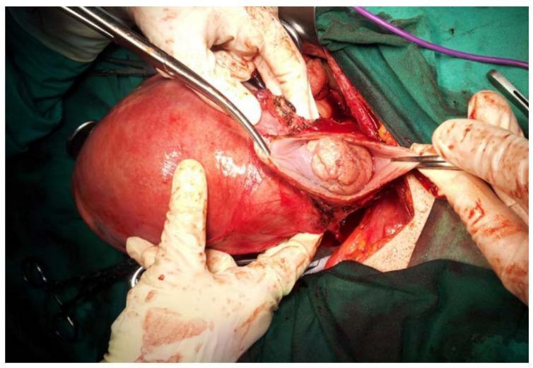
This clinical photograph captures an intraoperative view of an abdominal radical trachelectomy performed on a pregnant patient at 17 weeks of gestation for Stage IB2 cervical cancer. The primary anatomical focus is a significantly enlarged, gravid uterus, which displays a reddish-pink, smooth, glistening serosal surface and takes up the majority of the surgical field. To the right of the uterine corpus, surgical instruments and gloved hands retract tissues to expose the cervical region and paracervical structures. Visible within the dissected operative site is a prominent, pinkish-tan, lobulated mass consistent with the cervical tumor specimen. The image demonstrates the complex surgical anatomy involved in fertility-sparing oncological procedures during pregnancy, highlighting the relationship between the gravid uterus and the neoplastic cervical tissue being excised. The field includes surgical drapes, forceps, and evidence of tissue dissection and hemostasis typical of radical pelvic surgery in a gynecologic oncology context.
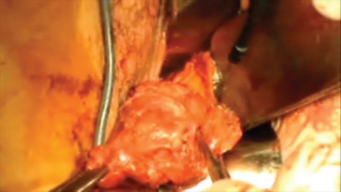
This clinical photograph captures an intraoperative view of a radical vaginal trachelectomy (RVT) for cervical cancer. The central focus is the surgical specimen, which consists of the cervix and attached parametrial tissue. The specimen is predominantly pinkish-red and fleshy, exhibiting a lobulated, irregular texture characteristic of vascularized uterine tissue. Visible surgical instruments, including a metal retractor and a grasping forceps or clamp, are positioned around and within the surgical field to provide exposure and stabilize the tissue for excision. The background shows the surrounding anatomical structures of the vaginal vault and pelvic cavity, which appear moist and hyperemic due to active surgical manipulation. This image illustrates a critical stage in fertility-sparing oncology surgery, emphasizing the resection margins required for treating early-stage cervical adenocarcinoma while preserving the uterine corpus.
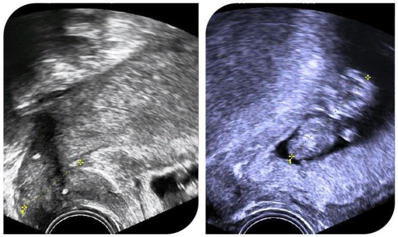
Diagnostic Image: Two side-by-side transvaginal ultrasound frames illustrating a successful intrauterine pregnancy in a patient following radical abdominal trachelectomy. The images demonstrate the pelvic anatomy with a focus on the cervix and lower uterine segment. Key visible features include a hyperechoic (bright) band representing a cerclage suture, positioned at the base of the uterus to provide structural support for the shortened cervix. The left frame shows a measurement of the cervical length (indicated by calipers and a dotted line). The right frame shows a gestational sac containing an embryo/fetus, visualized as a hypoechoic (dark) fluid-filled structure against the heterogeneous echotexture of the uterine myometrium. The educational focus is on the monitoring of cervical competence and the integrity of a prophylactic cerclage in a post-trachelectomy obstetric patient. Medical specialty: Obstetrics and Gynecology, Gynecologic Oncology. Clinical significance: Demonstrates the feasibility of pregnancy and the role of cerclage after radical fertility-sparing surgery for cervical cancer.
https://www.esgo.org/media/2025/05/Pocket-Guidelines_Fertili…
Conservative Methods of Carcinoma Cervix (Fertility-Sparing Treatment)
General Patient Selection Criteria
- Desire for future fertility
- Early FIGO stage (IA1 to IB2)
- Adequate surgical margins feasible
- No lymphovascular space invasion (LVSI) in lower-stage options
- Negative lymph node status (confirmed by sentinel node biopsy or pelvic lymphadenectomy)
- Negative imaging for metastatic disease (MRI/PET-CT)
- Tumor size within specified limits
1. Cryosurgery / Laser Ablation
- Cryosurgery uses a cold metal probe to freeze and destroy abnormal cells
- Laser ablation (CO2 laser) vaporizes abnormal tissue through the vagina
- These are ablative (not excisional), so no specimen for margin assessment
- Not suitable if there is any invasive component or glandular disease
- Recurrence surveillance with close follow-up colposcopy is mandatory
2. LEEP (Loop Electrosurgical Excision Procedure)
- A thin wire loop carries electric current to excise a cone-shaped tissue sample
- Provides a histological specimen (unlike ablation)
- Often diagnostic and therapeutic simultaneously for microinvasive disease
- Adequate when margins are negative; repeat excision needed if margins involved
3. Cervical Conization (Knife Cone Biopsy)
- Conization alone is definitive treatment
- Requires 3 mm negative resection margin
- Risk of pelvic node metastasis is <1%, so lymph node staging is not mandatory
- A large Japanese series of 200 stage IA1 SCC patients treated with laser conization showed zero recurrences at median 117 months follow-up
- Also applied to stage IA1 adenocarcinoma in selected centers
- Conization + pelvic lymph node staging (sentinel node biopsy or pelvic lymphadenectomy)
- If margins are negative and nodes are negative, conization can be sufficient
4. Simple (Non-Radical) Trachelectomy + Lymph Node Staging
- Squamous cell carcinoma (any grade) or adenocarcinoma grade 1-2
- Tumor size ≤2 cm
- No LVSI
- Depth of invasion ≤10 mm
- Conization margins and endocervical curettage negative for malignancy or high-grade dysplasia
- Negative imaging
- Surgical approach: vaginal or laparoscopic
- Isthmus-uterine anastomosis is performed + cerclage placement
- Obstetric outcomes: good, but preterm birth risk is elevated
5. Radical Trachelectomy + Pelvic Lymphadenectomy
Types:
| Approach | Notes |
|---|---|
| Vaginal Radical Trachelectomy (VRT) | Dargent's original technique (1994); vaginal excision of cervix + parametria + sentinel node biopsy |
| Abdominal Radical Trachelectomy (ART) | Recommended for IB2 (2-4 cm); laparotomy or mini-invasive (laparoscopic/robotic); wider parametrial resection |
| Laparoscopic/Robotic | Minimally invasive; used for IB2 via abdominal approach |
Surgical Steps (Radical Trachelectomy):
- Pelvic lymphadenectomy (or sentinel node biopsy) - intraoperative frozen section
- If nodes positive - abandon fertility-sparing plan, proceed to radical hysterectomy or chemoradiation
- Excision of cervix + 1-2 cm of vaginal cuff + parametria
- Anastomosis of uterine isthmus to vaginal cuff
- Permanent cerclage placement (Shirodkar or McDonald)
Mandatory margin requirement:
- Non-fragmented specimen with ≥1 mm histological tumor-free margin from carcinoma or dysplasia
Outcomes:
- Compared with women who underwent conization, radical hysterectomy leads to worse sexual function; radical trachelectomy preserves better quality of life - Berek & Novak's Gynecology
- Pregnancy is achievable post-trachelectomy; prophylactic cerclage supports cervical competence (ultrasound surveillance required)
6. Neoadjuvant Chemotherapy + Conservative Surgery
- Platinum-paclitaxel-based regimens used to downsize tumor
- Followed by conization or simple/radical trachelectomy
- Retrospective data suggest abdominal radical trachelectomy has the lowest recurrence rate for IB2
- Ongoing prospective trials are clarifying validity - not yet standard of care
Summary Table
| Method | FIGO Stage | LVSI | Tumor Size | Lymph Node Staging |
|---|---|---|---|---|
| Laser/Cryoablation | Stage 0 (CIN3/CIS) | No | N/A | No |
| LEEP | Stage 0 / IA1 | Absent | Microinvasive | No (IA1 no LVSI) |
| Conization alone | IA1 without LVSI | Absent | <3 mm invasion | Not required |
| Conization + node staging | IA1 with LVSI, IA2 | Present or absent | <5 mm | Yes |
| Simple trachelectomy | IB1 (low risk) | Absent | ≤2 cm, invasion ≤10 mm | Yes |
| Radical trachelectomy | IB1 (higher risk), IB2 | May be absent | ≤4 cm | Yes (mandatory) |
| NACT + surgery | IB2 | Absent | 2-4 cm | Yes |
Follow-Up After Conservative Treatment
- Pap smear + HPV co-testing every 6 months for 2 years, then annually
- Colposcopy for any abnormality
- MRI if recurrence suspected
- Obstetric surveillance: cervical length monitoring, early delivery planning (most deliver at 34-37 weeks by cesarean)
- Goldman-Cecil Medicine, Chapter 184: "In limited situations in which preservation of fertility is desired, a radical trachelectomy (removal of the cervix only) can be performed." Conization or LEEP may be sufficient for women with microscopic disease.
- Berek & Novak's Gynecology: Nerve-sparing and conservative approaches associated with better sexual function outcomes vs radical hysterectomy.
- ESGO Pocket Guidelines on Fertility-Sparing Treatment in Gynaecological Cancers (2025)
- CancerNetwork - Fertility Preservation in Cervical Cancer
Conization in ca cervix
cervical conization cone biopsy outcomes
cervical conization cone biopsy procedure diagram
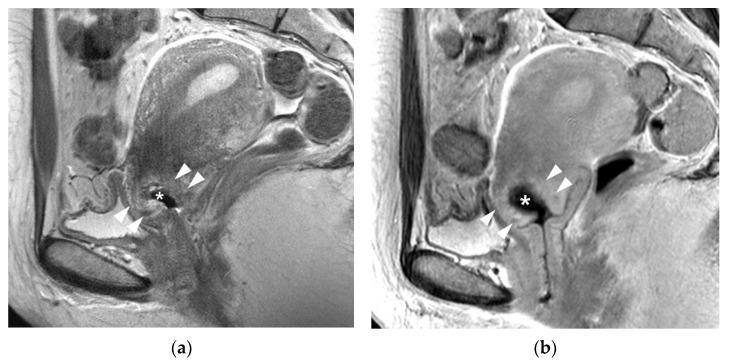
This diagnostic image consists of two sagittal MRI views of the female pelvis in a 42-year-old patient with IA1 cervical cancer, demonstrating post-conization changes. Panel (a) is a T2-weighted sagittal image showing a characteristic cone-shaped tissue defect (marked with an asterisk) at the exocervix following a cervical biopsy. Surrounding the defect, there are poorly demarcated hyperintense regions (indicated by arrowheads) representing post-biopsy inflammation within the cervical stroma. Panel (b) is a delayed contrast-enhanced sagittal image of the same region. It reveals a hematoma (marked with an asterisk) filling the biopsy defect, which appears hypointense compared to the surrounding tissue. The arrowheads in this panel highlight a poorly demarcated zone of enhancement surrounding the defect, further illustrating reactive inflammation. These images are used in oncological radiology to differentiate post-surgical changes, such as inflammation and hematoma, from residual invasive tumor, which in this case was not detected. The visual focuses on gynecological oncology and the utility of multi-sequence MRI in staging and post-operative assessment of early-stage cervical malignancies.
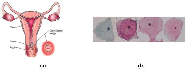
This composite educational figure illustrates the anatomical and cytological basis of cervical screening. Panel (a) is an anatomical diagram of the female reproductive system, displaying the uterus superiorly, the cervix at the mid-level, and the vagina inferiorly. It highlights a 'cone-shaped wedge' representing a cervical biopsy or diagnostic excision area, with a corresponding inset showing the cervical os in a transverse view. Panel (b) presents diagnostic cytology images from the Herlev dataset, showing individual cervical cells under microscopy. The cells demonstrate varying Papanicolaou-style staining patterns ranging from cyan/pale blue to magenta. Key visible features include centrally located, dark-stained nuclei surrounded by expansive, irregularly shaped cytoplasm with distinct polygonal or folded borders. This visual is designed for pathology and gynecology education, emphasizing the transition from macroscopic cervical anatomy to microscopic cellular morphology used in cancer screening and classification.
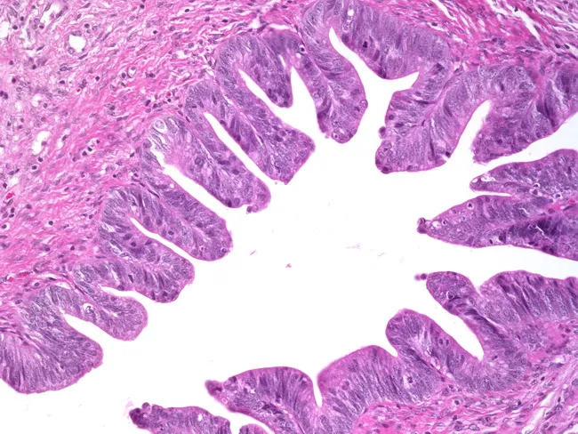
Histopathology, Light microscopy, H&E stained cervical tissue, high-power view. The specimen is a cone biopsy from the cervix containing endocervical glandular epithelium exhibiting adenocarcinoma in situ (AIS) characterized by neoplastic cells lining proliferative glandular papillae, with marked cytologic atypia and frequent apoptotic bodies. The architectural pattern shows slender, arborizing papillary fronds with glandular back-to-back arrangement but without destructive invasion into the stroma. Notable features include nuclear enlargement, hyperchromasia, along with stratified epithelium and mitotic activity confined to the epithelium. In addition, there is an associated squamous intraepithelial lesion (CIN) coexisting with AIS, reflecting multi-lineage cervical neoplasia in a single specimen. The differential diagnosis includes invasive adenocarcinoma, in situ adenocarcinoma with gland crowding, and high-grade CIN with gland involvement; however, stromal invasion is not evident. Clinical significance: AIS with concurrent CIN carries risk of multifocal disease and progression; cone biopsy margins are prognostically important, and negative margins do not eliminate recurrence risk, requiring close follow-up and possibly additional treatment. This image is relevant for pathology education, cervical cancer screening, colposcopy correlation, and research on neoplastic progression in cervical epithelium. Clinical implications include assessing margins, staging risk, guiding surveillance intervals, and discussing fertility-sparing options when appropriate. Educational value for residents and fellows.
Conization in Carcinoma Cervix
Definition
- The entire transformation zone (TZ)
- The squamocolumnar junction (SCJ)
- A portion of the endocervical canal (ECC)
- The entire lesion surrounded by uninvolved (negative) margins
Types of Conization
| Type | Instrument | Notes |
|---|---|---|
| Cold-Knife Conization (CKC) | Scalpel (No. 11 blade) | Gold standard; least thermal artifact; best histological margins |
| LEEP Cone (Loop Conization) | Electrosurgical loop | More accessible; some thermal artifact at margins |
| Laser Conization | CO2 laser | Precise, less bleeding; moderate artifact |
| Cryoconization | Freezing | Rarely used for conization |
Variation in Cone Shape
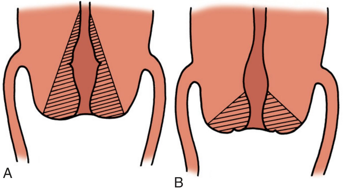
- Premenopausal women: Lesions are mostly ectocervical. Cone should have a broad base and wide angle at the top (Fig A)
- Postmenopausal women: SCJ has retreated into the ECC; lesions are more endocervical. Cone should be long, narrow, with an acute angle at the top (Fig B)
Indications
For Diagnosis:
- Inadequate colposcopic evaluation (lesion not fully visualized)
- Lesion not seen on colposcopy but Pap smear is significantly abnormal
- Incomplete visualization - lesion extends into the ECC
- Squamocolumnar junction not fully visualized (retreated into os)
- Positive endocervical curettings (dysplasia or cancer on ECC)
- Discrepancy between cytology, histology, and colposcopic impression (Pap smear ≥2 grades worse than biopsy)
- Inability to exclude invasive cancer on colposcopy
For Therapy (and/or Staging):
- Cytology/biopsy suggests microinvasive carcinoma (must rule out frank invasion - also potentially therapeutic)
- Stage IA1 without LVSI - conization alone is definitive treatment; <1% lymph node risk
- Stage IA1 with LVSI / Stage IA2 - conization + lymph node staging
- High-grade dysplasia (CIN 2/3) >2 cm or involving >2 quadrants
- Cryotherapy contraindicated or lesion too large for cryotip
- Markedly irregular cervical surface that cryotherapy won't reach
- Glandular involvement on biopsy (relative indication)
- Lesion extends >5 mm into the os
- Non-compliance (patient unlikely to follow up after lesser treatment)
- Correction of cervical stenosis
Contraindications
Absolute:
- Known frank invasive carcinoma (Stage IB+) - conization is insufficient; radical surgery/chemoradiation needed
- Note: Carcinoma in situ is NOT frank invasion - it is severe dysplasia and conization IS appropriate
- For microinvasive cancer, conization is needed to RULE OUT frank invasion
- Contraindications to general or regional anesthesia
- Unstable medical conditions (rarely applicable)
Relative:
- Unstable bleeding disorders
- Inflammatory cervicitis (increases bleeding risk)
- Heavy menses at time of surgery
- Pregnancy - only trained physicians capable of managing complications should proceed
Equipment Required
- Povidone-iodine solution
- Colposcope with green filter
- Acetic acid and Lugol's solution (full strength)
- Vasopressin 20 U in 20 mL normal saline (for intrastromal injection)
- Long scalpel handle with No. 11 blade
- Long fine-tooth forceps
- Kevorkian endocervical curette
- Electrocautery unit
- O-chromic or O-Vicryl suture (Surgicel/Gelfoam/Avitene for hemostasis)
- Large Graves speculum
- Uterine sound
Step-by-Step Procedure (Cold-Knife Conization)
- Anesthesia - General, local (intrastromal), or regional; drain the bladder
- Lesion mapping - Apply full-strength Lugol's solution (Schiller's test); all Lugol's-negative (non-staining) areas must be included in the cone. OR use acetic acid + colposcopy with green filter
- Hemostasis preparation - Circumferentially infiltrate the cervical stroma with vasopressin 20 U in 20 mL saline (intracervical injection - Fig 134-2)
- Sound the uterus - Determine uterine position and size
- Circular incision - Start at the 6 o'clock position (prevents blood obscuring the incision line). Angle the blade centrally toward the cervical canal at the required depth
- Tissue elevation - Use fine-tooth forceps to elevate the cone away from its bed without damaging the epithelium
- Specimen orientation - Mark the 12 o'clock position with a single suture into the stroma for the pathologist
- Measure the specimen before fixation (it shrinks before the pathologist measures it)
- ECC curettage - Curette the remaining ECC above the upper margin of the cone to rule out residual disease
- D&C - If indicated, perform at this time
- Hemostasis - Superficial electrocoagulation (ball electrode), suture ligatures, or absorbable gelatin sponge (Gelfoam) packing
- Fix specimen - Send cone in fixative for pathologic examination
Margin Assessment and Oncological Adequacy
| Margin Status | Action |
|---|---|
| Negative margins (≥3 mm) | Conization is therapeutic for Stage IA1 without LVSI; close surveillance |
| Positive ectocervical margins (dysplasia only) | Close follow-up; repeat cone usually NOT required - inflammation during healing often resolves lesion |
| Positive ECC above excisional site (high-grade) | Repeat conization should be considered |
| Both margins AND ECC positive | Repeat conization strongly considered; especially in older, high-risk patients |
Role of Conization by FIGO Stage
| FIGO Stage | Role of Conization |
|---|---|
| CIN 2/3 (Stage 0 / preinvasive) | Therapeutic (primary treatment) |
| Stage IA1, no LVSI | Definitive therapy if margins clear; no node staging needed |
| Stage IA1 with LVSI | Conization + sentinel node biopsy/pelvic lymphadenectomy |
| Stage IA2 | Conization + lymph node staging; simple trachelectomy if conization margins inadequate |
| Stage IB1 (≤2 cm, ConCerv criteria) | Conization for staging; simple trachelectomy if margins involved |
| Stage IB1 (>2 cm) / IB2+ | Conization alone is INSUFFICIENT; radical trachelectomy or hysterectomy required |
| Stage IIB+ | Conization has NO therapeutic role; chemoradiation is treatment |
Complications
| Complication | Notes |
|---|---|
| Pain and cramping | Generally minimal |
| Immediate hemorrhage | Inject 1 mL lidocaine-epinephrine at bleeding site; electrocoagulation; rarely figure-of-8 suture |
| Delayed hemorrhage | Eschar sloughs at 7-10 days; some spotting expected for 10-14 days |
| Cervical stenosis | <3 mm opening; prevents menses or adequate Pap smear sampling |
| Uterine perforation | Rare |
| Pelvic cellulitis / cervicitis | Very rare |
| Bladder or rectal damage | Especially in significant vaginal atrophy with shallow fornices |
| Cervical incompetence | Risk of preterm birth in future pregnancies |
| Infertility | Loss of mucus-producing endocervical glands |
| Positive margins / missing lesion | Rare |
- Fetal loss rate: 10% (rupture of membranes, preterm labor, hemorrhage)
- Postoperative hemorrhage rate: 30%
Post-Procedure Management
- Follow-up appointment at 4-6 weeks
- Avoid intercourse, douching, tampon use until healing confirmed at follow-up
- Notify clinician of: fever, excessive vaginal bleeding, purulent discharge
- First follow-up Pap smear: 3-4 months after procedure if all margins are clear
- Subsequent surveillance: Pap + HPV co-testing every 6 months for 2 years, then annually
Histopathology Images
- Pfenninger & Fowler's Procedures for Primary Care, 3rd Ed - Full procedural protocol
- Creasy & Resnik's Maternal-Fetal Medicine - Conization in pregnancy
- Goldman-Cecil Medicine - Role of conization in cervical cancer staging/treatment
- Histology: A Text and Atlas (Mescher) - Cervical cancer and HPV context
- ESGO Pocket Guidelines: Fertility-Sparing Treatment in Gynaecological Cancers (2025) - Margin requirements and stage-specific recommendations