


Painful and Itchy flakes on hands dorsal and ventral sides for 6 months
chronic hand eczema hyperkeratotic scaling palmar
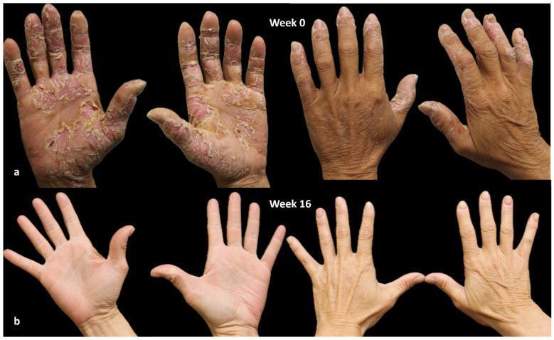
Clinical photograph comparison of chronic hand eczema (CHE) at baseline (Week 0) and 16 weeks post-treatment with dupilumab. Row A (Week 0) displays severe dermatological manifestations with a Hand Eczema Severity Index (HECSI) of 270. Key features include diffuse erythema, thick hyperkeratotic plaques, extensive scaling, and painful fissuring. The palmar surfaces are most affected, showing yellowish, cracked lesions and desquamation, particularly on the thumbs and central palms. The dorsal surfaces exhibit milder scaling and lichenification. Row B (Week 16) demonstrates a dramatic clinical response with a HECSI score of 0. There is a complete resolution of active lesions, erythema, and scaling. The skin texture has returned to a normal, supple appearance with uniform pigmentation and restored epidermal integrity across both palmar and dorsal aspects. This comparison illustrates the efficacy of biologic therapy in managing severe, refractory chronic hand eczema by targeting underlying inflammatory pathways.
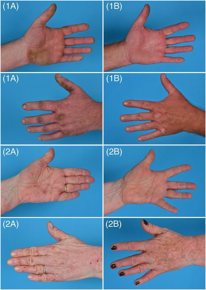
This set of clinical photographs demonstrates the therapeutic progression of two patients with chronic hand eczema (CHE) over a 16-week period. The images are organized into Case 1 and Case 2, displaying both palmar and dorsal views at baseline (A) and follow-up (B). Case 1 (1A) illustrates severe hyperkeratotic CHE, characterized by thick, yellowish, calloused plaques on the palm and fingers with associated erythema. By week 16 (1B), the hyperkeratosis has nearly resolved, showing smooth skin with minimal residual redness. Case 2 (2A) shows severe atopic CHE with diffuse erythema, scaling, and excoriations on the palmar surface, and erythematous papules and lichenification on the dorsal side. At week 16 (2B), there is a significant reduction in active inflammation and scaling on the palm, while the dorsal view shows post-inflammatory hyperpigmentation and clearance of acute lesions. The comparison highlights the efficacy of systemic treatment in managing severe, treatment-resistant hand eczema, focusing on the reduction of hyperkeratosis, erythema, and scaling to improve patient quality of life.
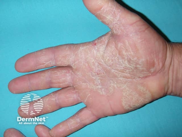
This clinical photograph documents a palmar dermatitic pattern characterized by diffuse xerosis with hyperkeratotic scales on the palmar surfaces of the hand. The epidermis shows thickened, white, scaly plaques concentrated along the mid- and thenar/hypothenar regions and intermetacarpal folds. Fine fissuring is present at several palmar creases, and the skin texture is rough with reduced pliability. The dorsum of the hand appears relatively unaffected. There is mild erythema absent or minimal, and no vesiculation or purpura is seen. The pattern is compatible with chronic hand dermatitis (eczema) or palmoplantar hyperkeratosis; psoriasis could mimic this presentation but typically features thicker plaques with erythema and more silvery scales, whereas eczema shows dryness, fissuring, and scaling. Secondary infection is not evident. The image highlights the importance of emollient-based therapy, barrier restoration, and topical anti-inflammatory agents in management. This image could be used to illustrate palmar eczema in dermatology education, serve as a reference for differential diagnosis in hand dermatoses, and support treatment response monitoring in clinical practice. It is relevant for teledermatology consultations and research on palmar skin diseases.
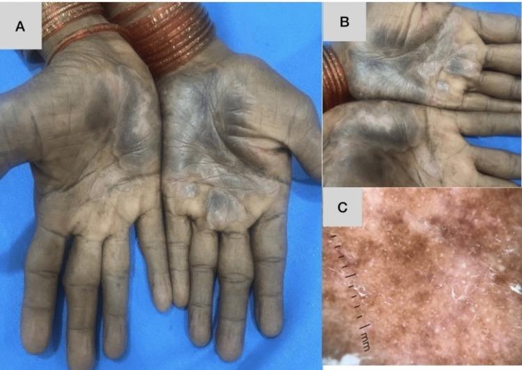
This composite educational image illustrates a case of chronic hand eczema with a clinical appearance mimicking palmoplantar psoriasis. Panel A (Clinical Photograph) displays the palmar surfaces of both hands, showing bilaterally symmetrical, hyperkeratotic, and hyperpigmented patches. The lesions are primarily concentrated in the central palmar region and extend along the flexural creases of the fingers, exhibiting a thick, scaly texture on a background of skin darkening. Panel B (Dermoscopic Image) provides a magnified view of the palmar skin, highlighting the texture and vascular alterations. It shows differentiated vascular structures, including linear and branched vessels, which are often found in psoriasiform eruptions. Panel C (High-Magnification Dermoscopy) reveals a light red-to-brownish background with regularly spaced red dots and occasional globules. Fine, white powdery scaling is visible across the surface. This case demonstrates the clinical and dermoscopic overlap between hyperkeratotic eczema and palmoplantar psoriasis, emphasizing the role of histopathology for definitive diagnosis in complex dermatological presentations.
tinea manuum fungal infection hand hyperkeratosis
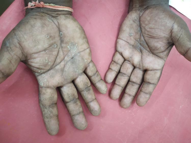
Clinical photograph showing a dermatological manifestation on the palmar and distal dorsal surfaces of bilateral hands, diagnosed as tinea manuum. The image reveals diffuse palmar involvement characterized by xerosis, hyperkeratosis, and fine scaling, particularly concentrated along the palmar creases and across the fingers. Multiple discrete, nummular, well-demarcated scaly plaques are scattered across the palms. There is evidence of mottled dyschromia, with intermixed areas of hyperpigmentation and hypopigmentation. The skin texture appears rough and thickened, with some loss of the normal dermatoglyphic pattern and accentuation of skin lines. The lesions extend from the central palmar region to the volar and lateral aspects of the digits, demonstrating a pattern consistent with chronic fungal infection or hyperkeratotic palmar dermatosis. This visual is relevant for medical education in dermatology, specifically for differentiating between tinea manuum, palmar psoriasis, and chronic hand eczema.
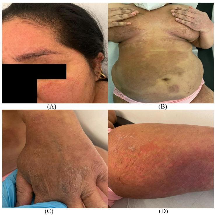
This composite clinical photograph displays widespread dermatophytosis (tinea) across four anatomical regions: the face (A), trunk (B), hand (C), and groin/thigh (D). The lesions are characterized by large, erythematous, scaly plaques with distinctive sharp, slightly raised borders. Image A (tinea faciei) shows confluent red patches on the forehead and cheeks. Image B (tinea corporis) demonstrates extensive, multi-colored annular plaques covering the abdomen and submammary regions. Image C (tinea manuum) illustrates diffuse hyperkeratosis and scaling on the dorsal hand. Image D (tinea cruris) reveals a prominent, intensely erythematous plaque with active scaling at the margins on the inner thigh. These clinical features represent a severe and extensive presentation of superficial fungal infection, typical of multidrug-resistant dermatophyte strains like Trichophyton indotineae. The distribution pattern highlights the progressive nature of the infection from localized to disseminated cutaneous involvement.
Clinical Assessment: Chronic Hyperkeratotic Hand Dermatosis (6 months)
Disclaimer: This is an AI-assisted educational analysis, not a substitute for in-person dermatological evaluation. The findings below should guide a consultation with a dermatologist.
What the Images Show
- Diffuse hyperkeratosis - marked thickening and leathery texture across the entire palm and digits
- Coarse scaling and desquamation - thick, yellowish-brown flakes peeling off
- Lichenification - accentuated skin creases from chronic rubbing/inflammation
- Painful fissures - linear cracks in palmar creases and finger joints
- Mild underlying erythema visible in cracked areas
- Bilateral and diffuse distribution involving both dorsal and ventral (palmar/volar) surfaces of the hands
Top Differential Diagnoses
"The eruption presents as hyperkeratotic, fissure-prone, erythematous areas of the middle or proximal palm. Vesicles are not seen. The volar surfaces of the fingers may also be involved. Males outnumber females by 2:1, and the patients are usually older adults. Histologically, the lesions show chronic spongiotic dermatitis."
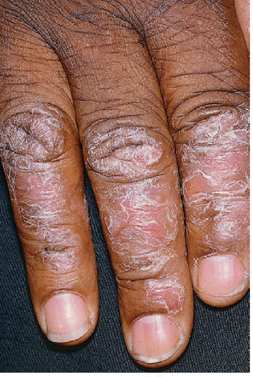
- Sharply demarcated plaques with silvery scale
- Nail pitting or onycholysis
- Occasional pustules (especially in the palmoplantar pustular variant)
- Lesions on elbows, knees, scalp, or navel
- Family history of psoriasis
- Irritant: from repeated exposure to water, detergents, soaps, chemicals (common in cleaners, healthcare workers, construction)
- Allergic: delayed hypersensitivity to rubber gloves, nickel, fragrances, epoxy resins
- The bilateral, symmetric distribution could suggest irritant exposure more than allergic
- Typically unilateral ("two feet, one hand" syndrome)
- Causes diffuse fine scaling along palmar creases
- Bilateral presentation (as seen here) makes this less likely, but cannot be excluded without a KOH prep

- Acute phase = deep-seated itchy vesicles on sides of fingers
- Chronic phase = exactly what is seen here: hyperkeratotic, scaling, fissured palms
- The predominant itch symptom supports this
Comparison with Chronic Hand Eczema Reference Image
Recommended Workup
| Test | Purpose |
|---|---|
| KOH skin scraping | Rule out tinea manuum (fungal) - quick, painless, done in clinic |
| Patch testing | If allergic contact dermatitis suspected - identifies specific chemical triggers |
| Skin biopsy | If psoriasis vs. eczema cannot be distinguished clinically |
| Nail examination | Look for pitting, onycholysis (favors psoriasis) |
Treatment (Pending Diagnosis Confirmation)
- Barrier repair: Apply heavy emollient (white petrolatum/Vaseline) after each handwash, especially at night. Occlude with cotton gloves or a plastic bag for better penetration - Andrews' Diseases of the Skin, p.95
- Avoid triggers: Minimize wet work, use vinyl gloves for cleaning, wear cotton gloves underneath
- Avoid soaps/detergents on hands; use mild, fragrance-free cleansers
- First-line: Potent/ultrapotent topical corticosteroids (e.g., clobetasol 0.05%) - enhanced by soak and smear technique
- For refractory cases: Coal tar preparations (20% LCD in ointment), oral retinoids (acitretin or alitretinoin - ~50% clearance in hyperkeratotic hand eczema at 6+ months), PUVA phototherapy
- If psoriasis confirmed: Topical calcipotriol, systemic agents (methotrexate, biologics)
- If fungal confirmed: Topical terbinafine or oral antifungal (fluconazole/itraconazole)
Key Questions for Your Dermatologist
- Does the rash affect your feet, scalp, elbows, or knees as well?
- Do you have nail changes (pitting, lifting, brown discoloration)?
- Do you work with chemicals, water, or wear gloves regularly?
- Did it start with small blisters on the sides of your fingers?
- Does anyone in your family have psoriasis or eczema?
- Have you tried any creams - and did they help?
- Andrews' Diseases of the Skin - Clinical Dermatology, pp. 94-96 (Hand Eczema section)