
Describe the structure and function of the vertebral column. Biomechanics essay
vertebral column structure function biomechanics anatomy
Structure and Function of the Vertebral Column: A Biomechanics Essay
1. Overview and Evolutionary Context
2. Gross Structure
2.1 Vertebral Regions and Numbers
| Region | Number | Key features |
|---|---|---|
| Cervical | 7 | Smallest, most mobile; transverse foramina for vertebral arteries |
| Thoracic | 12 | Articulate with ribs; costal facets on bodies and transverse processes |
| Lumbar | 5 | Largest bodies; bear the greatest compressive load |
| Sacral | 5 (fused) | Transmit body weight to the pelvic girdle |
| Coccygeal | 3-4 (fused) | Vestigial tail; attachment for pelvic floor muscles |
2.2 The Typical Vertebra
- Vertebral body (anterior): The weight-bearing cylinder of cancellous bone enclosed in a cortical shell. It bears the majority of axial compressive force and provides attachment for the intervertebral disc above and below.
- Vertebral arch (posterior): Formed by two pedicles and two laminae, it encloses the vertebral foramen. Together the stacked foramina create the vertebral (spinal) canal, housing and protecting the spinal cord.
3. The Intervertebral Disc
- Annulus fibrosus: An outer ring of collagen fibres surrounding a wider zone of fibrocartilage arranged in a lamellar configuration, with successive lamellae oriented at approximately ±30° to the disc's transverse plane. This alternating oblique arrangement resists torsional (rotational) forces in both directions.
- Nucleus pulposus: A gelatinous central core composed of type II collagen and proteoglycans that interact with water to resist compressive stress. Intradiscal pressure is highest during flexion, which explains why patients with disc herniation find forward flexion most uncomfortable (Firestein & Kelley's Textbook of Rheumatology).
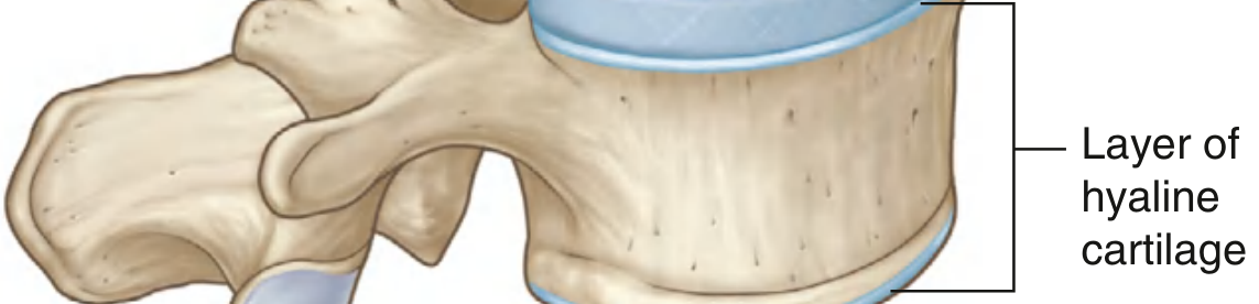
4. Joints of the Vertebral Column
4.1 Zygapophysial (Facet) Joints
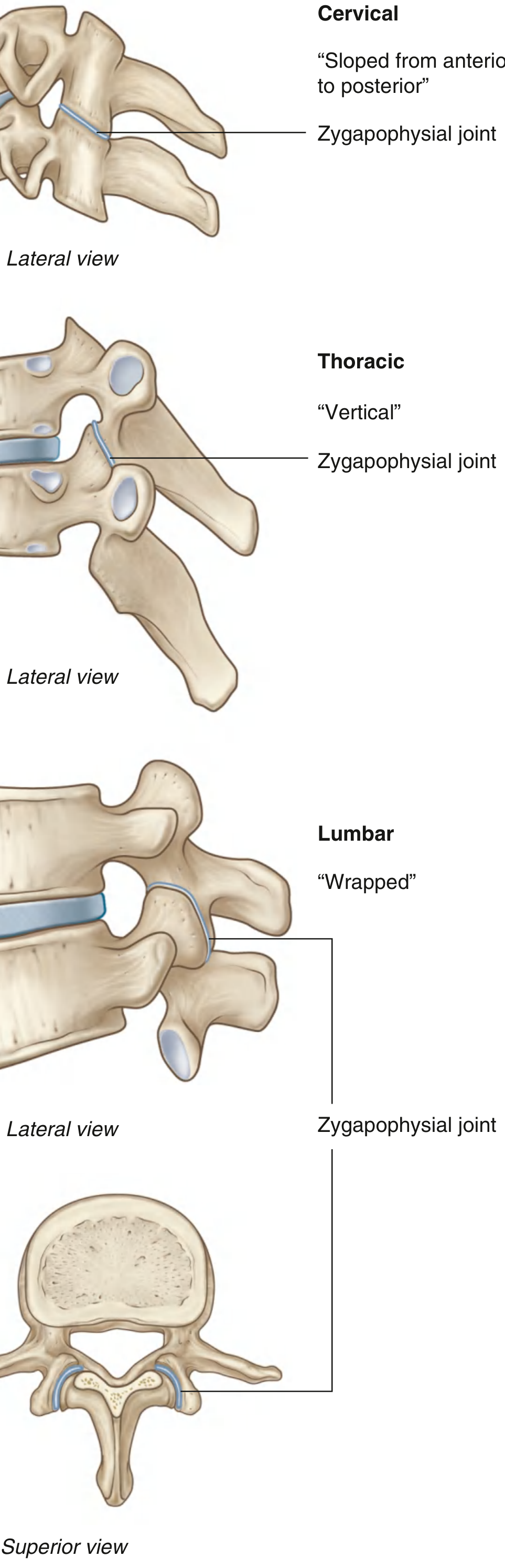
- Cervical: Slope inferiorly from anterior to posterior, facilitating flexion and extension.
- Thoracic: Oriented vertically (near the frontal plane), limiting flexion and extension but permitting rotation - biomechanically appropriate given the thoracic cage's role in respiration.
- Lumbar: Curved, "wrapped" surfaces where adjacent processes interlock, limiting rotation considerably but still permitting substantial flexion and extension.
4.2 Uncovertebral (Luschka) Joints
5. Ligamentous Support
| Ligament | Attachment | Biomechanical role |
|---|---|---|
| Anterior longitudinal ligament (ALL) | Anterior aspect of vertebral bodies and discs | Resists hyperextension |
| Posterior longitudinal ligament (PLL) | Posterior aspect of vertebral bodies within canal | Resists hyperflexion; reinforces disc centrally but absent posterolaterally |
| Ligamentum flavum | Joins laminae of adjacent vertebrae | Resists flexion; high elastin content restores neutral posture; buckles with degeneration causing canal stenosis |
| Interspinous ligament | Between spinous processes | Resists flexion separation |
| Supraspinous ligament | Tips of spinous processes (nuchal ligament in neck) | Limits extreme flexion |
6. Spinal Curvatures
- Cervical lordosis (concave posteriorly) - secondary curvature; develops when infants first lift their heads
- Thoracic kyphosis (concave anteriorly) - primary curvature; present at birth; compensatory mechanism to maintain a level line of sight and increase thoracic cavity volume
- Lumbar lordosis (concave posteriorly) - secondary curvature; develops when walking begins
- Sacral kyphosis - primary curvature; fixed
7. Kinematics and Range of Motion
- Flexion - anterior tilting and gliding of the superior vertebra; widens the intervertebral foramen; compresses the anterior disc and stretches the posterior ligaments
- Extension - reversal of the above; narrows the foramen; compresses the posterior disc and ligamentum flavum
- Lateral flexion - coupled with rotation in most spinal regions
- Axial rotation - most pronounced in the thoracic spine given vertical facet orientation
- Circumduction - composite movement combining all of the above
Regional movement summaries:
| Region | Dominant movement | Limited movement | Basis |
|---|---|---|---|
| Cervical | Flexion/extension, rotation | - | Angled facets, uncovertebral joints |
| Thoracic | Rotation | Flexion/extension | Vertical facets, rib cage constraint |
| Lumbar | Flexion/extension | Rotation | Interlocking facets |
8. Load Transmission and Biomechanical Forces
- Axial compression is borne primarily by the vertebral bodies and discs (approximately 80% through the anterior column at neutral posture). The nucleus pulposus, being nearly incompressible, converts axial load into radial tension within the annulus fibrosus - a hydrostatic pressure mechanism.
- Shear and torsion are resisted by the facet joints, the oblique lamellae of the annulus fibrosus, and the ligaments.
- Bending moments generated by loads anterior to the spine (e.g., carrying an object, forward bending) are resisted by the posterior ligamentous complex and the erector spinae group, which must generate a large counterforce due to their short moment arm relative to the spine's centre of rotation. This amplification means that a 10 kg load held at arm's length can generate hundreds of newtons of compressive force on lumbar discs - explaining the high prevalence of degenerative lumbar disease.
9. Protective Function: The Vertebral Canal
10. Clinical Correlations
| Condition | Structural basis | Biomechanical mechanism |
|---|---|---|
| Disc herniation | Annular tear, usually posterolateral | Sustained compression or repetitive flexion-torsion loading raises intradiscal pressure; nucleus pulposus extruded through weakest annular zone |
| Lumbar spinal stenosis | Ligamentum flavum hypertrophy, facet joint osteoarthritis, osteophytes | Degenerative narrowing of central canal or lateral recesses; aggravated by extension (decreased canal diameter) |
| Spondylolysis / spondylolisthesis | Stress fracture of pars interarticularis | Repetitive hyperextension with axial loading (common in gymnasts, fast bowlers); anterior vertebral body slippage when bilateral |
| Osteoporotic fracture | Loss of trabecular bone in vertebral body | Failure under normal compressive loads; risk quantified by DXA scanning |
| Scoliosis | Lateral curvature with rotational component | Abnormal coupling patterns; assessed clinically with Adams forward bending test |
Summary
- Gray's Anatomy for Students - Joints between vertebrae, zygapophysial joints, disc herniation
- Firestein & Kelley's Textbook of Rheumatology - Anatomy, disc mechanics, ligaments
- Rheumatology, 2-Volume Set - Mechanical function of the spine, spinal curvature and shock absorption
- General Anatomy and Musculoskeletal System (THIEME Atlas) - Evolutionary biomechanics, bipedal adaptation
- Grainger & Allison's Diagnostic Radiology - Ligament anatomy
Summarise and generate word document
ls -la /tmp/workspace/ 2>/dev/null || mkdir -p /tmp/workspace && echo "created"
mkdir -p /tmp/workspace/vertebral-column && cat > /tmp/workspace/vertebral-column/generate_doc.py << 'PYEOF' from docx import Document from docx.shared import Pt, Inches, RGBColor from docx.enum.text import WD_ALIGN_PARAGRAPH from docx.oxml.ns import qn from docx.oxml import OxmlElement doc = Document() # ── Page margins ────────────────────────────────────────────────────────────── from docx.shared import Cm section = doc.sections[0] section.top_margin = Cm(2.5) section.bottom_margin = Cm(2.5) section.left_margin = Cm(3) section.right_margin = Cm(2.5) # ── Default body style ──────────────────────────────────────────────────────── normal = doc.styles["Normal"] normal.font.name = "Calibri" normal.font.size = Pt(11) # ── Helper: add a styled paragraph ──────────────────────────────────────────── def body(text, bold=False, italic=False, space_before=0, space_after=6): p = doc.add_paragraph() p.paragraph_format.space_before = Pt(space_before) p.paragraph_format.space_after = Pt(space_after) p.paragraph_format.line_spacing = Pt(16) run = p.add_run(text) run.bold = bold run.italic = italic run.font.name = "Calibri" run.font.size = Pt(11) return p def add_table(headers, rows): table = doc.add_table(rows=1 + len(rows), cols=len(headers)) table.style = "Table Grid" # header row hdr_cells = table.rows[0].cells for i, h in enumerate(headers): hdr_cells[i].text = h run = hdr_cells[i].paragraphs[0].runs[0] run.bold = True run.font.size = Pt(10) run.font.name = "Calibri" # shading tc = hdr_cells[i]._tc tcPr = tc.get_or_add_tcPr() shd = OxmlElement("w:shd") shd.set(qn("w:val"), "clear") shd.set(qn("w:color"), "auto") shd.set(qn("w:fill"), "1F3864") tcPr.append(shd) for para in hdr_cells[i].paragraphs: for r in para.runs: r.font.color.rgb = RGBColor(0xFF, 0xFF, 0xFF) for row_data in rows: row_cells = table.add_row().cells for i, val in enumerate(row_data): row_cells[i].text = val for para in row_cells[i].paragraphs: for r in para.runs: r.font.size = Pt(10) r.font.name = "Calibri" doc.add_paragraph() # spacing after table # ══════════════════════════════════════════════════════════════════════════════ # TITLE PAGE # ══════════════════════════════════════════════════════════════════════════════ p_title = doc.add_paragraph() p_title.alignment = WD_ALIGN_PARAGRAPH.CENTER p_title.paragraph_format.space_before = Pt(60) p_title.paragraph_format.space_after = Pt(8) r = p_title.add_run("Structure and Function of the Vertebral Column") r.bold = True r.font.size = Pt(22) r.font.name = "Calibri" r.font.color.rgb = RGBColor(0x1F, 0x38, 0x64) p_sub = doc.add_paragraph() p_sub.alignment = WD_ALIGN_PARAGRAPH.CENTER p_sub.paragraph_format.space_after = Pt(6) r2 = p_sub.add_run("A Biomechanics Essay") r2.bold = True r2.italic = True r2.font.size = Pt(14) r2.font.name = "Calibri" r2.font.color.rgb = RGBColor(0x2E, 0x74, 0xB5) p_date = doc.add_paragraph() p_date.alignment = WD_ALIGN_PARAGRAPH.CENTER r3 = p_date.add_run("July 2026") r3.font.size = Pt(11) r3.font.name = "Calibri" r3.font.color.rgb = RGBColor(0x70, 0x70, 0x70) doc.add_page_break() # ══════════════════════════════════════════════════════════════════════════════ # SECTION 1 — Overview # ══════════════════════════════════════════════════════════════════════════════ doc.add_heading("1. Overview and Evolutionary Context", level=1) body( "The vertebral column is the defining structural feature of all vertebrates. In humans it has been " "fundamentally remodelled to support habitual bipedal locomotion. The chimpanzee spine follows a simple " "'arch and cord' construction; the human spine instead forms a double-S-shaped curve, allowing the axial " "skeleton to act as a shock-absorbing spring while shifting body weight over the feet. This redesign also " "required the pelvis to broaden and the iliac wings to spread, creating a basin for the abdominal viscera " "that were formerly supported by the anterior abdominal wall in a quadruped posture." ) body("Source: General Anatomy and Musculoskeletal System (THIEME Atlas of Anatomy)", italic=True) # ══════════════════════════════════════════════════════════════════════════════ # SECTION 2 — Gross Structure # ══════════════════════════════════════════════════════════════════════════════ doc.add_heading("2. Gross Structure", level=1) doc.add_heading("2.1 Vertebral Regions", level=2) body( "The adult vertebral column comprises approximately 33 vertebrae in five regions. Vertebral bodies " "progressively increase in cross-sectional area from C2 to L5, directly reflecting the cumulative " "increase in compressive load each segment must bear." ) add_table( ["Region", "Number", "Key Structural Features"], [ ["Cervical", "7", "Smallest; most mobile; transverse foramina transmit vertebral arteries"], ["Thoracic", "12", "Costal facets on bodies and transverse processes articulate with ribs"], ["Lumbar", "5", "Largest bodies; bear greatest compressive load"], ["Sacral", "5 (fused)","Transmit body weight to pelvic girdle via sacroiliac joints"], ["Coccygeal", "3-4 (fused)","Vestigial tail; attachment for pelvic floor muscles"], ] ) doc.add_heading("2.2 The Typical Vertebra", level=2) body( "Each vertebra has two functional parts:" ) body( "Vertebral body (anterior): A cylinder of cancellous bone within a cortical shell. It bears approximately " "80% of axial compressive load at neutral posture and provides the superior and inferior endplates for " "disc attachment.", bold=False ) body( "Vertebral arch (posterior): Two pedicles and two laminae enclose the vertebral foramen. The stacked " "foramina form the vertebral canal, housing the spinal cord. Seven processes project from the arch: " "one spinous (posteriorly), two transverse (laterally), and four articular processes that form " "zygapophysial joints with adjacent vertebrae.", bold=False ) # ══════════════════════════════════════════════════════════════════════════════ # SECTION 3 — Intervertebral Disc # ══════════════════════════════════════════════════════════════════════════════ doc.add_heading("3. The Intervertebral Disc", level=1) body( "The intervertebral disc (IVD) is the primary load-distributing structure between vertebral bodies. " "It is a fibrocartilaginous symphysis composed of two concentric zones:" ) body( "Annulus fibrosus: An outer ring of collagen, surrounding a wider zone of fibrocartilage arranged in " "concentric lamellae. Successive lamellae are oriented at approximately +30 degrees and -30 degrees " "to the disc's transverse plane. This alternating oblique arrangement resists torsional forces in both " "directions simultaneously - a key anti-rotation mechanism.", bold=False ) body( "Nucleus pulposus: A gelatinous core of type II collagen and proteoglycans. The highly hydrophilic " "proteoglycans attract and bind water, creating turgor pressure. The nucleus behaves as an incompressible " "fluid: axial compressive load is converted into radial tension (hoop stress) within the annulus fibrosus, " "distributing force across the entire disc rather than concentrating it centrally.", bold=False ) body( "Intradiscal pressure is highest during flexion, explaining why disc herniation patients find forward " "bending most painful. With ageing, the nucleus progressively loses water content, disc height decreases, " "annular tears occur, and herniation risk rises - typically posterolaterally where the posterior " "longitudinal ligament is absent and the annulus is thinnest.", space_before=4 ) body("Source: Gray's Anatomy for Students; Firestein & Kelley's Textbook of Rheumatology", italic=True) # ══════════════════════════════════════════════════════════════════════════════ # SECTION 4 — Joints # ══════════════════════════════════════════════════════════════════════════════ doc.add_heading("4. Joints of the Vertebral Column", level=1) doc.add_heading("4.1 Zygapophysial (Facet) Joints", level=2) body( "The synovial zygapophysial joints between superior and inferior articular processes of adjacent " "vertebrae are the primary determinants of segmental movement direction. Their orientation varies " "systematically by region:" ) add_table( ["Region", "Facet Orientation", "Movement Facilitated", "Movement Restricted"], [ ["Cervical", "Sloped infero-posteriorly", "Flexion and extension", "Minimal restriction"], ["Thoracic", "Vertical (near frontal plane)", "Axial rotation", "Flexion and extension"], ["Lumbar", "Curved, interlocking ('wrapped')", "Flexion and extension", "Axial rotation"], ] ) doc.add_heading("4.2 Uncovertebral (Luschka) Joints", level=2) body( "Unique to cervical vertebrae C3-C7, uncinate processes on the lateral margins of the superior " "vertebral surface articulate with the body above, forming small synovial uncovertebral joints. " "They guide lateral bending and are clinically important because osteophytes at these joints can " "narrow the intervertebral foramina and compress cervical nerve roots." ) body("Source: Gray's Anatomy for Students", italic=True) # ══════════════════════════════════════════════════════════════════════════════ # SECTION 5 — Ligaments # ══════════════════════════════════════════════════════════════════════════════ doc.add_heading("5. Ligamentous Support", level=1) body( "Five principal ligaments reinforce the vertebral column and constrain movement:" ) add_table( ["Ligament", "Location", "Biomechanical Role"], [ ["Anterior longitudinal ligament (ALL)", "Anterior surfaces of bodies and discs", "Resists hyperextension"], ["Posterior longitudinal ligament (PLL)", "Posterior surfaces of bodies within the canal", "Resists hyperflexion; absent posterolaterally at disc level"], ["Ligamentum flavum", "Joins laminae of adjacent vertebrae", "Resists flexion; high elastin content restores neutral posture; hypertrophy causes stenosis"], ["Interspinous ligament", "Between spinous processes", "Resists flexion separation"], ["Supraspinous ligament", "Tips of spinous processes (nuchal ligament in cervical region)", "Limits extreme flexion"], ] ) body("Source: Firestein & Kelley's Textbook of Rheumatology", italic=True) # ══════════════════════════════════════════════════════════════════════════════ # SECTION 6 — Spinal Curvatures # ══════════════════════════════════════════════════════════════════════════════ doc.add_heading("6. Spinal Curvatures", level=1) body( "The adult spine has four sagittal curvatures. The two primary curvatures (thoracic kyphosis and sacral " "kyphosis) are present at birth. The two secondary curvatures (cervical lordosis and lumbar lordosis) " "develop postnatally in response to loading:" ) body("- Cervical lordosis develops when infants first lift their heads.") body("- Lumbar lordosis develops when walking begins.") body("- Thoracic kyphosis is a compensatory mechanism to maintain a level line of sight and increase thoracic cavity volume.") body( "The S-shaped curvature confers a decisive mechanical advantage: a curved column resists axial " "compressive loads approximately ten times better than a straight column of equivalent mass, because " "bending is recruited to dissipate energy. During locomotion, at heel strike the cervical and lumbar " "lordoses both increase; at toe-off they decrease. This cyclical deformation stores and releases energy " "in the discs, ligaments, and paraspinal muscle tendons, functioning as a passive shock-absorption system.", space_before=4 ) body("Source: Rheumatology, 2-Volume Set (Elsevier, 2022)", italic=True) # ══════════════════════════════════════════════════════════════════════════════ # SECTION 7 — Kinematics # ══════════════════════════════════════════════════════════════════════════════ doc.add_heading("7. Kinematics and Range of Motion", level=1) body( "Movement at any single intervertebral segment is small, but the summation across all segments produces " "a large composite range of motion. The vertebral column performs five principal movements: flexion, " "extension, lateral flexion, axial rotation, and circumduction." ) body( "Coupling is a fundamental biomechanical principle: at the segmental level, motions are not fully " "independent. Lateral flexion and axial rotation are consistently coupled throughout the cervical and " "thoracic regions. The direction and magnitude of coupling depend on spinal posture, regional curvature, " "disc fluidity, and facet orientation.", space_before=4 ) add_table( ["Region", "Dominant Movement", "Restricted Movement", "Structural Basis"], [ ["Cervical", "Flexion/extension, rotation", "None major", "Angled facets; uncovertebral joints guide movement"], ["Thoracic", "Axial rotation", "Flexion/extension", "Vertical facets; rib cage adds circumferential stiffness"], ["Lumbar", "Flexion/extension", "Axial rotation", "Interlocking curved facets block rotation"], ] ) body("Source: Gray's Anatomy for Students", italic=True) # ══════════════════════════════════════════════════════════════════════════════ # SECTION 8 — Load Transmission # ══════════════════════════════════════════════════════════════════════════════ doc.add_heading("8. Load Transmission and Biomechanical Forces", level=1) body( "The vertebral column transmits cumulative superincumbent body weight from the skull to the sacrum. " "Load is shared between the anterior and posterior columns:" ) body( "Axial compression (~80% through the anterior column at neutral posture) is borne by the vertebral " "bodies and discs. The incompressible nucleus pulposus converts axial load into radial hoop tension " "within the annulus - a hydraulic pressure mechanism that distributes force uniformly." ) body( "Shear and torsional forces are resisted by the facet joints, the oblique lamellae of the annulus " "fibrosus, and the posterior ligamentous complex." ) body( "Bending moments generated by loads anterior to the spine (e.g., carrying a load at arm's length, " "forward bending) must be counteracted by the erector spinae muscles. Because these muscles have a " "very short moment arm relative to the spinal centre of rotation, they must generate forces many times " "the external load. A 10 kg object held at arm's length can produce hundreds of newtons of compressive " "force at L4/L5 - a key reason for the high prevalence of lumbar degenerative disease.", space_before=4 ) body( "The muscular stabilisation system operates on two levels: the global muscles (erector spinae, " "quadratus lumborum) control gross movement and resist large external moments; the deep local muscles " "(multifidus, transversus abdominis) co-contract to stiffen individual spinal segments, providing " "intersegmental stability independent of movement direction." ) # ══════════════════════════════════════════════════════════════════════════════ # SECTION 9 — Protective Function # ══════════════════════════════════════════════════════════════════════════════ doc.add_heading("9. Protective Function: The Vertebral Canal", level=1) body( "The stacked vertebral foramina form the vertebral (spinal) canal. The spinal cord descends to " "approximately L1-L2 in adults; below this, the canal contains the cauda equina. The canal's dimensions " "change with spinal movement: flexion widens the canal and intervertebral foramina, while extension " "narrows them. This explains two characteristic clinical postures:" ) body("- Lumbar stenosis patients adopt a flexed 'shopping trolley' posture to widen the canal and relieve neurogenic claudication.") body("- Disc herniation patients aggravate symptoms with flexion, as intradiscal pressure rises and the herniated material is pushed further posteriorly.") # ══════════════════════════════════════════════════════════════════════════════ # SECTION 10 — Clinical Correlations # ══════════════════════════════════════════════════════════════════════════════ doc.add_heading("10. Clinical Correlations", level=1) add_table( ["Condition", "Structural Basis", "Biomechanical Mechanism"], [ ["Disc herniation", "Annular tear, usually posterolateral", "Sustained compression or repetitive flexion-torsion raises intradiscal pressure; nucleus pulposus extruded through weakest annular zone"], ["Lumbar spinal stenosis", "Ligamentum flavum hypertrophy, facet OA, osteophytes", "Degenerative narrowing of central canal or lateral recesses; aggravated by extension"], ["Spondylolysis / spondylolisthesis", "Stress fracture of pars interarticularis", "Repetitive hyperextension with axial loading (gymnasts, fast bowlers); anterior vertebral slippage when bilateral"], ["Osteoporotic vertebral fracture", "Loss of trabecular bone in vertebral body", "Failure under normal compressive loads; risk quantified by DXA scanning"], ["Scoliosis", "Lateral curvature with rotational component", "Abnormal coupling; assessed with Adams forward bending test"], ] ) # ══════════════════════════════════════════════════════════════════════════════ # SUMMARY # ══════════════════════════════════════════════════════════════════════════════ doc.add_heading("Summary", level=1) body( "The vertebral column achieves a remarkable synthesis of seemingly contradictory demands: mechanical " "stiffness and flexible mobility, efficient load transmission and effective shock absorption, and rigid " "neural protection alongside wide range of motion. Its regional structural specialisation - in vertebral " "body cross-section, facet orientation, disc thickness, and ligamentous arrangement - precisely matches " "the kinematic and loading demands of each spinal level. The double-S curvature transforms pure axial " "compression into a spring-like bending system with far greater energy absorption capacity than any " "straight structure. The intervertebral disc functions as a sophisticated hydraulic shock absorber, " "and the ligamentum flavum's high elastin content passively restores posture after each movement. " "The deep stabilising musculature adds a dynamic, neuromuscular layer of intersegmental control. " "Understanding these biomechanical principles underpins the interpretation of all spinal pathology and " "informs the design of surgical, rehabilitative, and preventive interventions." ) # ══════════════════════════════════════════════════════════════════════════════ # REFERENCES # ══════════════════════════════════════════════════════════════════════════════ doc.add_heading("References", level=1) refs = [ "Gray's Anatomy for Students - Joints between vertebrae; zygapophysial joints; herniation of intervertebral discs.", "Firestein & Kelley's Textbook of Rheumatology - Anatomy; disc mechanics; spinal ligaments.", "Rheumatology, 2-Volume Set (Elsevier, 2022) - Mechanical function of the spine; spinal curvature and shock absorption.", "General Anatomy and Musculoskeletal System, THIEME Atlas of Anatomy - Evolutionary biomechanics; bipedal adaptation.", "Grainger & Allison's Diagnostic Radiology - Normal anatomy of spinal ligaments.", ] for ref in refs: p = doc.add_paragraph(style="List Bullet") p.paragraph_format.space_after = Pt(4) r = p.add_run(ref) r.font.size = Pt(10) r.font.name = "Calibri" # ── Save ─────────────────────────────────────────────────────────────────────── out_path = "/tmp/workspace/vertebral-column/Vertebral_Column_Biomechanics_Essay.docx" doc.save(out_path) print(f"Saved: {out_path}") PYEOF python3 /tmp/workspace/vertebral-column/generate_doc.py
python3 -c " from docx import Document doc = Document('/tmp/workspace/vertebral-column/Vertebral_Column_Biomechanics_Essay.docx') for p in doc.paragraphs[:20]: if p.text.strip(): print(f'[{p.style.name}] {p.text[:80]}') "
Summary: Structure and Function of the Vertebral Column
-
Evolutionary context - The human spine's S-shaped double curvature evolved specifically for bipedal gait, replacing the simple arch-and-cord structure of other primates.
-
Gross structure - 33 vertebrae in 5 regions (cervical, thoracic, lumbar, sacral, coccygeal). Bodies increase in size caudally to bear greater load. Each vertebra has a weight-bearing body anteriorly and a protective arch posteriorly.
-
Intervertebral disc - The annulus fibrosus (oblique lamellar collagen, resists torsion) surrounds the nucleus pulposus (hydrophilic proteoglycans that convert axial compression into hoop stress). Intradiscal pressure peaks in flexion; ageing causes dehydration and herniation risk.
-
Joints - Zygapophysial facet joints dictate regional movement: cervical (angled - flexion/extension), thoracic (vertical - rotation), lumbar (interlocking - flexion/extension, no rotation). Uncovertebral joints guide cervical lateral bending.
-
Ligaments - ALL resists hyperextension; PLL resists hyperflexion; ligamentum flavum (high elastin) restores neutral posture; interspinous and supraspinous ligaments limit extreme flexion.
-
Spinal curvatures - The S-curve resists compression ~10x better than a straight column; stores and releases energy during locomotion as a passive shock absorber.
-
Kinematics - Five movements (flexion, extension, lateral flexion, rotation, circumduction). Lateral flexion and rotation are coupled in the cervical and thoracic regions.
-
Load transmission - 80% of axial load through the anterior column. Short moment arm of paraspinal muscles amplifies lumbar disc compressive forces dramatically during forward bending.
-
Protective function - Canal dimensions change with movement; flexion widens the canal (stenosis patients flex), extension narrows it (disc herniation patients avoid flexion).
-
Clinical correlations - Table covering disc herniation, lumbar stenosis, spondylolisthesis, osteoporotic fracture, and scoliosis with structural and biomechanical explanations.
Vertebral Column Biomechanics Essay
Word Document · DOCX