
Transition of fetal circulation to neonatal circulation 5 marks
Reading File
Finding Sources
Finding Sources
Searching Images
fetal circulation transition to neonatal circulation diagram
Reading File
Reading File
Finding Sources
Reading File
Reading File
Reading File
Loading Image
Loading Image
I now have comprehensive information from authoritative textbooks and the diagrams. Let me compile a complete, well-structured answer.
Transition of Fetal Circulation to Neonatal Circulation
Fetal Circulation - Overview
The fetal circulation is designed to bypass the non-functional fetal lungs and receive oxygenated blood from the placenta instead. Three major shunts make this possible:
- Ductus venosus - bypasses the liver
- Foramen ovale - bypasses the pulmonary circuit (right-to-left atrial shunt)
- Ductus arteriosus - bypasses the lungs (right-to-left, pulmonary trunk to aorta)
Fetal Circulation Diagram:
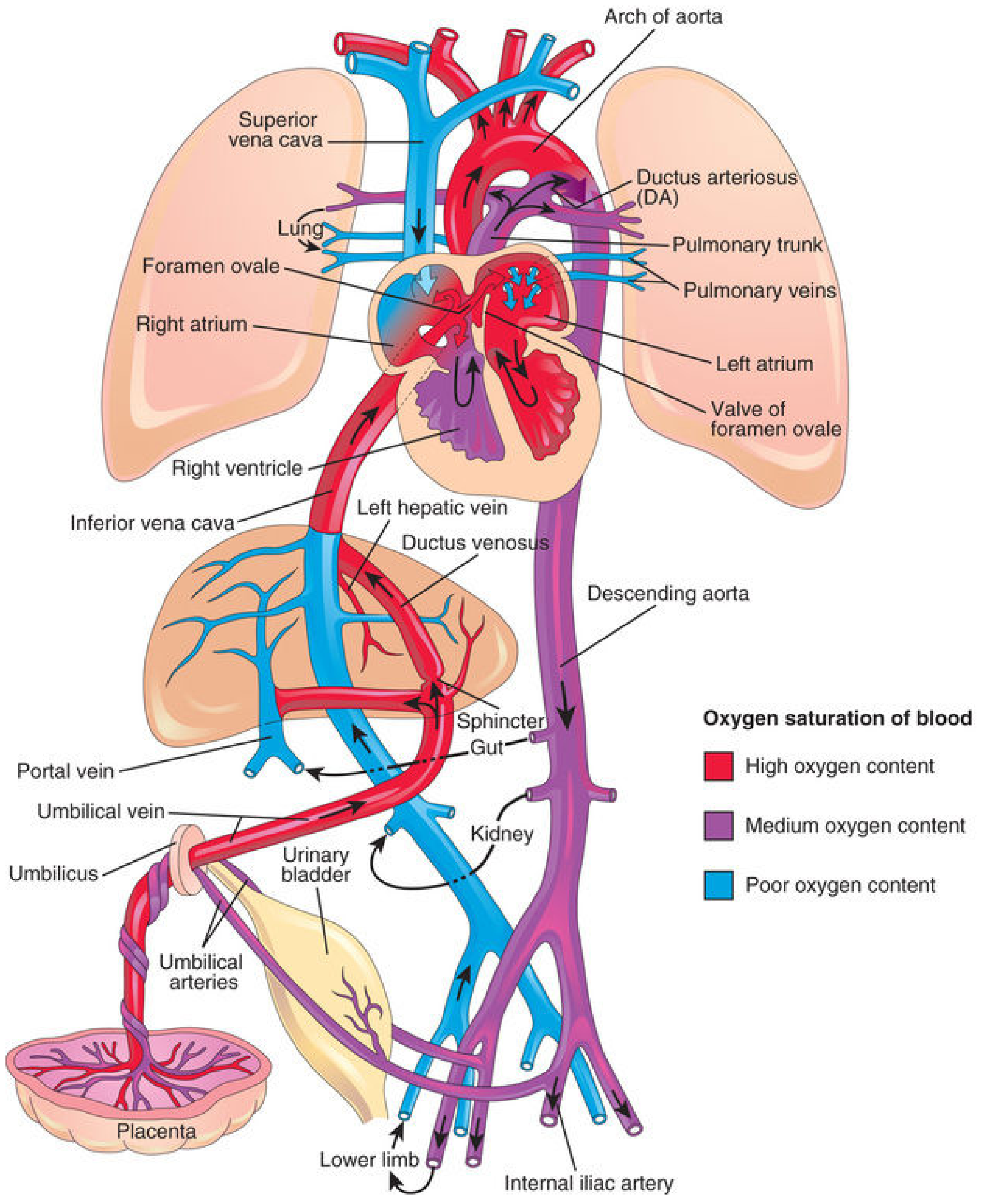
Fig. 13.46 - The Developing Human: Fetal circulation. Colors indicate oxygen saturation. Three shunts (ductus venosus, foramen ovale, ductus arteriosus) allow blood to bypass the liver and lungs.
Blood flow in fetal life:
- Highly oxygenated blood from placenta enters via the umbilical vein
- ~50% bypasses the liver via the ductus venosus directly into the IVC; the other 50% flows through liver sinusoids
- Blood from the IVC enters the right atrium; most is directed by the crista dividens through the foramen ovale into the left atrium
- From the left atrium → left ventricle → aorta → preferentially perfuses the brain and upper body
- Blood from the SVC and right atrium enters the right ventricle → pulmonary trunk
- Since pulmonary vascular resistance is HIGH (lungs collapsed, vessels vasoconstricted), most of this blood crosses the ductus arteriosus into the descending aorta
- Deoxygenated blood returns to placenta via two umbilical arteries (branches of internal iliac arteries)
Triggering Events at Birth
At birth, two pivotal changes occur almost simultaneously:
- Clamping of the umbilical cord / placental separation - removes the large low-resistance placental circuit, which doubles systemic vascular resistance, raising aortic, left ventricular, and left atrial pressures.
- Initiation of breathing / lung expansion - the lungs expand, pulmonary vessels are no longer compressed, and hypoxic pulmonary vasoconstriction is relieved. Pulmonary vascular resistance falls by as much as 5-fold, causing a massive increase in pulmonary blood flow.
Closure of the Three Shunts
1. Ductus Venosus
- With cord clamping, flow in the umbilical vein ceases
- The ductus venosus collapses due to loss of blood flow
- Functional closure: within hours of birth
- Anatomic closure: over the next few weeks
- Adult remnant: Ligamentum venosum (on the visceral surface of the liver)
2. Foramen Ovale
Mechanism:
- Increased pulmonary blood flow returns to the left atrium via pulmonary veins → raises left atrial pressure
- Simultaneously, reduced venous return to the right atrium (cord clamping) → lowers right atrial pressure
- This pressure reversal (LA > RA by 2-4 mmHg) pushes the flap-valve of the foramen ovale (derived from septum primum) against the septum secundum, causing functional closure
Timeline:
- Functional closure: with the first few breaths
- During a transitional phase, intermittent right-to-left flow may still occur
- Anatomical closure (fibrous fusion): over months to years; in 2/3 of people, permanent fusion occurs within months to a few years
- In ~20% of adults, the foramen ovale fails to fuse completely (patent foramen ovale, PFO), but remains functionally closed as long as LA pressure exceeds RA pressure
- Adult remnant: Fossa ovalis in the interatrial septum
3. Ductus Arteriosus
Mechanism:
- After birth, systemic vascular resistance rises (aortic pressure rises) while pulmonary vascular resistance falls (pulmonary artery pressure falls)
- Blood flow through the ductus reverses direction: from right-to-left (fetal) to left-to-right (aorta → pulmonary artery) transiently
- The key trigger for constriction is a rise in PaO₂: fetal ductal PO₂ is only 15-20 mmHg; after lung inflation, it rises to ~100 mmHg
- Increased O₂ directly contracts smooth muscle in the ductal wall, and also reduces production of prostaglandin E₂ (PGE₂) and prostacyclin (PGI₂), which were keeping the ductus open in fetal life
- Bradykinin released from inflated lungs also contributes to ductal smooth muscle contraction
- TGF-β mediates anatomic fibrotic closure
Timeline:
- Functional closure: By 24 hours - 20% closed; by 48 hours - ~80% closed; by 96 hours - 100% closed in full-term neonates
- Anatomic closure: 1-4 months after birth (fibrosis of lumen)
- Adult remnant: Ligamentum arteriosum
Clinical note: In premature neonates or with persistent hypoxia, the ductus remains open (Patent Ductus Arteriosus - PDA). Indomethacin (prostaglandin synthesis inhibitor) is used to pharmacologically close a PDA.
Umbilical Vessels
| Vessel | Fate after birth | Adult remnant |
|---|---|---|
| Umbilical vein | Obliterates | Ligamentum teres hepatis (round ligament of liver) |
| Umbilical arteries (proximal) | Persists | Superior vesical arteries |
| Umbilical arteries (distal) | Obliterates | Medial umbilical ligaments |
| Ductus venosus | Obliterates | Ligamentum venosum |
Neonatal Circulation Diagram
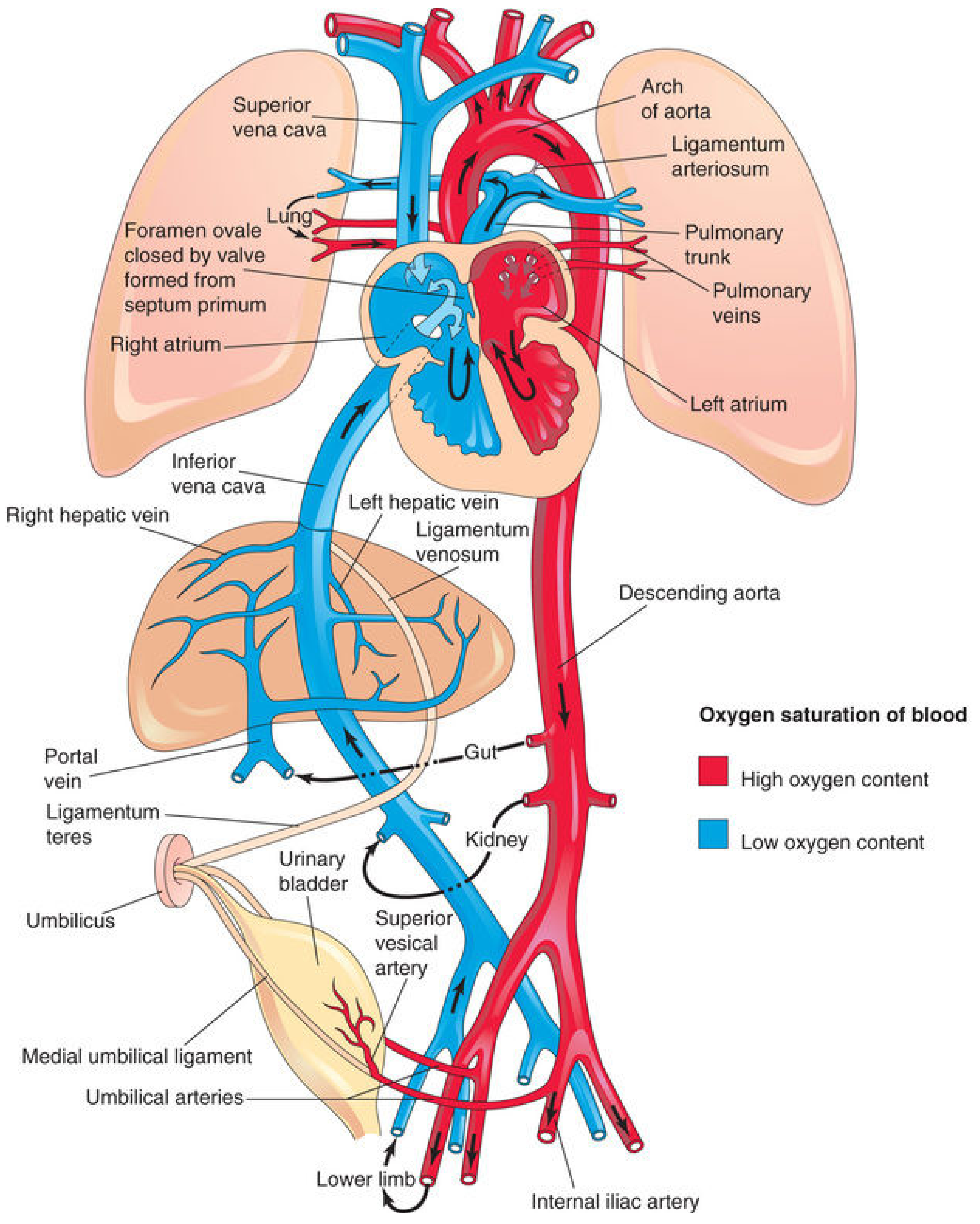
Fig. 13.47 - The Developing Human: Neonatal circulation. All three shunts are now closed. Right and left heart now pump in series.
Ventricular Wall Changes
In the fetus and neonate, the right ventricular wall is thicker than the left (because the RV pumps against high pulmonary vascular resistance). After birth, as pulmonary resistance falls and systemic resistance rises, the left ventricle begins doing more work. By the end of the first month, the LV wall becomes thicker than the RV wall, and the RV undergoes relative atrophy from its reduced workload.
Summary Table
| Feature | Fetal | Neonatal |
|---|---|---|
| Gas exchange site | Placenta | Lungs |
| Pulmonary vascular resistance | High | Low |
| Systemic vascular resistance | Low (placental) | High |
| Ductus venosus | Patent | Closes → ligamentum venosum |
| Foramen ovale | Open (R→L shunt) | Functionally closed (LA>RA) |
| Ductus arteriosus | Patent (R→L) | Closes → ligamentum arteriosum |
| Right & left heart | Pump in parallel | Pump in series |
| Umbilical vein | Carries O₂-rich blood | Obliterates → lig. teres |
Sources:
- The Developing Human: Clinically Oriented Embryology (Moore et al.), pp. 869-876
- Guyton and Hall Textbook of Medical Physiology, pp. (Changes in Fetal Circulation at Birth)
- Ganong's Review of Medical Physiology, 26th ed.
This is a shared conversation. Sign in to Orris to start your own chat.