
A 40 years old female presents with melasma from past 4 years
Reading File
Now let me search the medical library for melasma comprehensively.
Finding Sources
Searching PubMed
melasma treatment management
Excellent. Now let me read the most comprehensive melasma sections in parallel.
Reading File
Reading File
Let me read more of the Andrews section and the treatment section from Dermatology 2-Volume Set.
Reading File
Reading File
Let me get the clinical image and read more from the Fitzpatrick section on pathogenesis/treatment.
Loading Image
Reading File
I now have comprehensive information. Let me compose the full clinical answer.
Melasma - Clinical Overview
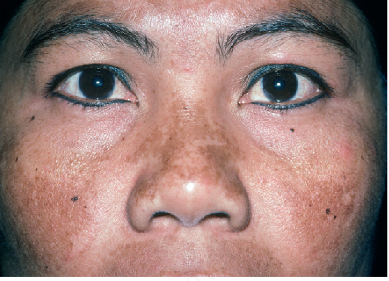
Classic melasma showing symmetric hyperpigmented patches over the malar prominences (Andrews' Diseases of the Skin)
Definition
Melasma (syn. chloasma, "mask of pregnancy") is a common acquired disorder of symmetric, hyperpigmented patches with irregular outlines, occurring most commonly on the face. It represents dysregulated melanogenesis driven by UV exposure and sex hormones, with a chronic relapsing course.
- Andrews' Diseases of the Skin, p. 993
- Dermatology 2-Volume Set 5e, p. 1343
Epidemiology & Risk Factors
| Factor | Detail |
|---|---|
| Sex | >90% female; F:M ~9:1 |
| Age | Young to middle-aged adults (peak 30s-40s) |
| Skin type | Fitzpatrick IV-V most affected; Hispanics, East/Southeast/South Asians, Black, Middle Eastern |
| Triggers | Sun exposure (primary), pregnancy, oral contraceptives (OCP), HRT |
| Other | Phenytoin, finasteride therapy, endocrinologic disorders |
In this 40-year-old female, key questions include OCP use, pregnancy history, sun exposure history, and Fitzpatrick skin type.
Pathogenesis
- UV exposure is the primary trigger: melasma affects sun-exposed areas, worsens in summer, patients have lower MED (minimal erythema dose), and solar elastosis is more marked in melasma-affected skin
- Wnt signaling: Melasma skin has reduced WIF-1 (Wnt antagonist) expression → increased Wnt → stimulates melanogenesis
- Hormonal influence: OCP, HRT, and pregnancy exacerbate via estrogen/progesterone-driven melanocyte stimulation; OCP-induced melasma often persists even after discontinuation
- Genetic predisposition: Correlation with number of melanocytic nevi
Andrews' Diseases of the Skin, p. 993
Classification
By Location (Clinical Patterns)
- Centrofacial - forehead, nose, upper lip, chin (most common)
- Malar - cheeks, nose
- Mandibular - jaw line
By Histological Depth
| Type | Melanin Location | Wood's Lamp | Treatment Response |
|---|---|---|---|
| Epidermal | Basal & suprabasal epidermis | Enhanced (accentuated) | Better |
| Dermal | Within melanophages | Not enhanced | Poor |
| Mixed | Both layers | Variable | Intermediate |
Note: Most cases show both epidermal and dermal melanin on histology and confocal microscopy - pure classification is unreliable. Dermal deposits respond poorly to topical agents.
Investigations / Workup
- Wood's lamp examination - helps broadly categorize (though unreliable for definitive typing)
- Dermatoscopy - useful to assess depth and rule out other pigmented lesions
- Dermoscopy findings: pseudonetwork of hyperpigmentation, telangiectasias in sun-damaged skin
- History: OCP use, pregnancy, sun habits, topical agents used
- Consider: ANA, thyroid antibodies (associated autoimmune conditions occasionally reported)
- Biopsy: Rarely needed; if diagnosis uncertain or exogenous ochronosis suspected
Differential Diagnosis
| Condition | Distinguishing Features |
|---|---|
| Drug-induced hyperpigmentation | Less patterned, less irregular outline; history of offending drug (doxycycline, amiodarone) |
| Post-inflammatory hyperpigmentation (PIH) | History of inflammation; may not follow facial distribution |
| Riehl melanosis (Pigmented contact dermatitis) | Sites of cosmetic contact; brown-grey, lichenoid on histology |
| Ota's nevus / Horii nevus | Asian women; grey-blue macules; dermal melanocytes on histology |
| Lichen planus pigmentosus | Temples/preauricular; coexistent LP lesions (~20%) |
| Erythema dyschromicum perstans | Slate-grey, involves sun-protected areas too |
| Exogenous ochronosis | Progressive darkening after HQ use; banana-shaped deposits on histology |
| Actinic lichen planus | Fine scale, violaceous, lichenoid infiltrate |
Treatment
All Patients (Foundation)
- Broad-spectrum sunscreen SPF ≥30 daily (with physical blockers: zinc oxide or titanium dioxide; iron oxide added to block visible light via makeup/sunscreen)
- Avoidance of sun exposure and tanning beds
- Sun-protective hats and clothing
- Discontinue OCP if possible
- Camouflage makeup with iron oxide
Dermatology 2-Volume Set 5e, p. 1343
First-Line Topical Therapy
Kligman's Formula (Triple Combination) - gold standard:
- Hydroquinone 4% + Tretinoin 0.05-0.1% + Mild topical corticosteroid (class 5-7)
- Applied at bedtime
- Used daily for 2-4 months, then reduced to 1-2x/week for maintenance
- Most effective topical regimen available
Alternatives/Adjuncts:
- Hydroquinone 4% alone (bedtime)
- Azelaic acid 15-20%
Cautions: Overuse of the triple combo → fixed erythema, telangiectasias, acneiform eruptions, hypertrichosis. Prolonged HQ use → exogenous ochronosis. Corticosteroid overuse → perioral dermatitis, atrophy.
Adjunctive Topical Therapies
| Agent | Concentration | Mechanism |
|---|---|---|
| L-ascorbic acid (Vitamin C) | 10-15% | Inhibits tyrosinase, antioxidant |
| Kojic acid | 1-4% | Inhibits tyrosinase |
| Tranexamic acid | 2-5% | Inhibits plasminogen activator in keratinocytes → reduces prostaglandin-driven melanogenesis |
| Niacinamide | 4% | Inhibits melanosome transfer |
Adjunctive Oral Therapy
Tranexamic acid 250 mg BID for 8-12 weeks
- Screen patients for thromboembolism risk factors before prescribing
- Mechanism: inhibits UV-induced plasminogen activator activity in melanocytes and keratinocytes, reducing melanin synthesis
Recent meta-analyses confirm efficacy: a 2024 systematic review and a 2025 meta-analysis of intradermal tranexamic acid both support tranexamic acid (oral and intradermal) as effective for melasma management with good safety profiles. A 2025 meta-analysis also supports microneedling combined with tranexamic acid.
Second-Line (Procedural)
Chemical Peels (every 4-6 weeks):
- Glycolic acid (start 30%, increase as tolerated)
- Salicylic acid 20-30%
- Note: Peels alone have not reproducibly enhanced 4% HQ efficacy and risk PIH if irritation occurs
Third-Line (Light & Laser)
| Modality | Notes |
|---|---|
| Intense Pulsed Light (IPL) | Can improve melasma but high relapse rate |
| Non-ablative fractional laser | Best laser option; 60% of skin types III-V achieve 75-100% improvement; improves HQ penetration |
| Pulsed dye laser | May enhance topical combination therapy |
| Picosecond lasers | Emerging evidence |
| Q-switched Nd:YAG (low fluence) | Risk of increased pigmentation/punctate leukoderma - use cautiously |
| Er:YAG | Temporary improvement followed by PIH - generally avoided |
Light-based therapies carry risk of PIH, hypopigmentation, and scarring in darker skin types (Fitzpatrick IV-V). Always do a test site first.
Long-Term Maintenance
- Daily broad-spectrum sunscreen (non-negotiable)
- Topical retinoid (continues to suppress recurrence)
- Topical alpha-hydroxy acid (e.g. glycolic acid cream)
- L-ascorbic acid, azelaic acid, kojic acid, or topical tranexamic acid
Prognosis & Counselling Points
- Melasma is a chronic, relapsing condition - treatment suppresses but does not cure
- OCP-induced melasma often persists despite discontinuation (unlike melasma of pregnancy, which usually clears within months of delivery)
- Dermal melasma responds much more poorly than epidermal type
- Realistic expectations are essential - partial improvement is more common than complete clearance
- Even a Wood's lamp "epidermal" pattern often has some dermal component on histology
- Sun protection alone can modestly improve melasma and is indispensable for any treatment to work
Dermatology 2-Volume Set 5e, p. 2859 | Andrews' Diseases of the Skin, p. 993-994
Summary for this patient: A 40-year-old female with 4 years of melasma is a classic presentation. Assess Fitzpatrick type, OCP use, sun exposure, and prior treatments. Start with rigorous photoprotection, Kligman's formula (if tolerated), and consider adding oral tranexamic acid 250 mg BID for 8-12 weeks (screen for VTE risk). If inadequate response after 3-6 months, escalate to chemical peels or non-ablative fractional laser. Counsel on chronic relapsing nature and need for maintenance therapy.
This is a shared conversation. Sign in to Orris to start your own chat.