
Anatomy of Knee
I'll look up the anatomy of the knee from the medical library for you.
Reading File
Now let me search the medical library for knee anatomy content in parallel.
Finding Sources
Finding Sources
Excellent sources found. Let me read the most comprehensive ones in parallel - Gray's Anatomy for Students, S Das Manual, Bailey & Love, and the THIEME Atlas.
Reading File
Reading File
Reading File
Reading File
Now let me read more from Gray's Anatomy (the most comprehensive anatomy source) and also fetch the key diagrams.
Reading File
Loading Image
Loading Image
Now let me fetch more key images and read more anatomy details from Gray's.
Reading File
Loading Image
Loading Image
Now let me read the ligaments and movements sections from Gray's Anatomy.
Reading File
Loading Image
I now have rich content. Let me read the cruciate ligament section and movements section to complete the picture.
Reading File
Loading Image
I now have comprehensive content from Gray's Anatomy for Students plus excellent diagrams. Let me compile the complete answer.
Anatomy of the Knee Joint
The knee is the largest synovial joint in the body. It is a complex hinge joint formed by two articulations sharing a single articular cavity:
- Tibiofemoral joint - weight-bearing articulation between femoral condyles and tibial plateau
- Patellofemoral joint - articulation between the patella and the anterior femoral trochlea, redirecting the pull of the quadriceps anteriorly
- Gray's Anatomy for Students, p. 709
1. Articular Surfaces
The articular surfaces are covered by hyaline cartilage and include:
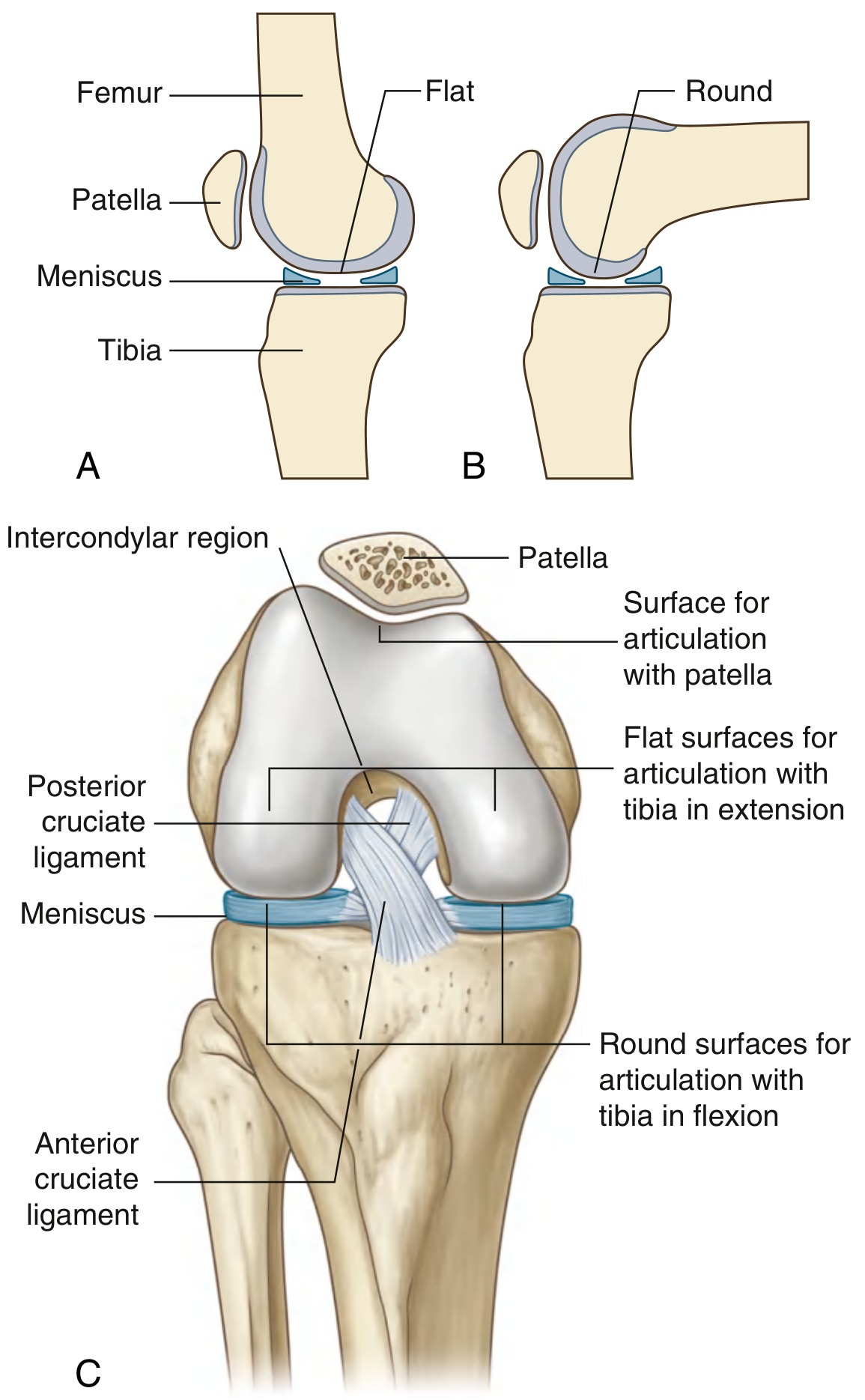
- Two femoral condyles - the surfaces are curved/round in flexion and become flat in full extension, accommodating the changing contact pattern during movement
- Superior tibial condyles (tibial plateau)
- Posterior surface of the patella - articulates with the V-shaped trochlear groove on the anterior distal femur
2. Menisci
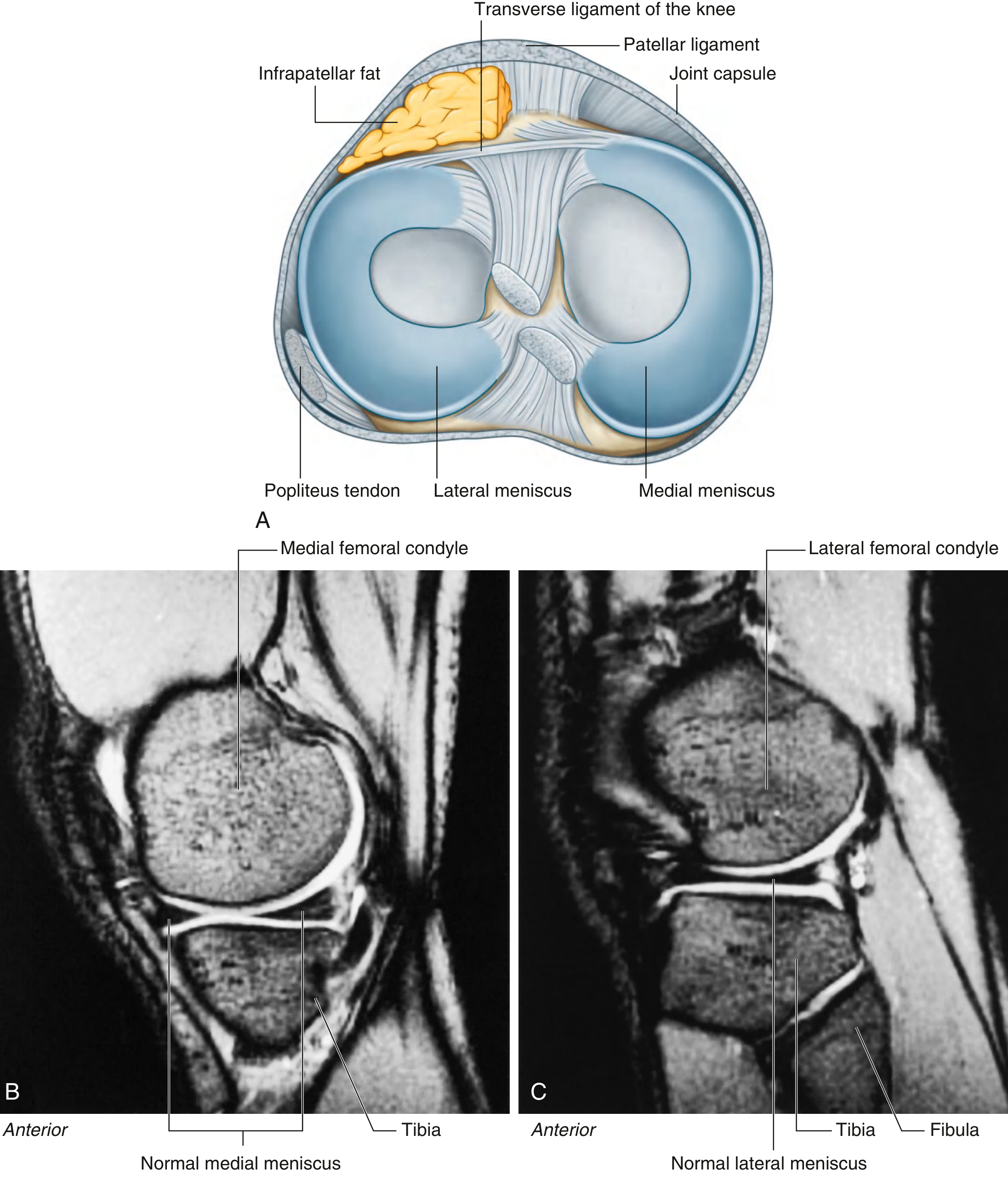
There are two fibrocartilaginous C-shaped menisci between the femoral condyles and the tibial plateau.
| Feature | Medial Meniscus | Lateral Meniscus |
|---|---|---|
| Shape | C-shaped, larger | Almost circular, smaller |
| Capsule attachment | Firmly attached to capsule & tibial collateral ligament | NOT attached to capsule |
| Mobility | Less mobile | More mobile |
| Injury risk | More commonly torn | Less commonly torn |
Both menisci are interconnected anteriorly by the transverse ligament of the knee. The lateral meniscus is connected to the popliteus tendon, which passes between it and the capsule.
Function: They improve congruency between femoral and tibial surfaces, act as shock absorbers, and facilitate lubrication.
3. Ligaments
Overview Diagram
Patellar Ligament
- The distal continuation of the quadriceps femoris tendon below the patella
- Runs from the apex and margins of the patella to the tibial tuberosity
- Functions as a pulley, increasing the lever arm of the quadriceps
Collateral Ligaments
Fibular (Lateral) Collateral Ligament (FCL/LCL):
- Cord-like structure
- Attaches from the lateral femoral epicondyle above to the lateral surface of the fibular head below
- Not attached to the lateral meniscus or capsule
- Taut in extension; resists varus stress
Tibial (Medial) Collateral Ligament (TCL/MCL):
- Broad and flat
- Attaches from the medial femoral epicondyle (just below adductor tubercle) to the medial surface of the tibia (above the pes anserinus)
- Deep surface blends with the fibrous capsule and attaches to the medial meniscus - hence medial meniscus injuries often accompany MCL tears
- Resists valgus stress
Cruciate Ligaments
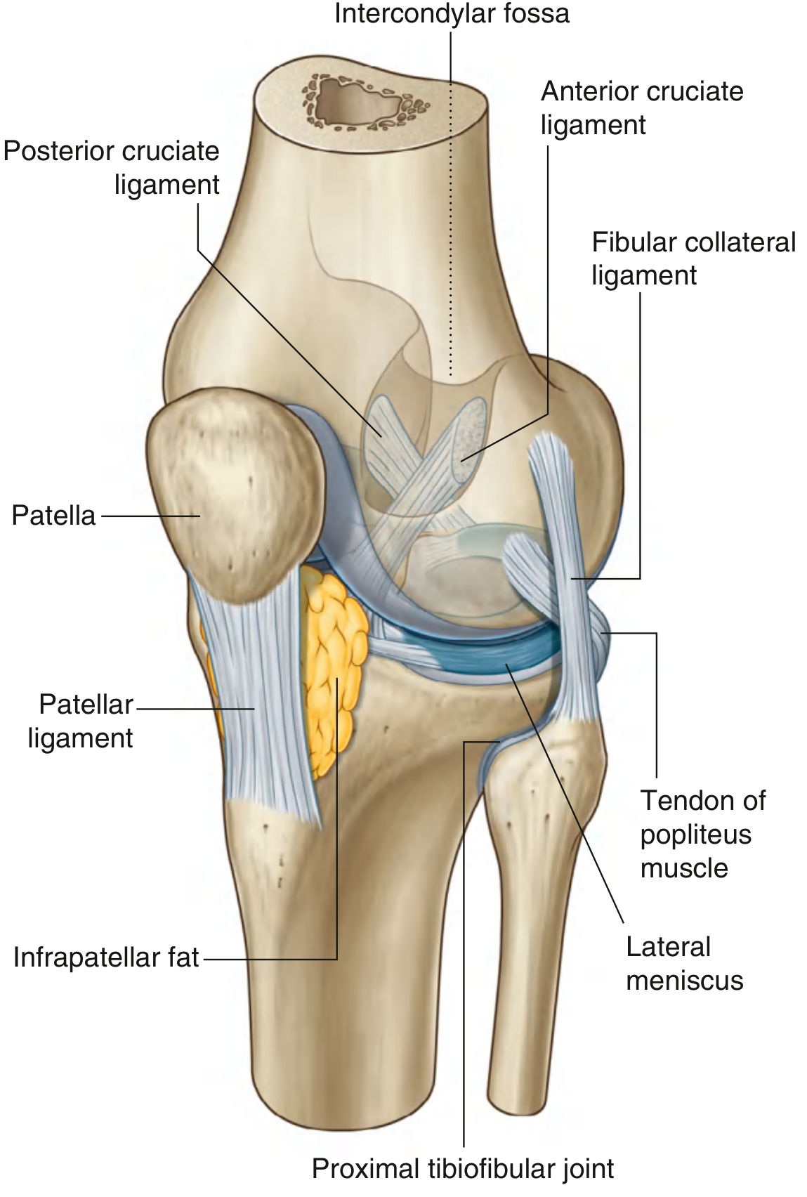
These are intra-articular but extra-synovial ligaments in the intercondylar region. They cross each other in the sagittal plane (hence "cruciate" = cross-shaped).
| ACL | PCL | |
|---|---|---|
| Tibial attachment | Anterior intercondylar area | Posterior intercondylar area |
| Femoral attachment | Posterior lateral wall of intercondylar fossa | Medial wall of intercondylar fossa |
| Direction | Passes posterosuperolaterally | Passes anterosuperiorly |
| Function | Prevents anterior tibial displacement; resists hyperextension | Prevents posterior tibial displacement |
| Clinical test | Anterior drawer test, Lachman test | Posterior drawer test |
The ACL crosses lateral to the PCL as they pass through the intercondylar region.
4. Joint Capsule
The fibrous membrane of the knee is extensive and reinforced by muscular extensions:
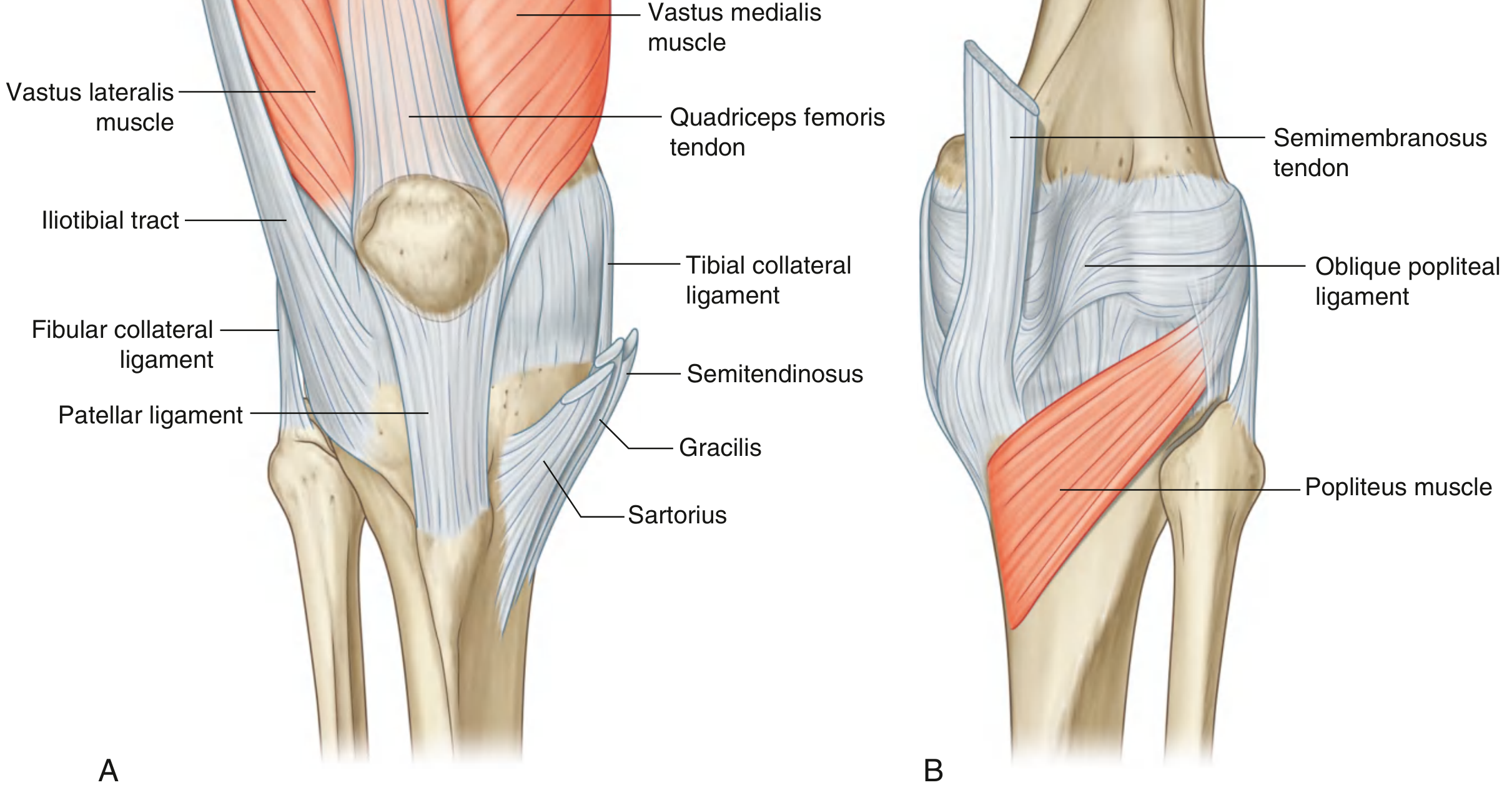
- Anteriorly: reinforced by the quadriceps expansion (vastus medialis and lateralis), which merges with the patellar ligament
- Anterolaterally: reinforced by the iliotibial tract
- Posteromedially: reinforced by the oblique popliteal ligament (a reflection of semimembranosus tendon)
- Posterolaterally: also reinforced by the arcuate popliteal ligament
The popliteus tendon passes through an aperture in the posterolateral capsule.
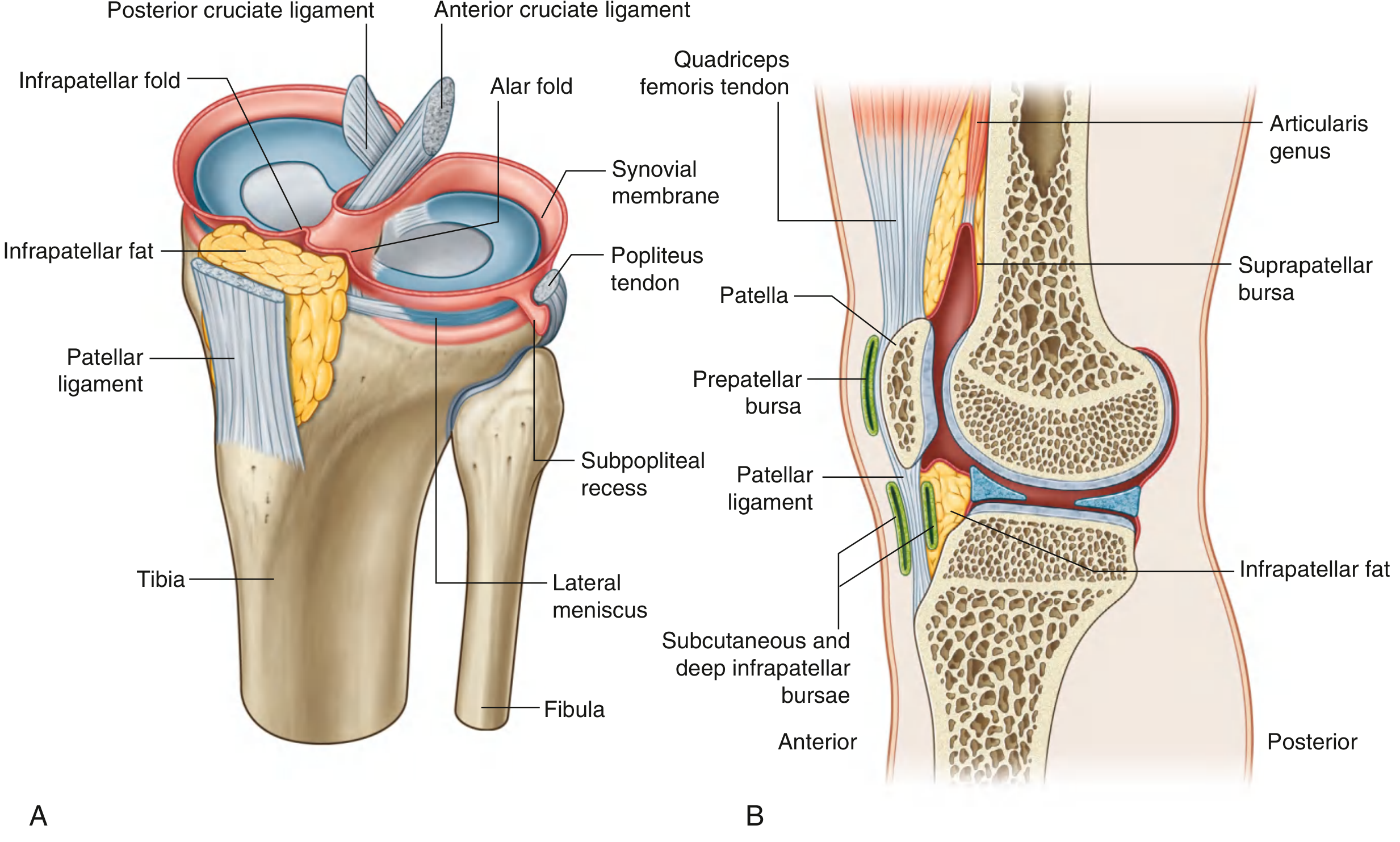
5. Synovial Membrane and Bursae
The synovial membrane lines the fibrous capsule and attaches to the margins of articular surfaces and menisci. The cruciate ligaments lie outside the articular cavity but inside the fibrous membrane.
Key Bursae:
| Bursa | Location | Communication with Joint |
|---|---|---|
| Suprapatellar bursa | Between distal femur and quadriceps tendon | Yes (continues with articular cavity) |
| Subpopliteal recess | Between popliteus tendon and lateral meniscus | Yes |
| Prepatellar bursa (subcutaneous) | Anterior to patella | No |
| Deep infrapatellar bursa | Deep to patellar ligament | No |
| Subcutaneous infrapatellar bursa | Superficial to patellar ligament | No |
| Semimembranosus bursa | Posteriorly | Sometimes |
The articularis genus muscle (a small muscle from the distal femur) attaches to the suprapatellar bursa to pull it away from the joint during extension.
6. Posterior Knee (Popliteal Fossa)
The posterior capsule is strengthened by the oblique popliteal ligament and arcuate popliteal ligament, plus attachments from gastrocnemius heads.
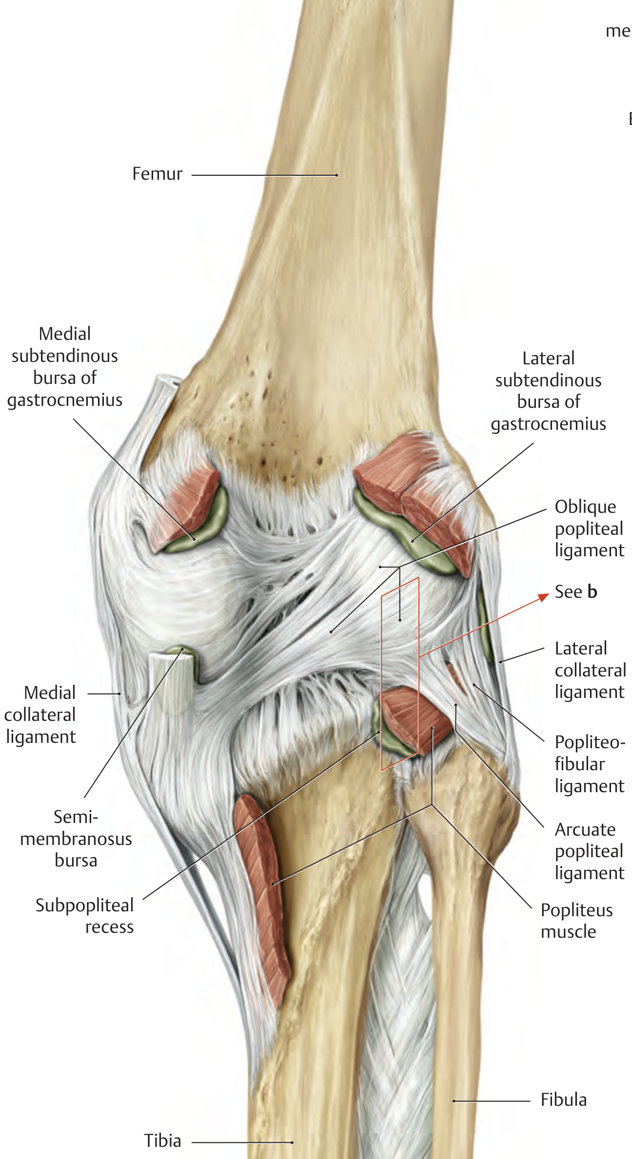
Key posterior structures:
- Oblique popliteal ligament - extension of semimembranosus tendon running superomedial to superolateral
- Arcuate popliteal ligament - arches over the popliteus muscle
- Popliteofibular ligament - connects popliteus tendon to fibular head
- Popliteus muscle - "unlocks" the fully extended knee by internally rotating the tibia
7. Movements
The knee is primarily a hinge joint allowing:
| Movement | Range | Muscles |
|---|---|---|
| Extension | 0° (neutral) to 5° hyperextension | Quadriceps femoris |
| Flexion | 0° to 135° | Hamstrings (biceps femoris, semitendinosus, semimembranosus), gastrocnemius |
| Medial rotation (of tibia, when flexed) | Limited | Popliteus, semitendinosus, gracilis, sartorius |
| Lateral rotation (of tibia, when flexed) | Limited | Biceps femoris |
Locking Mechanism ("Screw Home")
At full extension, the femur medially rotates on the fixed tibia (or the tibia laterally rotates on the femur), which "locks" the knee into a close-packed, stable position. This reduces the muscular energy needed to maintain standing. Popliteus is the key muscle that "unlocks" the knee by laterally rotating the femur (internally rotating the tibia).
8. Blood Supply
- Genicular anastomosis - a network formed by branches of:
- Femoral artery (descending genicular)
- Popliteal artery (superior medial & lateral, inferior medial & lateral, and middle genicular)
- Anterior tibial recurrent artery
- Circumflex fibular artery
9. Nerve Supply
- Femoral nerve (via branches to vastus muscles) - anterior
- Obturator nerve - medial (also supplies hip, explaining referred pain)
- Common fibular (peroneal) nerve - lateral and posterior
- Tibial nerve - posterior
10. Clinical Correlates
| Condition | Anatomy Involved |
|---|---|
| ACL tear | Most common sports injury; anterior drawer test / Lachman test positive |
| Medial meniscus tear | Medial meniscus is fixed to capsule; bucket-handle tear causes locking |
| MCL tear (valgus stress) | Tibial collateral ligament; may also tear medial meniscus (O'Donoghue triad with ACL) |
| Prepatellar bursitis ("Housemaid's knee") | Inflammation of subcutaneous prepatellar bursa |
| Baker's cyst | Distension of semimembranosus bursa posteriorly |
| Unhappy triad | ACL + MCL + medial meniscus (from valgus + rotation force) |
Sources:
- Gray's Anatomy for Students, 4th Ed., pp. 709-720
- THIEME Atlas of General Anatomy and Musculoskeletal System, pp. 462-465
- Bailey and Love's Short Practice of Surgery, 28th Ed., p. 4162
- Campbell's Operative Orthopaedics, 15th Ed., 2026
This is a shared conversation. Sign in to Orris to start your own chat.