
Unit structures of superior mediastinum
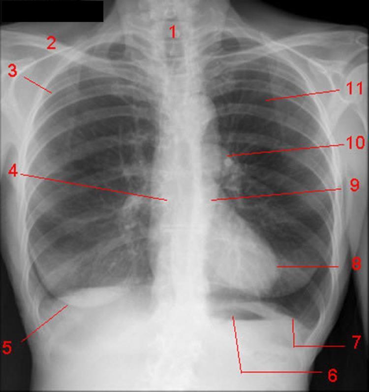
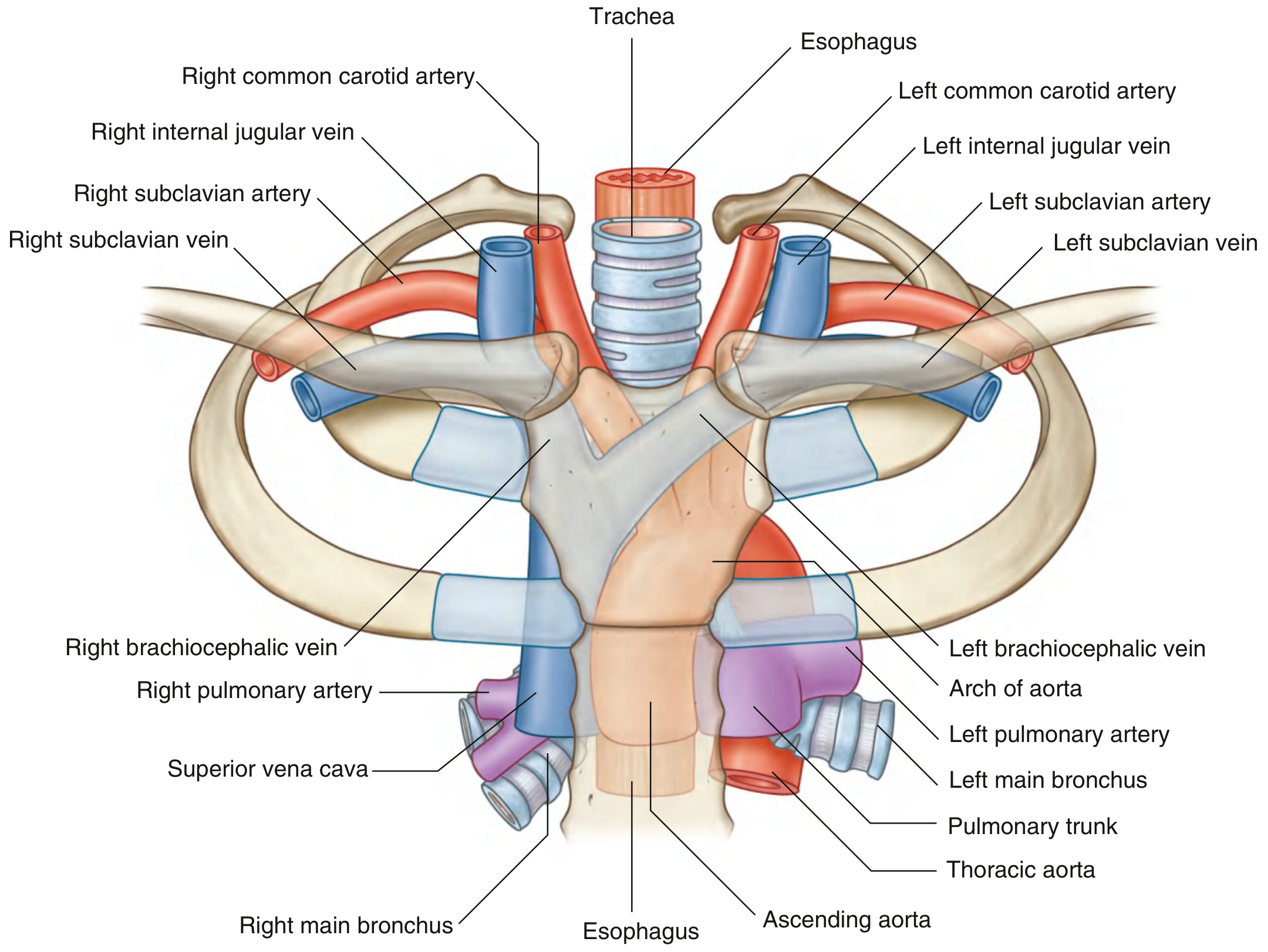
Superior Mediastinum – Contents and Arrangement
Boundaries (for context): posterior to the manubrium of the sternum, anterior to the bodies of vertebrae T1-T4; superiorly it opens into the neck via an oblique plane from the jugular notch to the superior border of T1; inferiorly a transverse plane from the sternal angle to the T4/T5 disc separates it from the inferior mediastinum; laterally bounded by mediastinal parietal pleura on each side.
Major structures
| Category | Structures |
|---|---|
| Lymphoid organ | Thymus (most anterior structure) |
| Veins | Right and left brachiocephalic veins, left superior intercostal vein, superior vena cava (SVC) |
| Arteries | Arch of the aorta and its three large branches (brachiocephalic trunk, left common carotid artery, left subclavian artery) |
| Airway/foregut | Trachea, esophagus |
| Nerves | Both phrenic nerves, both vagus nerves, left recurrent laryngeal nerve (branch of the left vagus) |
| Lymphatics | Thoracic duct |
| Other | Small nerves, blood vessels, and lymphatics |
General anteroposterior layering
Working from front to back, the superior mediastinum is organized roughly as:
- Thymus - most anterior
- Great veins - right and left brachiocephalic veins uniting to form the SVC
- Arterial layer - arch of the aorta with its three branches
- Nerves related to the arch - phrenic and vagus nerves run along the great vessels; the left recurrent laryngeal nerve hooks under the aortic arch
- Airway - trachea, in the midline
- Esophagus - just posterior to the trachea, with the thoracic duct ascending along its left side toward the neck
Clinically relevant relationships
-
The left recurrent laryngeal nerve loops under the aortic arch just lateral to the ligamentum arteriosum, making it vulnerable in aortic arch pathology or surgery (hoarseness from nerve injury).
-
The thoracic duct passes through the superior mediastinum ascending along the left side of the esophagus before arching to drain into the left subclavian/internal jugular venous junction - injury here causes chylothorax.
-
The SVC is formed by the union of the two brachiocephalic veins in the superior mediastinum; compression here (e.g., by tumor) produces SVC syndrome.
-
The thymus, being the most anterior structure, is the classic site of anterior mediastinal masses (thymoma) that can compress underlying great vessels.
-
Gray's Anatomy for Students (9780323934237), Superior Mediastinum section, p. 251-252
If you'd like, I can go deeper into any specific structure (e.g., relations of the aortic arch, the thymus in different age groups, or nerve pathways) or pull additional labeled diagrams.
Analyse the previous year questions of RGUHS Anatomy question papers and suggest the most expected questions from mediastinum
Searching the Web
RGUHS MBBS anatomy previous year question papers mediastinum thorax
Searching the Web
RGUHS first year MBBS anatomy question paper PDF "mediastinum" OR "thoracic duct" OR "superior mediastinum"
Reading Web Page
https://www.rguhs.ac.in/Exam/2025/qp%20blueprint.pdf
Searching the Web
RGUHS anatomy question bank thorax "mediastinum" long essay short essay list repeated questions
Reading Web Page
https://anatomyqa.com/important-exam-questions-thorax-anatom…
RGUHS Anatomy - Mediastinum: Most Expected Questions (Based on PYQ Pattern Analysis)
What the analysis is based on
- RGUHS official Question Paper Blueprint (2024-25 batch, issued 2025) - confirms Thorax including Diaphragm carries 20 marks in Anatomy Paper I (out of 100), one of the higher-weighted systems after Head & Neck (27) and Upper Limb (17).
- RGUHS exam pattern breakdown (faculty-reported, Dec 2024 supplementary paper): Thorax typically contributes 1 Long Essay (10 marks) + short essays/short answers, and mediastinum/heart is explicitly called out as the recurring "either-or" long essay theme for Thorax across paper versions.
- Cross-referencing actual PYQ snippets (2019-2023 RGUHS papers) which list "Posterior mediastinum" and "Inlet of thorax" as questions that have appeared.
- Standard Indian MBBS anatomy question banks aligned to this pattern show mediastinum content recurring as both long-essay and short-note items every cycle.
Exam pattern for Thorax (as per current blueprint)
- MCQs: 20 marks total (case-scenario based, last 10 questions of the paper) - mediastinum contents/relations are classic MCQ fodder (e.g., "structure NOT part of superior mediastinum," "nerve at risk during mediastinal mass excision")
- Remainder of Thorax's 20 marks split across 1 Long Essay (10 marks) and 1-2 Short Essays/Short Answers (5 marks each)
- Mediastinum competes with heart, lungs, pleura, and diaphragm for this space, but it is consistently one of the 2-3 most repeated themes
Most expected questions from Mediastinum
Long Essay (10 marks) - highest probability
- Describe the superior mediastinum - boundaries, contents (thymus, great veins, arch of aorta and branches, trachea, esophagus, phrenic/vagus nerves, thoracic duct), and applied anatomy
- Describe the posterior mediastinum - boundaries, contents (esophagus, descending thoracic aorta, azygos/hemiazygos veins, thoracic duct, sympathetic trunk, splanchnic nerves), applied anatomy
- Describe the thoracic duct - origin (cisterna chyli), course through posterior and superior mediastinum, tributaries, termination, applied aspect (chylothorax, thoracic duct injury)
Short Essay / Short Note (5 marks) - very likely
- Contents of the superior mediastinum (list + brief description)
- Contents of the posterior mediastinum
- Azygos system of veins (course, tributaries, communication with hemiazygos, clinical relevance in SVC obstruction as collateral pathway)
- Boundaries and subdivisions of the mediastinum (superior, anterior, middle, posterior - the classic four-part division)
- Arch of the aorta - branches and relations within the superior mediastinum
- Inlet/root of the neck and its continuity with the superior mediastinum (has appeared as a direct PYQ topic)
Short Answer / Applied Anatomy (2-3 marks or viva-style)
- Left recurrent laryngeal nerve - course and why it is vulnerable in aortic arch pathology (hoarseness)
- Formation of the superior vena cava and SVC syndrome
- Relations of the esophagus in the posterior mediastinum, constrictions of the esophagus
- Thymus - location, age changes, and relevance to anterior mediastinal masses
MCQ-style facts (20 marks pool, case-scenario based)
- Vertebral levels marking mediastinal boundaries (T1, sternal angle/T4-T5 disc)
- Which structures are NOT part of a given mediastinal compartment
- Nerve/vessel injured in specific clinical scenarios (mediastinal mass, aortic aneurysm, penetrating chest trauma)
Study priority suggestion
Given the marks weightage and repetition pattern, prioritize in this order:
- Superior mediastinum contents and boundaries (most repeated long essay/short essay topic)
- Thoracic duct (course spans posterior + superior mediastinum, frequently asked standalone)
- Posterior mediastinum contents
- Azygos venous system
- Applied/clinical one-liners (recurrent laryngeal nerve, SVC syndrome, chylothorax)
Caveat: This is a probability-based estimate from blueprint weightage, faculty commentary on recent papers, and recurring PYQ themes, not an official leak or guarantee of the next paper. RGUHS explicitly states topic distribution is "indicative only" and can vary. Use this to prioritize revision, not to skip other Thorax topics (heart, lungs, pleura, diaphragm) which share the same 20-mark pool.
This is a shared conversation. Sign in to Orris to start your own chat.